Bacterial DNA-recognition and DNA-modifying systems provide ways to mark, map and perturb eukaryotic chromosomes. Their value comes from separating the targeting module from the reporter or effector, so a chromosome becomes experimentally observable without assuming that its sequence alone specifies its behaviour.
For fluorescent operator-repressor chromosome labelling, an integrated array of bacterial operator sequences is bound by a fluorescent repressor fusion. A LacI fusion bound to a lac operator array makes a selected genetic locus visible in a living nucleus. Two differently labelled loci can report separation of sister chromatids, movement relative to the nuclear periphery, or distributions of distances between chromosome regions. Carmen C. Robinett and colleagues demonstrated operator-based imaging in mammalian cells and yeast. The tag reports the marked locus, not every part of the chromosome, and repeated operators or excessive bound repressor can perturb DNA replication and chromatin structure.[ref]
Lac-operator chromosome imaging study by Carmen C. Robinett and others. Live GFP-repressor imaging and targeted yeast insertion
The bacterial CRISPR-Cas9 recognition mechanism makes targeting programmable. A fluorescent dCas9 fusion and a guide RNA can visualize native repeated sequences; multiple guides can improve detection of a nonrepetitive locus. Baohui Chen and colleagues used this approach to follow human genetic loci. Catalytically inactive targeting avoids making the DNA double-strand break that would itself alter chromosome behaviour. Appropriate controls include guide specificity, signal from an unlabelled locus, and whether tagging changes growth or the measured dynamics.[ref]
CRISPR imaging of human genomic loci by Baohui Chen and others. Live imaging of endogenous human loci
For DamID, bacterial Dam methyltransferase is fused to a nuclear protein. Methylated GATC sites record neighbourhoods visited by the fusion. A fusion to a nuclear-envelope component can identify lamina-associated domains, whereas one to a chromatin-binding protein maps its genomic neighbourhoods. Comparing with untethered Dam helps control accessibility-dependent background. The accumulated methylation signal is a time-integrated proximity record: it need not establish direct binding to every recovered sequence, and resolution depends on GATC distribution and local spreading.[ref]
Bacterial enzymes also make structural measurements possible. In Hi-C, restriction enzymes generate DNA fragments whose proximity-dependent ligation junctions can be sequenced. In ATAC-seq, a modified bacterial Tn5 transposase inserts sequencing adapters preferentially into accessible DNA. Neither assay directly photographs a fixed chromosome structure: contact frequency and accessibility are different observables. Enzyme sequence preferences, fixation, and cell-to-cell heterogeneity must be controlled. Together, live labelling, proximity recording and sequencing-based assays connect chromosome movement, nuclear neighbourhood and chromatin accessibility, rather than substituting one measurement for all three.
The first task is to map the DNA associated with the centromeric histone variant CENP-A; the second is to determine how that occupancy is maintained. These require different evidence. A shared DNA sequence is not sufficient evidence for an inherited centromeric state.
Use validated anti-CENP-A chromatin immunoprecipitation sequencing with input DNA and an appropriate negative immunoprecipitation control. Check antibody recognition separately in human, horse and chicken; an endogenous epitope-tagged allele can provide an orthogonal control if its chromosome-segregation function is retained. Native digestion can recover CENP-A-containing nucleosomes, whereas crosslinking can retain indirect associations. CUT&RUN provides a complementary occupancy assay with different recovery biases. Establish centromeric function by colocalization with kinetochore proteins and by normal chromosome segregation, not merely by calling a sequencing peak.
Mapping repetitive DNA is the main analytical obstacle. Use centromere-resolved assemblies where possible, retain information from multimapping reads, and analyse satellite-family enrichment as well as uniquely mappable sequence. Absence of a peak in an incomplete reference is not evidence of absent CENP-A. Long-read sequence assemblies, informative single-nucleotide polymorphisms, and DNA fluorescence in situ hybridization can connect a recovered sequence to a particular chromosome and distinguish homologues.
For precise positioning, compare enriched domains with input-normalized background at several resolutions. Map nucleosome-protected fragments and use fibre immunofluorescence or sequence probes to check domain extent and heterogeneity. A broad population peak may represent alternative positions in different cells or homologues, rather than one broad CENP-A domain on every chromosome.
The expected outcome is a common centromeric function without a universal centromeric DNA sequence. Human CENP-A commonly occupies selected portions of alpha-satellite arrays, but human neocentromeres show that such arrays are not obligatory. Horse chromosome 11 provides a normal satellite-free centromere. Chicken chromosomes include both repeat-rich centromeres and comparatively short non-tandem-repetitive centromeric regions.[ref]
Chickens possess centromeres with both extended tandem repeats and short non-tandem-repetitive sequences by Wei-Hao Shang and others. Comparative chicken centromere mapping
[ref]Birth, evolution, and transmission of satellite-free mammalian centromeric domains by Solomon G. Nergadze and others. Satellite-free mammalian centromeres and inherited positional variation
To test mitotic stability, start from single-cell clones and remap CENP-A on the same haplotype after successive divisions, while checking that the underlying DNA has not changed. Pulse labelling of old versus newly incorporated CENP-A can distinguish retention from replenishment. Follow both chromosome identity and domain position: stable centromeric function does not require an invariant nucleotide boundary. To test meiotic transmission, use informative pedigrees or gamete-and-offspring material and allele-resolved mapping, with matched cell types where possible. An inherited positional state, or bounded movement of that state on unchanged DNA, supports epigenetic centromere inheritance. Artificial centromere deletion and neocentromere recovery provide an additional perturbation test, but establishment after deletion is different from ordinary meiotic inheritance.[ref]
Chromosome Engineering Allows the Efficient Isolation of Vertebrate Neocentromeres by Wei-Hao Shang and others. Chicken neocentromere formation and CENP-A domain mapping
Essential cell cycle genes cannot generally be discovered by selecting viable complete deletions alone. A temperature-sensitive mutation solves this problem: its product supports growth under permissive conditions but fails under restrictive conditions. Screen conditional mutants for a reproducible division defect, then distinguish a specific cycle block from nonspecific loss of viability. A cdc mutant is defined by its cycle phenotype, not simply by poor growth at high temperature.
In Saccharomyces cerevisiae, bud size and nuclear morphology provide convenient stage markers. After shifting an asynchronous culture, mutants defective at a particular step tend to accumulate with a characteristic morphology. Leland H. Hartwell and colleagues used time-lapse observations to distinguish the time a function must act from the stage at which its absence becomes visible. Temperature-shift timing, DNA-content measurements and recovery after return to permissive conditions strengthen the interpretation. A delayed terminal phenotype need not identify the moment of the original molecular defect.[ref]
Genetic control of the cell-division cycle in yeast. I. Detection of mutants by Leland H. Hartwell and others. Conditional mutants, execution points and termination phenotypes
In Schizosaccharomyces pombe, elongated cells that continue growing without dividing are especially useful candidates; nuclear number, septum formation and DNA content separate blocks in replication, mitosis and division. Paul Nurse, Pierre Thuriaux and Kim Nasmyth isolated conditional mutants defining multiple essential functions. Complementation testing groups recessive mutations affecting the same function, and segregation analysis distinguishes linked from independent loci. Molecular identification, rescue and biochemical tests then establish what the affected products do.[ref]
Genetic control of the cell division cycle in the fission yeast Schizosaccharomyces pombe by Paul Nurse and others. Conditional mutants affecting DNA synthesis, nuclear division and septation
Three contrasting examples explain why similar-looking division blocks need not have the same cause. In fission yeast, Cdc2, the cyclin-dependent kinase 1 catalytic subunit, is required for key cycle transitions. A restrictive loss-of-function allele can leave cells growing without entering mitosis; some alleles also reveal the earlier requirement at Start. Cdc25 is an activating protein phosphatase that removes inhibitory phosphorylation from Cdc2. Loss of Cdc25 therefore leaves the mitotic kinase inadequately activated and commonly produces an elongated G2-arrested phenotype. In budding yeast, Cdc6 participates in DNA replication licensing: failure to establish competent origins prevents successful genome duplication and hence normal division. Species and allele must be stated: identically numbered CDC names in different yeasts are not automatically homologous proteins.
The importance for cancer is that an unbiased genetic screen exposed conserved mechanisms connecting growth, DNA duplication and division. The yeast cross-species complementation experiment showed that central regulatory functions could be conserved across large evolutionary distances. Human cancers can alter cyclin-CDK regulation, DNA-replication control and checkpoint responses, providing a mechanistic connection to uncontrolled proliferation and genomic instability. This does not mean that every yeast cdc gene is a human oncogene, nor that any observed arrest is necessarily a checkpoint response: direct loss of an essential activity can itself stop progression.
Both checkpoints couple cell-cycle timing to spatial organization, but they act at different transitions. The budding-yeast spindle position checkpoint prevents premature mitotic exit when the anaphase spindle has not been appropriately placed across the mother-bud axis. The fly centrosome orientation checkpoint delays entry into mitosis until interphase centrosomes have the orientation needed for a properly oriented division.
In Saccharomyces cerevisiae, one product of division must enter the bud. If both spindle poles and chromosome masses remain in the mother, cytokinesis would generate the wrong nuclear distribution. The mitotic exit network promotes sustained activation of Cdc14 and the decline of mitotic CDK activity. Its upstream GTPase Tem1 is restrained by the Bub2-Bfa1 complex. When the spindle is mispositioned, Kin4 helps preserve that inhibition and opposes the checkpoint-inactivating action of Cdc5 on Bfa1. Thus spindle mispositioning is connected to continued mitotic arrest rather than simply to an attempt to pull harder on chromosomes.[ref]
The Protein Kinase Kin4 Inhibits Exit from Mitosis in Response to Spindle Position Defects by Katharine E. D'Aquino and others. Kin4-dependent inhibition of mitotic exit
Spatial regulation makes this pathway sensitive to which compartment a spindle pole occupies. Kin4 activity is associated with the mother-cell side, whereas the bud environment favours release of the inhibitory state when a pole enters appropriately. Elm1 contributes to activation of Kin4. This is a regulated network involving localization and phosphorylation, rather than an infallible single switch at the bud neck. It is also distinct from the spindle checkpoint, which monitors kinetochore attachment and restrains anaphase; a spindle can have attached chromosomes yet be in the wrong position.[ref]
Elm1 kinase activates the spindle position checkpoint kinase Kin4 by Ayse Koca Caydasi and others. Kin4 activation and regulation of Bub2-Bfa1
In male Drosophila melanogaster germline stem cells, contact with the hub defines the stem-cell niche. Normally one centrosome is retained toward the hub and the other moves away, preparing a division in which one daughter retains niche contact. Live imaging showed that misoriented centrosomes can delay division until orientation is restored, including in aged or dedifferentiated cells. Counting misoriented interphase centrosomes alone therefore overestimates the frequency of misoriented mitoses.[ref]
Centrosome misorientation reduces stem cell division during aging by Jun Cheng and others. Interphase misorientation, delayed cycling and reorientation before mitosis
The fly checkpoint involves Centrosomin, Par-1, and regulation of cyclin A localization. Par-1 and the germline spectrosome help restrain the mitotic programme; perturbing this organization can uncouple orientation from mitotic entry. Bazooka provides a cortical platform associated with centrosome anchoring. These observations support spatial control of mitotic competence, not identity of the molecular pathway with yeast Tem1 signalling.[ref]
Cyclin A localization and the centrosome orientation checkpoint by Hebao Yuan and others. Par-1, spectrosome-associated cyclin A and checkpoint defects
[ref]A decisive experimental comparison measures both position and transition timing in live cells. A pure orientation defect raises misorientation and prolongs the relevant pause; loss of the checkpoint permits the transition despite misorientation. Rescue with an appropriate wild-type component helps establish specificity. In yeast, score mitotic exit and nuclear inheritance; in fly stem cells, score entry into mitosis and daughter contact with the niche. This separates the mechanism that constructs the correct geometry from the mechanism that waits for it.
Genomic instability means an elevated propensity to acquire genetic alterations. It includes changes in nucleotide sequence, repeat length, chromosome structure and chromosome number. A genome carrying one fixed alteration is not, by that fact alone, genomically unstable: instability concerns the process that generates further alterations.
At the sequence level, DNA replication introduces mismatches and small insertion-deletion errors. Polymerase proofreading and DNA mismatch repair normally suppress their fixation. Failure of mismatch repair is particularly evident as microsatellite instability. Damaged bases, stalled replication forks and collisions with transcription can increase replication stress; unrepaired or improperly repaired lesions can produce substitutions and rearrangements. Homologous recombination and non-homologous end joining solve different repair problems, and inappropriate use or incomplete repair can alter genome structure.
At the chromosome level, failed chromosome-end protection can allow end fusion. Subsequent separation of fused chromosomes can initiate breakage-fusion-bridge cycles, producing repeated rearrangement and copy-number change. Faulty kinetochore attachment, weakened sister chromatid cohesion or abnormal spindle organization can cause chromosome missegregation and aneuploidy. The resulting imbalances may themselves disturb cell physiology and DNA replication. DNA confined in an aberrant micronucleus can experience defective replication and fragmentation; repair of clustered fragments is one route to chromothripsis, rather than every rearrangement arising through that mechanism.[ref]
Chromothripsis from DNA damage in micronuclei by Cheng-Zhong Zhang and others. Live-cell tracking linked to sequencing of descendants
The experimental signatures distinguish mechanisms. Mutation spectra and mismatch-repair status address sequence errors; repeat-length distributions address microsatellite instability; karyotyping, copy-number profiles and live chromosome imaging address numerical or structural chromosome instability. Single-cell or lineage-resolved measurements can show ongoing production of new states. A mixed endpoint population alone cannot establish whether variation arose rapidly during the experiment or was selected from pre-existing diversity.
In cancer, instability can generate selectable combinations that promote growth, escape from controls or resistance to treatment. It is neither automatically beneficial to the tumour nor sufficient for malignant transformation: excessive instability can impair proliferation or trigger apoptosis and cellular senescence. Examples of mechanistic connections include mismatch-repair deficiency in Lynch syndrome, homologous-recombination defects involving BRCA1 or BRCA2, and inherited replication or repair disorders. Chromosome missegregation can also contribute to developmental disease, pregnancy loss and infertility; instability in long-lived somatic tissues can impair function without producing a tumour.
A useful causal analysis asks what fails first, what alterations it produces, and which cellular responses restrain or select the affected cells. Restoring a repair factor or correcting a segregation defect should reduce the appropriate new-error rate, not merely change which clone grows best. The same reasoning explains why a genomic-instability phenotype can be informative in disease while remaining insufficient to diagnose a particular condition from morphology alone.
Susceptibility of an isolated bacterium and failure to clear an infection measure different things. The scenario does not establish a particular resistance gene or a real species-specific mechanism. First distinguish antibiotic resistance, an increased ability to grow at inhibitory drug concentrations, from antibiotic tolerance, slower killing without the corresponding increase in the growth-inhibiting concentration, and bacterial persistence, survival of a subpopulation in an otherwise susceptible population.
One explanation is a reversible physiological state. Slowly growing or metabolically altered bacteria can survive an antibiotic whose lethal action depends on the active process it targets. Regrown survivors can then recover the original susceptibility. Nathalie Q. Balaban and colleagues directly followed individual bacteria and showed switching between growth states associated with persistence. The important inference is phenotypic heterogeneity, not that all survivors must share one dormant state or one toxin-antitoxin pathway.[ref]
Bacterial persistence as a phenotypic switch by Nathalie Q. Balaban and others. Single-cell observations and susceptibility after regrowth
A second explanation is the local infection environment. Biofilms can generate heterogeneous growth conditions, altered penetration and protected reservoirs; intracellular localization can separate bacteria from otherwise adequate extracellular drug exposure. Nutrient limitation, oxygen conditions, pH and host stress can change physiology or drug activity. Conversely, a laboratory sample may miss a resistant minority, mixed infection or an inducible phenotype, so apparent susceptibility should not be treated as proof that every bacterium at every site has the same response.
Triggers can include changes imposed by the host environment, entry into a slow-growth state, or stress associated with prior exposure. These are alternative hypotheses: the question supplies no data selecting one. Bacteria surviving because exposure never reached an effective level are also different from bacteria surviving an adequately delivered exposure. Persistence may pre-exist exposure rather than being induced by it.
At the level of investigation, compare inhibition of growth with survival over time, examine whether the phenotype persists after regrowth, and assess population heterogeneity and protected infection sites. A high-level response to treatment failure is to identify the reservoir and exposure problem, establish which organism and phenotype are actually present, and consider approaches addressing that mechanism rather than assuming that simply increasing the same drug will solve it. Removal of a colonized foreign-body reservoir, where relevant, addresses a different cause from a change in antimicrobial choice. The information supplied is insufficient to name an effective drug regimen or infer a single molecular trigger.
A useful schematic is the upper critical solution temperature form of a binodal: a dome in temperature versus chromatin concentration. Below the dome, an intermediate overall concentration separates into a dilute phase and a chromatin-rich phase; above it the liquid is homogeneous. At a fixed temperature, the two intersections of a horizontal tie line give the compositions of the coexisting phases, not the concentrations of two different initial samples.
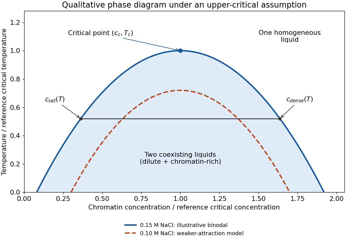
Schematic upper-critical-solution-temperature model for chromatin demixing and weaker attraction at lower salt
. Details. 
The diagram is a model, not measured phase-boundary data. The stated observation at one salt concentration does not by itself establish whether heating suppresses or promotes demixing. A lower critical solution temperature system would instead demix on heating, and would need the corresponding inverted geometry. The following answers make the usual enthalpically favourable-demixing assumption explicit.
The saturation concentration is the dilute-phase coexistence concentration at the specified temperature and solution conditions. On increasing the overall concentration through this boundary, a macroscopic condensed phase can first coexist with the dilute phase. The other branch gives the dense-phase concentration .
At the thermodynamic critical point, the two branches meet: their concentration difference vanishes. Its coordinates define the critical concentration and critical temperature . In the upper-critical model, admits no two-liquid coexistence. Thus is a temperature-dependent branch, whereas is the concentration at the endpoint.
Between coexistence concentrations, the lever rule gives the dense-phase volume fraction , assuming consistent concentration and volume definitions. Increasing total chromatin then primarily changes phase amounts rather than allowing the dilute phase to increase without bound.
At fixed volume and temperature, equilibrium minimizes the Helmholtz free energy . Mixing increases translational entropy, whereas favourable contacts between chromatin segments can lower the interaction contribution to internal energy. At low enough temperature, favourable contacts can compensate the entropy lost on concentrating the arrays; at higher temperature, the mixing contribution can dominate. This gives the upper-critical dome assumed in (a).
A simple illustrative Flory–Huggins solution theory writes the dimensionless mixing free energy per site asHere is an effective polymer size, not automatically the number 12, and represents an effective energetic incompatibility of polymer and solvent. The common-tangent construction determines coexisting compositions. Solving givesSince decreases on heating, the coexistence interval narrows and closes at . For fixed pressure the corresponding potential is Gibbs free energy, with the enthalpic term in ; enthalpy and internal energy should not be interchanged without stating the mechanical constraint. Hydration, ion release or temperature-dependent effective interactions can change this simple temperature trend.
In the regime where salt promotes chromatin condensation by screening repulsion between DNA-rich surfaces, reducing salt from 0.15 M to 0.1 M weakens the effective attraction. In the schematic upper-critical model this means a lower critical temperature and a smaller coexistence region; at a fixed suitable temperature the dilute-phase saturation concentration rises. The second curve is therefore drawn inside and below the first. The magnitude of the shift is not specified by the question.
This direction has a microscopic basis: reducing ionic strength increases the range of electrostatic repulsion between negatively charged DNA segments. Modelling of interacting arrays supports a role for screening and nucleosome contacts in stabilizing chromatin-rich phases.[ref]
Nucleosome plasticity is a critical element of chromatin liquid-liquid phase separation and multivalent nucleosome interactions by Stephen E. Farr and others. Salt-dependent coexistence and the role of nucleosome breathing
In the idealized limit of unscreened DNA-DNA repulsion, the ordinary condensed phase can disappear, rather than simply acquiring an arbitrarily small positive critical temperature. However, zero added NaCl is not literally zero counterions: charged chromatin requires charge compensation, and buffer ions and counterions associated with the preparation remain. Salt also screens attractive interactions, and very high salt can alter nucleosome stability. The proposed shift applies to the stated low-salt screening regime, not a universal monotonic law at every salt concentration.
Chromatin is a composite of charged DNA, folded histone cores, flexible histone tails and associated proteins. These parts differ in charge, hydrophobicity, flexibility, interaction partners and chemical modifications. It is therefore not a chemically uniform chain with one identical contact energy at every position.
Diversity also occurs along a chromosome: nucleosome occupancy, linker DNA length, histone variants, histone acetylation and other histone modifications change the local interaction pattern. Positively charged tails can contact DNA or neighbouring nucleosome surfaces; an acetylated lysine has different electrostatic properties and can recruit a modification-reading protein. The number, arrangement and lifetime of contacts matter as well as their average strength. A coarse-grained interaction parameter can summarize one preparation but should not be mistaken for a universal chemical constant of all chromatin.
The structural implication is a spectrum of arrangements, not a universal regular fibre. Local nucleosome packing, irregular accessible chains, compact domains and interactions between differently modified regions can coexist, and their balance depends on preparation and cellular context. Nucleosome breathing changes the sites available for intermolecular contacts and hence the connectivity of an array assembly; it is not merely a uniform change in the size of a rigid bead.[ref]
Nucleosome plasticity is a critical element of chromatin liquid-liquid phase separation and multivalent nucleosome interactions by Stephen E. Farr and others. Comparison of breathing and artificially constrained nucleosomes
Reconstituted-array experiments separate contributions that are difficult to disentangle in nuclei. Fluorescent arrays, microscopy, droplet fusion and molecular exchange measurements test condensation and dynamics; changing tails, linker organization or acetylation tests the interaction mechanism. Bryan A. Gibson and colleagues found that histone H1 and nucleosome spacing modulate condensation, while acetylation and reader proteins can produce different condensed states.[ref]
Organization of Chromatin by Intrinsic and Regulated Phase Separation by Bryan A. Gibson and others. Reconstituted arrays and regulation of condensate properties
In cells, combine imaging with contact maps and accessibility assays. Hi-C can reveal preferential contacts and chromatin compartments, but does not establish liquid behaviour. Fluorescence recovery after photobleaching measures recovery kinetics, but recovery alone does not prove equilibrium liquid-liquid demixing. Conversely, an in-vitro droplet is not proof that every nuclear domain forms by the same mechanism. The strongest case combines material behaviour with controlled molecular perturbations and measurements of the resulting organization.
Physicochemical diversity provides ways to regulate the same DNA without changing its sequence. Histone modifications, variants and recruited proteins can alter local contacts, accessibility and the partitioning of regulatory factors. This can couple chromatin organization to transcription, DNA replication and DNA repair, while allowing distinct genomic regions to behave differently in one nucleus.
For example, changing histone acetylation can weaken some intrinsic contacts while favouring binding of specific acetylation readers. A local state can therefore change both how strongly a region associates with itself and which partners it recruits. Reversible, multivalent interactions allow organization to respond to signals without requiring an irreversible solid assembly.
These mechanisms also allow memory: feedback between a chromatin-associated mark, its reader and modifying machinery can help restore a local state after replication. Such epigenetic inheritance still needs evidence for maintenance and re-establishment; a stable-looking condensed region alone does not establish it. Likewise, correlations between compaction and reduced transcription do not prove that condensation is always the cause of repression. Perturbations must separate altered material properties from direct effects on transcriptional machinery.
Articles by others on the same topic
There are currently no matching articles.