Topics (384k) Articles (398k) Users (410) Discussions (242) Comments (383) Files (13k) New article

Coronary oxygen uptake is the net rate at which a specified myocardial region removes oxygen from its supplying blood. For matched inflow and outflow with negligible oxygen storage change, the regional Fick principle balance isHere is volume flow, arterial oxygen content and venous oxygen content. The arteriovenous oxygen content difference measures removal per blood volume; multiplying by flow gives removal per time.
In an original example, , and give uptake . Coronary oxygen delivery is instead in the same amount-per-time units. The remaining oxygen leaves in the venous outflow.
The complete control-volume statement makes the assumptions explicit:where is stored oxygen in the represented region. If input and output carry and , while storage increases at , tissue uptake is , not . Thus an inflow–outflow difference during accumulation is not automatically a steady metabolic rate.
The required quantities also come from different observations. The primary coronary thermodilution study estimated volumetric flow using saline infusion and pressure/temperature sensing.[ref]
Direct volumetric blood flow measurement in coronary arteries by thermodilution by Wilbert Aarnoudse and others. Abstract, Methods
A separate primary whole-blood oximeter used red and infrared illumination, a photodetector and computation to obtain hemoglobin concentration and saturation.[ref]An oximeter for measuring hemoglobin concentration and oxygen content by A. P. Shepherd and others. Abstract, optical apparatus and separate computed quantities
Neither instrument alone supplies every term of the balance.The left coronary artery is a principal coronary artery arising from the proximal aorta. Its typical major branches are the left anterior descending artery and the circumflex artery, which distribute blood to different regions of the heart.[ref]
Anatomy and Physiology 2e by J. Gordon Betts and others. Section 19.1, Coronary Arteries and Figure 19.15
A main trunk and one of its branches are different anatomical levels; a finding in one should not be silently attributed to the other.The adjective left does not partition the entire heart into two exclusive arterial territories. Coronary dominance and branching variations alter posterior supply, and a right-coronary system can supply substantial parts of the left ventricle. Injected-heart and X-ray observations demonstrate variation even within right-dominant specimens.[ref]
Functional dominance of the right coronary artery: incidence in the human heart by C. E. Nerantzis and others. Abstract, variation within the right-dominant group and ventricular territories
An original two-branch conservation of mass model makes the trunk-versus-branch distinction quantitative. If the modeled trunk has only two outlets and negligible accumulation,Chosen branch flows and give trunk flow . Measuring only the first branch would not supply the total. If other branches or alternate routes carry flow, their terms must be included rather than forced into this two-outlet model.
Likewise, the consequences of a narrowed main segment and a narrowed distal branch need not be interchangeable. They occupy different positions in a network. A physiological inference requires the actual routes, their resistances and the represented tissue, not merely the shared word coronary.
The left coronary artery is therefore a useful anatomical reference for interpreting coronary angioplasty, branch-level measurements and oxygen-supply models. A measured diameter identifies geometry at a selected location; coronary oxygen delivery additionally requires flow and arterial oxygen content. These observations must refer to compatible territories before they are combined.
The right coronary artery is one of the two principal coronary arteries arising from the proximal aorta. It supplies portions of the heart's tissues and commonly gives a right marginal branch supplying the right ventricle.[ref]
Anatomy and Physiology 2e by J. Gordon Betts and others. Section 19.1, Coronary Arteries and Figure 19.15
The word right identifies the vessel; it does not imply that all of its supplied tissue lies in the right half of the heart.Coronary dominance concerns the origin of the posterior descending artery. A right-dominant pattern places that branch in the right-coronary system. It does not mean that the right ventricle is the main systemic pump or that the left coronary artery is unimportant.
A primary anatomical investigation injected radiopaque material into 200 human hearts and combined direct observation with X-ray analysis. The investigators classified 178 specimens as right dominant using posterior arterial origin, and identified additional branching variation within that group.[ref]
Functional dominance of the right coronary artery: incidence in the human heart by C. E. Nerantzis and others. Abstract, preparation, dominance criterion and observed branching variation
Injection makes the arterial route visible; examining the prepared heart then relates the image to anatomical branches.The observed proportion is a statement about those specimens under that definition. It is not a universal value that can replace an individual anatomical observation. The study also illustrates why a category such as right dominant does not specify every branch or the exact tissue territory supplied.
Vessel structure and functional supply are complementary questions. Coronary blood flow depends on the pathway's resistance and changing driving conditions, while coronary oxygen delivery also depends on blood oxygen content. A visible, patent right coronary artery establishes a route; it does not independently quantify the flow or oxygen available to every downstream tissue region. Anatomical mapping and physiological measurements therefore answer different parts of the same supply problem.
The posterior descending artery, also called the posterior interventricular artery, follows the posterior interventricular sulcus toward the apex of the heart. Its branches contribute to posterior ventricular and septal supply.[ref]
Anatomy and Physiology 2e by J. Gordon Betts and others. Section 19.1, Coronary Arteries and Figure 19.15
Its parent route is central to coronary dominance. In a right-dominant pattern it arises from the right coronary artery; a left-dominant route involves the circumflex artery. The branch's descriptive name identifies a course, while its origin identifies a connection within the coronary network.
Tracing the two possible routes makes the distinction concrete. One goes from the aorta into the right coronary artery and then to this branch. The other passes from the aorta into the left coronary artery, then the circumflex artery, before reaching it. The same distal branch name can therefore sit downstream of different proximal segments.
Primary direct and X-ray observations of injected hearts demonstrate that branching varies even within a dominance class.[ref]
Functional dominance of the right coronary artery: incidence in the human heart by C. E. Nerantzis and others. Abstract, preparation and branching variants
A textbook route is a reference for identifying the observed anatomy, rather than a reason to overwrite a different observed connection.The vessel should also be distinguished from the neighboring venous route. The middle cardiac vein follows the posterior interventricular region toward the coronary sinus. Arterial delivery and venous collection describe different directions and different sampled blood pools, even when the structures lie close together.
Coronary dominance classifies coronary arterial branching according to a stated anatomical criterion, commonly the origin of the posterior descending artery. A right-coronary origin is termed right dominant; a left-coronary origin through the circumflex artery is termed left dominant. The label does not state which ventricle is the systemic pump.
A primary anatomical study injected radiopaque medium into 200 human hearts and examined the preparation directly and by X-ray. It classified 178 as right dominant using posterior arterial origin, and described additional branching variation within that category.[ref]
Functional dominance of the right coronary artery: incidence in the human heart by C. E. Nerantzis and others. Abstract, method, criterion and observed variants
The sample proportion is . The remaining 22 specimens cannot be assigned to more detailed categories merely by subtracting from the total; their individual routes and the classification rules must be examined. A study using an additional shared-supply category can also require a more detailed rule than a two-way label.
The useful interpretation is topological. The route to the posterior descending artery can pass through the right coronary artery or through the left coronary artery and its circumflex artery. A finding upstream of that branch therefore has different downstream connections in the different patterns.
Even within a dominance category, branch size and supplied territory can vary. The term does not specify every arterial diameter, flow rate or tissue region. For physiological interpretation, the actual route must be combined with coronary blood flow and an appropriate territory definition rather than substituted for them.
Doppler range ambiguity means that a measured Doppler ultrasound signal does not uniquely identify the depth of the scatterers producing it. In continuous-wave Doppler echocardiography, simultaneous transmission and reception collect motion signals along the beam without a unique pulse travel time. A high-speed spectral component therefore does not, by itself, label an anatomical site.[ref]
Recommendations for Quantification of Doppler Echocardiography by Miguel A. Quiñones and others. General Principles, printed p. 167, and CW Doppler, p. 169
Repeated pulses can introduce a different range ambiguity. Let the pulse period be , with pulse repetition frequency . If reception is associated only with its delay within a pulse period, the possible round-trip travel times include . The corresponding depths arewhere is speed of sound. Identifying which transmitted pulse generated an echo requires additional information or a restriction on the possible depth.
For an original example, Hz gives microseconds. With m/s and observed within-period delay microseconds, possible depths are 7.7, 23.1 and 38.5 cm. Successive possibilities are separated by 15.4 cm. An anatomical restriction to depths below 15.4 cm would identify the first value; the echo timing alone would not.
Range ambiguity and aliasing of the Doppler ultrasound frequency are distinct. The first confuses position along the beam; the second confuses motion frequencies sampled at a given pulse rate. Increasing the pulse rate can improve the unaliased motion range while reducing unique depth coverage, producing the pulsed Doppler depth-velocity tradeoff. High-pulse-rate methods deliberately accept multiple possible sampling depths.[ref]
Recommendations for Quantification of Doppler Echocardiography by Miguel A. Quiñones and others. General Principles, pp. 167–168, high-PRF modification
Anatomical imaging and acquisition geometry supply constraints; a plausible waveform is not itself a unique range label.A linear bounded automaton is a Turing machine whose usable tape on an input of length is bounded by cells for a fixed constant . A common equivalent model places end markers around the input and permits rewriting only between them. The standard language-class characterization uses a nondeterministic machine: these machines recognize exactly the nonempty-word languages generated by context-sensitive grammars, with an explicit convention needed for the empty word.[ref]
For , repeatedly mark one unmarked , one and one , rejecting if the order is wrong or a matching symbol is absent. Accept when no symbols remain unmarked. This uses the original input cells and repeated sweeps, showing how bounded space can check three linked counts. “Linear bounded” limits space, not running time.
Accumulated contact slip measures the total distance slid at a specified contact, counting motion in either direction positively. For an absolutely continuous signed contact slip displacement in a consistently tracked local frame,It is the total variation of a function applied to the slip history. It differs from net slip , which can lose information through cancellation. Contact slip displacement explains the contact-frame convention and the connection to frictional dissipation from contact slip.
For an original piecewise-linear trajectory, suppose successive slip values are . Its net slip is , whileThe middle reversal contributes even though it moves toward smaller signed coordinates. Recording only the first and last values would report and miss eighty percent of the travelled distance in this stipulated history.
For exact samples at ordered times, the sum of absolute increments is a lower bound on the true accumulated motion; unobserved reversals can add distance between samples. The triangle inequality explains why inserting additional exact observations cannot decrease this sum. It equals the integral when the observed intervals contain no unresolved reversals and the path model is appropriate.
Measurement noise changes that conclusion about observed data. A truly stationary contact read as would acquire an apparent distance for positive . More samples can reveal real reversals while also accumulating readout noise. A useful imaging study therefore reports time resolution, length measurement calibration and the treatment of noise, rather than interpreting every absolute increment as physical sliding.
Frictional dissipation from contact slip is the irreversible loss of mechanical energy associated with relative sliding at a contact. In the ideal kinetic friction model, friction opposes the local slip velocity and has magnitude , with nonnegative coefficient and normal force . Its dissipated energy isThe work calculation and the distinction between signed and accumulated motion are developed in contact slip displacement. The kinetic-friction force law is an ideal physical model, rather than a universal description of every interface.[ref]
An original two-stage example keeps the loading history explicit. Let . Slide forward while , then backward while . The final signed slip is , the accumulated contact slip is , andThe two stages contribute and . Using the unweighted mean of the two normal loads would instead give ; the correct weighting follows sliding distance.
A physical measurement records relative motion close to the contact together with tangential force and normal loading. Integrating the opposing tangential force over the resolved sliding increments estimates the lost mechanical energy under the stated model. Force transmitted during ideal sticking, where local slip rate is zero, contributes no slip dissipation in that model, even if other parts of the apparatus deform. Grip travel and fixture mechanical compliance therefore must be distinguished from local sliding.
Confocal transverse-mode degeneracy occurs when distinct spatial modes of an ideal confocal optical cavity have the same resonance frequency. It follows from the rational Gouy phase advance of the equal-radius, equal-separation geometry, rather than from the modes having identical transverse field patterns.
For mirror separation , the paraxial resonance frequencies areThe primary MIT notes derive the confocal phase shift and mode-frequency families.[ref]
Fundamentals of Photonics and Quantum Electronics lecture notes by Franz X. Kärtner. §2.6.2, printed p. 116, equation 2.303 and Figure 2.82
Let total transverse order be . Replacing by leaves the frequency unchanged whenever the resulting indices describe permitted modes. Orders that are even integers therefore share the fundamental-order family, while orders that are odd integers form an interleaved family halfway between.There is also degeneracy among patterns with the same total order. At , the pairs have the same ideal frequency while their transverse distributions differ. A transverse optical cavity mode needs its spatial field as well as its resonance frequency to be identified.
An original frequency example uses and . Holding one transverse order fixed gives spacing . Relative to a chosen fundamental resonance, the mode with and longitudinal index one smaller has offset zero; the mode at the original longitudinal index has offset . Thus a spectrum containing both families can show half the fixed-order longitudinal spacing. It does not follow that every visible peak is a separate laser frequency.
The excitation strength depends on spatial coupling. In an ideal orthonormal cavity-mode basis, the incident field can be expanded with coefficients , with consistent field normalization. Matching the fundamental field suppresses overlap with orthogonal modes, even if those modes share its frequency. Conversely, transmission at the common resonance can contain several patterns when they are excited; frequency selection alone cannot separate a degenerate subspace.
A practical test scans the cavity length while recording both transmitted power and spatial beam shape, then changes alignment or mode matching. Luhs’s primary apparatus discussion identifies unwanted extra peaks as transverse modes of the analyzer and distinguishes them from the probe’s own longitudinal spectrum.[ref] A photodetector integrating the whole output can conceal spatial differences that a beam image reveals.
Mirror-spacing error, unequal curvatures, aberration and finite apertures perturb the ideal degeneracy. For a general one-way Gouy phase , the corresponding phase model gives . The nominally paired orders and then differ by when their longitudinal indices differ by one. This makes near-degenerate peaks a sensitive geometry probe, but attributing a measured splitting requires separating those perturbations from source structure and polarization effects.
The kification of a topological space has the same points and the finest topology making every original continuous map from a compact Hausdorff space continuous. Equivalently, is closed exactly when is closed in for every such test map. This topology is at least as fine as the original one, so the identity is continuous.[ref]
If is already a compactly generated space, its topology is recovered unchanged. In particular this holds for metric spaces. The operation is idempotent: . It lets one form products and mapping spaces within compactly generated spaces: the categorical product is the kification of the ordinary product. Compact test maps retain their original continuity, while the new topology supplies the closed sets they detect.
Protocol-dependent accessibility of lung gas means that the volume reached by a tracer gas depends on how long and how the tracer gas is allowed to mix. Gas that communicates slowly can be partly represented during one observation window and more fully represented during another. This differs from assuming that one fixed volume is permanently isolated.
An original compartmental model makes the dependence quantitative. A reservoir of volume initially contains helium fraction and rapidly mixes with a fast lung region . Their combined volume is , with initial fraction . A slow region of volume initially contains no tracer gas and exchanges equal gas volumes in each direction at rate . Assuming constant volumes and well-mixed compartments,Total tracer gas is conserved, while the concentration difference decays withApplying an equilibrium dilution formula before equilibration gives an apparent lung volume . It starts at and tends toward in this ideal model.
For stipulated L, L, L, and s, the recorded concentration is . After 10 s, the apparent volume is about 4.13 L; after full equilibration it is 5 L. The change occurs without adding lung gas: the tracer gas is reaching more of the existing volume. These are constructed mixing calculations, not a clinical waiting-time recommendation.
The primary respiratory text describes a closed-circuit helium apparatus with continuous concentration measurement using a thermal-conductivity meter, together with gas-volume corrections. It also states that poor communication and short dilution time can limit the represented gas volume.[ref]
Pulmonary Function Testing by Warren M. Gold and Laura L. Koth. Closed Circuit Methods, Figure 25-10, and discussion of poorly communicating gas
Leaks, changing gas conditions and unequal starting volumes can produce additional differences. A protocol-defined apparent volume therefore needs its mixing and measurement conditions attached to the result.The mitral coaptation index is a dimensionless normalization of the mitral valve's coaptation area. In the primary method of Frederick C. Cobey and colleagues, it is the three-dimensional end-systolic coaptation-zone area divided by the three-dimensional mitral-valve area used in that protocol:The denominator and surface-reconstruction convention are part of the definition.[ref]
The study used intraoperative transesophageal echocardiography in 25 patients with functional mitral regurgitation, followed by offline analysis of the end-systolic geometry.[ref]
Quantitative assessment of mitral valve coaptation using three-dimensional transesophageal echocardiography by Frederick C. Cobey and others. Abstract, Methods
This supplies an experimental acquisition context for the ratio; it does not make every area called annular area interchangeable with the protocol's valve-area measurement.An original normalization example uses cm and cm, giving . A second constructed valve with areas 3 and 20 cm has the same index despite twice the absolute contact area. Conversely, keeping contact area 1.5 cm while changing the denominator to 15 cm gives . Absolute area and normalized coverage answer different questions.
Uniformly scaling every length by a positive factor multiplies both areas by , leaving unchanged. Inconsistent surface conventions do not cancel: counting two apposed faces in the numerator while counting their interface once in another reconstruction can change the ratio without a biological change.
Finally, the index does not describe the connectivity of any remaining gaps. Two contact patches with equal coaptation area and equal denominator can have different shapes and positions, as illustrated under coaptation area. The mitral coaptation index is a geometric descriptor to interpret alongside the reconstructed valve and blood flow, rather than a complete description of valve sealing.
Surgical complication count versus patient incidence distinguishes the number of postoperative events from the number of people who experience at least one event. A person can have several postoperative complications. Counting events and counting affected patients therefore answer different questions, even with the same follow-up period.
Let be the number of operated patients with complete observation through a specified time, and let be the number of qualifying events for patient . ThenHere is an event count, estimates the proportion experiencing at least one complication during the specified period, and is mean events per patient. Unlike , the last quantity can exceed one.
For an original example, one 100-patient cohort has 40 events concentrated among 20 people. Another has 40 events, each in a different person. Both have events per patient, but their patient incidences are and . Neither summary alone reveals severity, persistence or how closely events cluster in time.
The Clavien-Dindo classification adds severity information, but a highest-grade-per-patient summary still discards multiplicity. In the original validation paper, the most severe complication was registered when a patient had more than one. That is an explicit patient-level severity convention, rather than a count of every complication.[ref]
Classification of Surgical Complications: A New Proposal With Evaluation in a Cohort of 6336 Patients and Results of a Survey by Daniel Dindo and others. Patients and Methods, Validation of the Modified Classification in a Cohort of 6336 Patients
Time and denominators must be specified. With 40 events over patient-days, the event rate is per patient-day; it is not a probability for each individual day. With incomplete follow-up, simple affected-patient proportions require further assumptions or methods accounting for observation time and competing events. Recurrent postoperative event analysis also has to account for dependence between several events in the same patient, rather than treating them as independent extra patients.
A reproducible comparison therefore defines the qualifying postoperative complication, observation window, patient denominator, event-counting rule and severity summary together. The original classification separately distinguishes complications from inherent sequelae and failure to cure, so those categories should not silently be added to the same numerator.[ref]
Classification of Surgical Complications: A New Proposal With Evaluation in a Cohort of 6336 Patients and Results of a Survey by Daniel Dindo and others. Methods and Definitions of Complications, Definition of Negative Outcome
A mathematical plane cutting an infinite circular cylinder at a nonzero angle to its axis produces an ellipse, unless it is parallel to that axis. More precisely, let the cylinder radius be , and let be the acute angle between the cylinder axis and the plane's normal vector, with . The semi-major and semi-minor axes and area areThe angle is measured from the normal, not from the cutting plane itself. At , the profile is a circle.
To prove the result, choose Cartesian coordinates so the cylinder is , with axis along . Translate the cut so it crosses the axis at . After rotating about the axis if necessary, an orthonormal basis in the cutting plane isA point in the plane is . Substitution into the cylinder equation gives , orBecause the basis is orthonormal, and measure actual lengths within the plane. The area of an ellipse then gives the stated area.
For an original example, and give , and , twice the perpendicular-profile area. As approaches , the ellipse grows without bound. At the excluded parallel orientation, a plane through the axis instead meets the lateral cylinder in two generator lines; the limiting formula does not describe a finite ellipse there.
Microtomy applies this geometry to thin physical sections of a circular material fibre. Its worked example and figure show how an elongated profile can arise from section orientation without deformation. Applying the ideal lemma requires accounting for finite thickness, a finite specimen and departures from a straight circular cylinder.
A Euclidean circle with center and radius is the set of points in the Euclidean plane at Euclidean distance exactly from :It is the boundary curve, while the closed disk includes all points with . The center belongs to that closed disk but not to the Euclidean circle.
For example, center and radius3 give . The point lies on the Euclidean circle; lies inside the closed disk because its distance from the center is2. Such an equation distinguishes a distance constraint from a region-filling inequality.
Every Euclidean circle is a circle in the topological sense. The map from the unit circle is a homeomorphism, with inverse . Topology forgets the particular center and radius, whereas the Euclidean distance retains them. The assumption matters: radius zero gives a point, which is not a circle.
The historical terminology differs slightly. Euclid's definition describes the plane figure contained by a circumference; the modern set above corresponds to that boundary. His equal-distance condition and distinguished center appear in BookI, Definitions15–18.[ref]
Euclid's Elements by Euclid. BookI, Definitions15–18, Joyce transcription and guide distinguishing Euclid's figure from the modern boundary curve
At a prime number and height , Morava K-theory is a periodic generalized homology theory represented by a topological spectrum with coefficient graded ringHere multiplication by is invertible and implements periodicity. Every nonzero homogeneous coefficient is invertible, so this is a graded field, although it is not a field as an ungraded ring.[ref]
Morava E-Theory and Morava K-Theory by Jacob Lurie. Lecture 22, Definition 5 and the ensuing coefficient computation, page 3
For , the period is two: the coefficient group is in each even degree and zero in odd degrees. Distinct heights supply different tests for topological spectra; vanishing of means that this particular probe misses , not that is necessarily trivial. This is the organizing idea of chromatic homotopy theory.
Co-sited chroma places a retained colour-difference sample at the same spatial coordinate as a selected luma sample along the relevant axis. With horizontal reduction by two, an illustrative co-sited grid occupies when luma samples occupy all integer positions. The Linux video documentation distinguishes this from centered chroma.[ref]
Co-siting does not require simply discarding alternate original samples. An original symmetric three-tap filter can produceFor a linear ramp , the terms simplify to . This filter's output is therefore aligned with the middle luma position for the ramp, even though it combines neighbouring samples.
An alternating input shows why filtering matters. Let . Keeping only even-indexed samples without filtering gives the constant148, losing the alternating pattern through aliasing. The stated three-tap filter instead gives at every retained position. This demonstrates suppression of this particular pattern; it does not prove rejection of every frequency that could alias.
Horizontal and vertical alignment are separate choices. A 4:2:0 chroma subsampling grid can be co-sited vertically while centered horizontally. During chroma reconstruction, using the correct coordinates and an appropriate low-pass filter are related but distinct requirements: correct placement cannot undo detail already discarded during sampling.
Pinned article: Introduction to the OurBigBook Project
Welcome to the OurBigBook Project! Our goal is to create the perfect publishing platform for STEM subjects, and get university-level students to write the best free STEM tutorials ever.
Everyone is welcome to create an account and play with the site: ourbigbook.com/go/register. We belive that students themselves can write amazing tutorials, but teachers are welcome too. You can write about anything you want, it doesn't have to be STEM or even educational. Silly test content is very welcome and you won't be penalized in any way. Just keep it legal!
Intro to OurBigBook
. Source. We have two killer features:
- topics: topics group articles by different users with the same title, e.g. here is the topic for the "Fundamental Theorem of Calculus" ourbigbook.com/go/topic/fundamental-theorem-of-calculusArticles of different users are sorted by upvote within each article page. This feature is a bit like:
- a Wikipedia where each user can have their own version of each article
- a Q&A website like Stack Overflow, where multiple people can give their views on a given topic, and the best ones are sorted by upvote. Except you don't need to wait for someone to ask first, and any topic goes, no matter how narrow or broad
This feature makes it possible for readers to find better explanations of any topic created by other writers. And it allows writers to create an explanation in a place that readers might actually find it.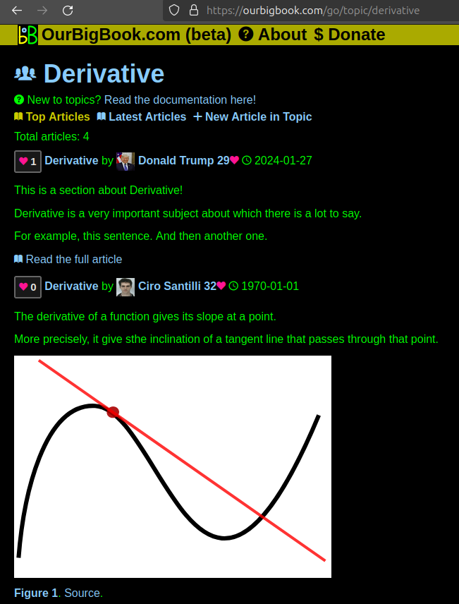
Figure 1. Screenshot of the "Derivative" topic page. View it live at: ourbigbook.com/go/topic/derivativeVideo 2. OurBigBook Web topics demo. Source. - local editing: you can store all your personal knowledge base content locally in a plaintext markup format that can be edited locally and published either:This way you can be sure that even if OurBigBook.com were to go down one day (which we have no plans to do as it is quite cheap to host!), your content will still be perfectly readable as a static site.
- to OurBigBook.com to get awesome multi-user features like topics and likes
- as HTML files to a static website, which you can host yourself for free on many external providers like GitHub Pages, and remain in full control

Figure 2. You can publish local OurBigBook lightweight markup files to either OurBigBook.com or as a static website.
Figure 3. Visual Studio Code extension installation.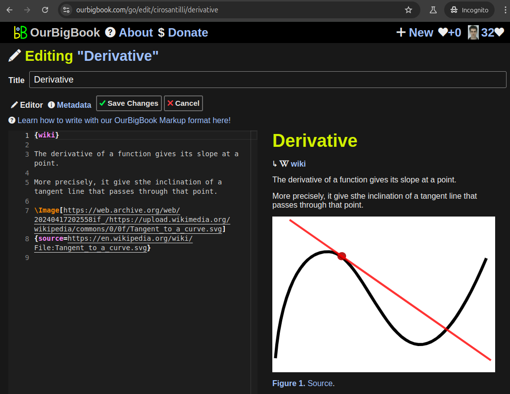
Figure 5. . You can also edit articles on the Web editor without installing anything locally. Video 3. Edit locally and publish demo. Source. This shows editing OurBigBook Markup and publishing it using the Visual Studio Code extension. 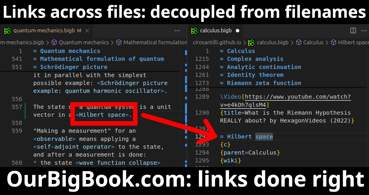
- Infinitely deep tables of contents:
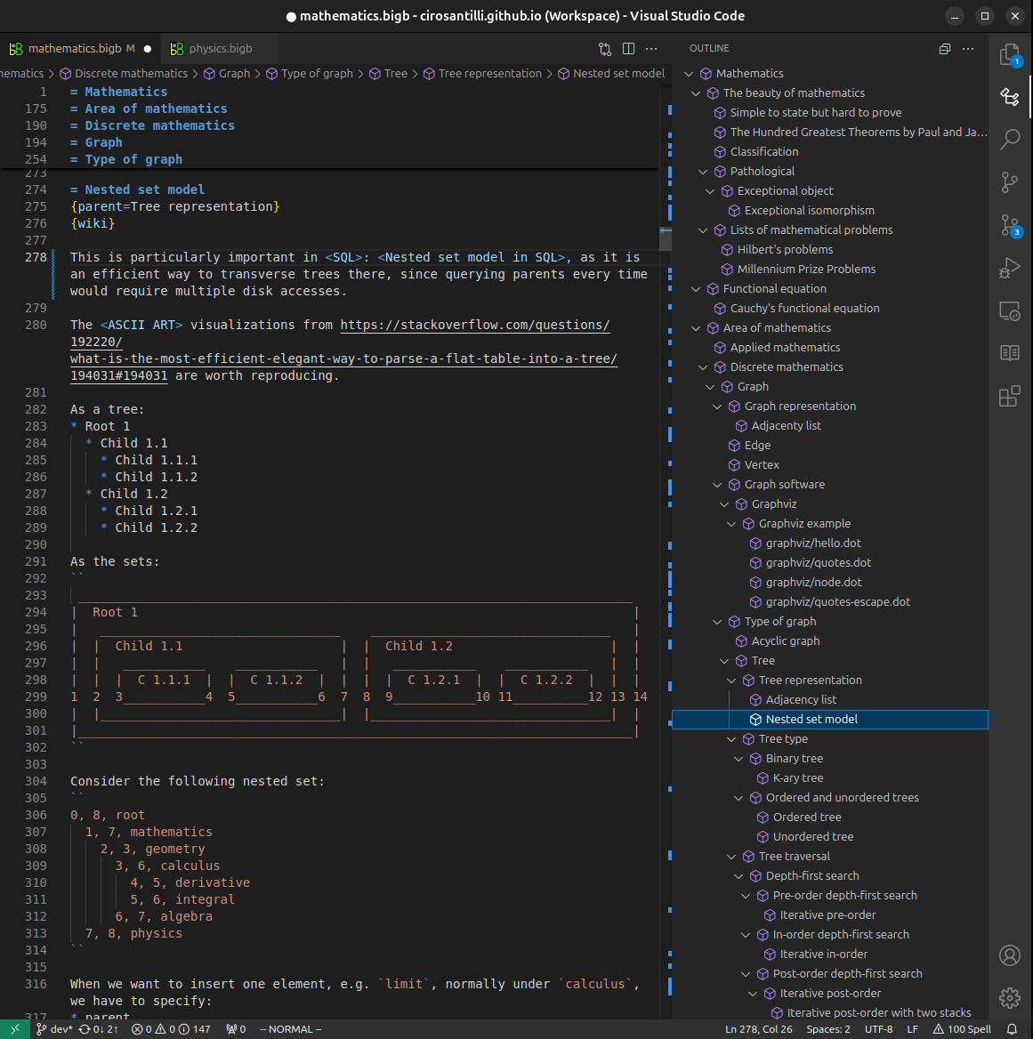

All our software is open source and hosted at: github.com/ourbigbook/ourbigbook
Further documentation can be found at: docs.ourbigbook.com
Feel free to reach our to us for any help or suggestions: docs.ourbigbook.com/#contact