Topics (384k) Articles (398k) Users (410) Discussions (242) Comments (383) Files (13k) New article

Hyaluronic acid, also called hyaluronan, is a linear glycosaminoglycan composed of alternating glucuronic acid and N-acetylglucosamine residues. It is nonsulfated, and unlike the sugar chains of conventional proteoglycans, it does not require covalent attachment to a protein core. Its carboxyl groups are largely ionized in physiological conditions, so the polymer is often encountered as hyaluronate rather than as the fully protonated acid. [ref]
https://www.ncbi.nlm.nih.gov/books/NBK579913/. Essentials of Glycobiology, chapter 16: composition, naming and physicochemical properties
The repeating sequence makes a useful bookkeeping example. A chain segment with complete two-sugar repeats contains glucuronic acid residues and N-acetylglucosamine residues. Its length can change enormously without changing that alternating composition. Composition alone, however, does not determine viscosity: chain length, concentration and solution conditions also matter.
Synthesis tests the two-building-block description experimentally. P. L. DeAngelis characterized the hyaluronic acid synthase from Pasteurella multocida using activated sugar donors, UDP-glucuronic acid and UDP-N-acetylglucosamine. The enzyme incorporates the two distinct sugars into the growing chain; activity also depends on divalent ions. This turns an abstract repeating formula into a laboratory reaction whose substrates and conditions can be varied.[ref]
Enzymological characterization of the Pasteurella multocida hyaluronic acid synthase by P. L. DeAngelis. Abstract and enzymological characterization: two sugar donors, divalent-ion effects and synthesis of alternating hyaluronan.
Hyaluronic acid is therefore both a molecular species defined by its repeat chemistry and a material whose bulk behavior depends on its molecular population. This distinction helps explain why identifying the sugar repeat does not by itself specify the properties of every preparation.
Keratan sulfate is a sulfated glycosaminoglycan with a backbone of alternating galactose and N-acetylglucosamine residues. Unlike hyaluronic acid and many other glycosaminoglycans, its characteristic two-sugar repeat does not contain a uronic acid. Backbone identity and the extent of sulfation are separate structural questions.[ref]
Isolation and partial characterization of keratan sulphate from human brain by I. Eronen and others. Abstract: sulfated galactose–N-acetylglucosamine disaccharide repeat
An ideal backbone segment with 30 complete repeats has 30 galactose residues and 30 N-acetylglucosamine residues, for 60 sugar residues altogether. This count does not fix its number of sulfate groups or the molecular mass of an actual preparation.
A 1985 study isolated a sulfated fraction from human brain and combined chemical analysis with susceptibility to a keratan-sulfate-cleaving enzyme. Agreement between the repeat structure and cleavage behavior supported its identification as keratan sulfate; size measurements also showed heterogeneity. Several different observations make identification stronger than reliance on a sulfated material's charge alone.[ref]
Isolation and partial characterization of keratan sulphate from human brain by I. Eronen and others. Abstract: isolation, gel filtration, repeat structure and endo-beta-galactosidase susceptibility
Heparin is a strongly sulfated glycosaminoglycan containing alternating glucosamine-derived and uronic acid residues, including iduronic acid and glucuronic acid. Chains vary in length and chemical substitution, so a preparation is not adequately described by one small-molecule molecular formula. Sulfation and carboxylate groups contribute substantial negative electric charge.[ref]
Heparin lyase in IUBMB Enzyme Nomenclature. EC 4.2.2.7: uronate and sulfated glucosamine-containing polysaccharide substrate
A defined fragment can test which structural details matter. In 1983, J. Choay and collaborators synthesized related four- and five-sugar fragments and verified their structures with nuclear magnetic resonance. The five-sugar fragment bound antithrombin and enhanced its inhibition of factor Xa, while the tested four-sugar fragment did not.[ref]
Structure-activity relationship in heparin: a synthetic pentasaccharide with high affinity for antithrombin III and eliciting high anti-factor Xa activity by J. Choay and others. Abstract: structural verification, antithrombin binding and anti-factor Xa activity
The experiment shows why counting sulfate groups or measuring total charge cannot by itself specify biological activity: a particular arrangement of residues also matters. This is a molecular structure–function example, distinct from selecting a clinical dose or predicting every property of an unfractionated preparation.
Dermatan sulfate is a family of sulfated glycosaminoglycans containing repeating iduronic acid and N-acetylgalactosamine units. The sugar linkage pattern and sulfate substitutions help define the material; chain length and substitution vary, so the name does not identify one small molecule with a unique molecular mass.[ref]
Its uronic-acid stereochemistry can be created after a chain has been assembled. Chondroitin-glucuronate 5-epimerase converts D-glucuronate residues in a chondroitin chain into L-iduronate residues in a dermatan chain. This enzyme is distinct from heparosan-N-sulfate-glucuronate 5-epimerase, which acts in a different polysaccharide context.[ref]
Chondroitin-glucuronate 5-epimerase in IUBMB Enzyme Nomenclature. EC 5.1.3.19, Reaction and Comments
An ideal segment of 20 complete two-sugar repeats contains 20 uronic-acid residues and 20 amino-sugar residues, or 40 sugar residues altogether. Epimerizing an eligible uronic-acid residue changes its configuration without increasing that total. Adding a sulfate group changes substitution without adding another sugar residue.
These distinctions guide an experiment: measuring chain extension, uronic-acid configuration and sulfate content asks three separate questions. A total carbohydrate measurement alone cannot determine which operation occurred. To compare two preparations meaningfully, record the property actually measured rather than assuming every dermatan-sulfate chain has identical composition.
Heparan sulfate is a family of linear, variably sulfated glycosaminoglycan chains. Its repeating framework contains uronic-acid and glucosamine-derived positions, but the modifications are not identical at every position. Heparan sulfate can form the carbohydrate component of a proteoglycan; the chain and the entire protein–carbohydrate assembly are distinct objects.[ref]
One source of diversity is epimerization. Heparosan-N-sulfate-glucuronate 5-epimerase converts eligible D-glucuronate positions into L-iduronate after suitable neighboring N-sulfation of glucosamine. A labelled-polymer experiment followed that configurational change using nuclear magnetic resonance, demonstrating why polymer context matters to the reaction.[ref]
Consider an original schematic pair of equal-length chains. Each has 20 uronic-acid and 20 amino-sugar positions. Give the two chains equal total numbers of sulfate substitutions, but put those substitutions at different eligible positions. Equal length and equal sulfate count do not specify the same sequence of local chemical environments. A bulk measurement of either quantity therefore leaves structural information unresolved.
This is why a single repeated drawing should not be mistaken for a complete description of every heparan sulfate preparation. Comparing chains requires specifying which features were measured: length, nitrogen substitution, oxygen substitution or uronic-acid configuration. These distinctions connect carbohydrate structure to molecular recognition without assuming all chains have one uniform composition.
Chondroitin is a glycosaminoglycan backbone built from alternating glucuronic acid and N-acetylgalactosamine residues with a defined glycosidic bond pattern. Chondroitin sulfate adds sulfate substitutions to that framework; the backbone name alone does not specify a sulfation pattern.[ref]
Identification and molecular cloning of a chondroitin synthase from Pasteurella multocida type F by P. L. DeAngelis and A. J. Padgett-McCue. Abstract: chondroitin disaccharide repeat and enzymatic synthesis
A 2000 experiment cloned chondroitin synthase from Pasteurella multocida type F. A recombinant soluble enzyme extended acceptor chains using the corresponding activated sugar donors. Related donor molecules did not substitute in the tested reaction. The product was susceptible to a chondroitin-cleaving enzyme but resistant to a hyaluronan-cleaving enzyme, providing a structural check beyond simply observing a large carbohydrate product.[ref]
Identification and molecular cloning of a chondroitin synthase from Pasteurella multocida type F by P. L. DeAngelis and A. J. Padgett-McCue. Abstract: donor specificity, size-exclusion chromatography and contrasting lyase susceptibility
For an original comparison, a segment with 25 complete repeats contains 25 uronic-acid and 25 amino-sugar residues. Adding sulfate substitutions leaves that sugar count unchanged. Converting eligible D-glucuronate positions into L-iduronate also leaves the count unchanged, but leads toward a dermatan-type chain through chondroitin-glucuronate 5-epimerase.[ref]
Chondroitin-glucuronate 5-epimerase in IUBMB Enzyme Nomenclature. EC5.1.3.19: chondroitin D-glucuronate to dermatan L-iduronate
A composition measurement, an enzyme cleavage test and a measurement of chain length therefore answer different questions. Agreement among them helps establish polymer identity without equating every negatively charged polysaccharide with the same molecule.
D-glucopyranose is the six-membered-ring hemiacetal form of D-glucose. The ring contains five carbon atoms and one oxygen atom; the sixth carbon, C6, lies outside the ring. Its name specifies both the glucose configuration and the ring form, but without an α or β prefix it does not select a single anomer.[ref]
Nomenclature of Carbohydrates (Recommendations 1996) by Alan D. McNaught. Sections2-Carb-5 and2-Carb-6: cyclic representations, α-D-glucopyranose and anomeric descriptors
Ring closure connects the C5 hydroxy group to the C1 aldehyde carbon of open-chain D-glucose. C1 becomes the anomeric carbon. No chemical atoms are lost in forming this cyclic hemiacetal, so both anomers retain the molecular formula .
In the conventional Haworth projection of D-glucose, the anomeric hydroxy group and the C5 hydroxymethyl substituent are on opposite faces in α-D-glucopyranose and on the same face in β-D-glucopyranose. This relative relationship is more useful than saying that α always means “down”: how a structure is drawn and which sugar configuration is involved must also be specified.[ref]
An original information-counting comparison uses three labels. gives composition; D-glucopyranose additionally specifies a particular sugar configuration and six-membered ring; α-D-glucopyranose further fixes the anomeric configuration. The α and β forms therefore provide an immediate pair of structures with the same elemental count. Neither the molecular formula nor the number of ring atoms alone determines the stereochemistry.
Replacing the anomeric hydroxy group by gives the corresponding methyl glycoside; choosing the α configuration yields methyl α-D-glucopyranoside. Free D-glucopyranose and that acetal derivative differ in chemical identity even though their names share the same sugar-derived root.[ref]
Nomenclature of Carbohydrates (Recommendations 1996) by Alan D. McNaught. Section2-Carb-33.1, named methyl glycoside example and mixed-acetal definition
A slit diaphragm is a specialized junctional structure spanning the filtration slit between adjacent podocyte foot process structures. It forms one component of the glomerular filtration barrier, alongside the glomerular basement membrane and capillary endothelium. The slit diaphragm bridges a gap; it is distinct from the cell membrane of either neighboring podocyte.
Nephrin provides a molecular example. Antibodies against its extracellular region, followed by gold-coupled secondary antibodies, localized the recognized protein to podocyte slits in the 1999 study. A model of how neighboring nephrin molecules might assemble was then proposed. The observed label location and the proposed molecular arrangement are different levels of evidence.[ref]
Nephrin is specifically located at the slit diaphragm of glomerular podocytes by V. Ruotsalainen and others. Figure3: localization; Figure5: hypothetical assembly
For a useful comparison, imagine two barriers with the same total slit length but different junctional organization. Equal length does not establish equal passage of water or proteins: molecular organization and the other barrier layers also matter. A localization image therefore helps identify components but cannot alone supply the glomerular filtration rate.
The minimum sink speed is the airspeed at which a glider has its smallest steady downward speed relative to the air, for a specified configuration, loading and flight condition. It maximizes time aloft from a given height in still air. It does not generally maximize horizontal range, which defines best glide speed.[ref]
For a differentiable glider sink polar , an interior minimum satisfies , with a positive second derivative sufficient for a strict local minimum. For the original shallow-glide model developed in best glide speed,With , this givesIndeed for . The speed ratio is about , within this stipulated parabolic drag polar approximation. A real polar need not follow that numerical ratio, especially near aerodynamic stall.
Using the same original , and gives and . From a stipulated height, the model's times are about at minimum sink and at best glide. Yet the approximate range ratios are and , respectively. The slower optimum buys time rather than maximum distance.
A horizontal wind changes range without changing air-relative sink at a fixed condition. Vertical air motion changes ground-relative height loss as well: descending at through air rising at gives ground-relative descent . Observing constant height therefore does not establish zero air-relative sink. A bank angle also changes the required lift and hence the appropriate polar; the straight-glide optimum cannot simply be reused for a tight turn.
The best glide speed is the airspeed giving the greatest horizontal distance per unit height lost in a specified steady glide condition. In still air this is the speed for maximum lift-to-drag ratio. It differs from the minimum sink speed, whose objective is longest time aloft for a given height loss.[ref]
Write a glider sink polar as downward rate . The horizontal air-relative speed is , so the exact still-air range per unit height is . For a shallow glide angle, and maximizing range is approximately minimizing . At an interior optimum,Geometrically, a line from the origin is tangent to the positive-downward-rate polar. This is not the horizontal tangent defining minimum sink speed.
For an original calculation, take a parabolic drag polar and approximate . At fixed air mass density , wing area and gravitational weight ,Minimizing givesStipulate , , , and . Then , in the corresponding SI units, , and the model gives . Its shallow-glide range ratio is . These parameters are invented; an attainable lift coefficient and a valid drag polar are prerequisites, not consequences of the optimization.
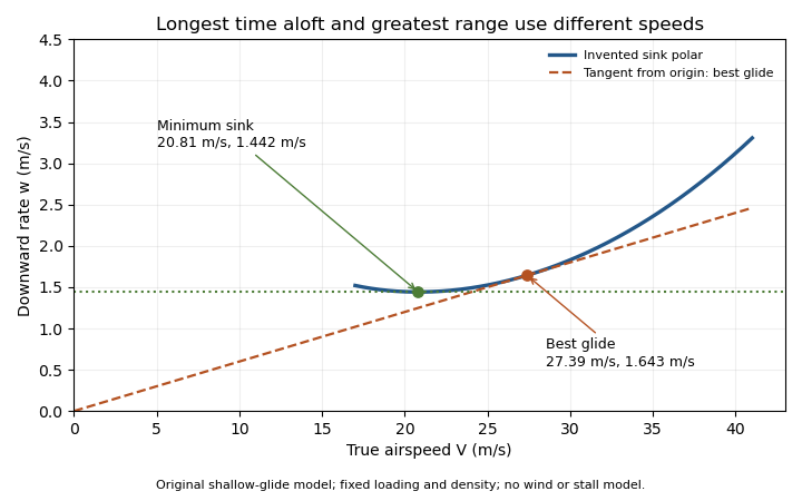
Minimum sink and best glide are different optima of an original shallow-glide polar. Positive vertical values denote downward speed; the curve is an invented model, not measured aircraft performance.
Details. Wind changes the ground-range objective to for an along-track wind component and no vertical air motion. Therefore the still-air optimum need not maximize ground distance. The FAA constructs the corresponding shifted tangents on its performance polars.[ref]
Glider Flying Handbook by Federal Aviation Administration. Chapter 5, printed pp. 5-10–5-11, wind and sinking-air polar constructions
A controlled performance measurement records steady airspeed and height loss at several speeds; wind, vertical air motion and changes of loading must be distinguished from the polar itself.A glider sink polar relates a glider's steady downward speed relative to the air to its true airspeed, for specified loading, configuration and atmospheric conditions. Write it as with downward motion positive. Some plots instead put descent below the horizontal axis; the sign convention must accompany the curve. The minimum sink speed and best glide speed select different points on this same performance relation.[ref]
The curve is a measurement or model of performance, rather than a drawing of the flight path. At steady speed, its horizontal air-relative component is . Thus time per height lost is , while horizontal distance per height lost is . The best glide speed article derives the tangent construction and an original parabolic drag polar example; those results need not be recomputed to interpret a measured polar.
A useful extension is glider ballast scaling. Suppose the gravitational weight changes from to at unchanged air mass density and geometry, and suppose the same lift coefficient and drag coefficient remain applicable. At corresponding steady glide conditions, the lift-to-drag ratio and glide angle are unchanged. Since ,For an original stipulated point and , increasing gravitational weight by gives and . The corresponding distance-to-height ratio is unchanged, but the same height is lost sooner. This scaling assumes the aerodynamic coefficients transfer; changes of Reynolds number, surface condition or trim can invalidate it. The FAA illustrates the loading-dependent displacement of glider polars.[ref]
To measure a polar, record steady true airspeed and height change at several operating points while documenting configuration and loading. Ground-relative descent includes vertical motion of the air: with upward air velocity , . For example, an air-relative sink of in air rising at appears as ground-relative descent. Repetition can reveal variability, but one trajectory cannot separate these two contributions without additional information. A measured groundspeed is likewise not automatically the horizontal component of the air-relative polar.
Nonuniform-density erosion inference estimates a polymer erosion front speed when local density varies through a specimen. Total mass divided by total volume gives an average density; the material being removed at the boundary can have a different density. Substituting the average into a surface-removal formula can therefore misestimate the speed.
For an original spherically symmetric model, let be the surviving radius and the local mass density. ThenDifferentiating the integral and defining positive inward speed givesThis radial form of the Reynolds transport theorem separates loss at a moving boundary from density change inside the surviving volume. All quantities must refer to the same material and conditioning state; a water-filled wet volume and a subsequently dried polymer mass do not automatically define the local density used here.
If the interior density profile is stationary in spatial coordinates over the measurement interval, the integral term vanishes. The appropriate boundary conversion is then , with . Writing average density as , a uniform-density substitution instead givesThe ratio is measurable information about the profile, not a shape correction already supplied by knowing the outer radius.
An illustrative fixed profile is , with a fixed initial radius, not the changing current radius. At , the boundary density is and the mean is . The uniform-density inference overestimates the instantaneous front speed by 25%. For cm, and true , mass leaves at ; dividing by the mean-density surface factor gives . These are stipulated profile values, not measured properties of a named polymer.
Time-dependent hydration, porosity or chemical change can make the interior integral nonzero. One mass slope cannot independently identify both that contribution and boundary speed. Boundary-density calibration for erosion fronts therefore needs spatial or compositional evidence in addition to a bulk mass measurement. NIST's mass-density definition is mass divided by volume; it does not equate a whole-specimen average with an unknown boundary value.[ref]
A discriminating experiment measures outer dimensions, conditioned mass and density or composition at several depths, using matched specimens or accounting for destructive sampling. Kureha's polyglycolic acid protocol filters and dries the remaining solids after water exposure.[ref] That observation supplies retained dry mass, not a radial density field. Establish an inward-moving degradation boundary before interpreting the inferred speed as polymer erosion, rather than another cause of material loss.
Correspondence analysis represents a nonnegative contingency table geometrically, displaying how row and column profiles depart from independence. Let be the table divided by its grand total, and let , be positive marginal proportions. Its standardized residual matrix isA singular value decomposition of provides principal axes; the sum of squared singular values equals the Pearson Pearson chi-squared statistic for contingency tables divided by the grand total. Thus weighted departures from independence become geometric variation.[ref]
For counts , the total is 80 and both margins are . Then has one nonzero singular value . The total inertia is therefore and the Pearson chi-squared statistic for contingency tables is 20. The association has one geometric dimension. Zero margins require removal or separate treatment before using the inverse square roots.
Pinned article: Introduction to the OurBigBook Project
Welcome to the OurBigBook Project! Our goal is to create the perfect publishing platform for STEM subjects, and get university-level students to write the best free STEM tutorials ever.
Everyone is welcome to create an account and play with the site: ourbigbook.com/go/register. We belive that students themselves can write amazing tutorials, but teachers are welcome too. You can write about anything you want, it doesn't have to be STEM or even educational. Silly test content is very welcome and you won't be penalized in any way. Just keep it legal!
Intro to OurBigBook
. Source. We have two killer features:
- topics: topics group articles by different users with the same title, e.g. here is the topic for the "Fundamental Theorem of Calculus" ourbigbook.com/go/topic/fundamental-theorem-of-calculusArticles of different users are sorted by upvote within each article page. This feature is a bit like:
- a Wikipedia where each user can have their own version of each article
- a Q&A website like Stack Overflow, where multiple people can give their views on a given topic, and the best ones are sorted by upvote. Except you don't need to wait for someone to ask first, and any topic goes, no matter how narrow or broad
This feature makes it possible for readers to find better explanations of any topic created by other writers. And it allows writers to create an explanation in a place that readers might actually find it.
Figure 1. Screenshot of the "Derivative" topic page. View it live at: ourbigbook.com/go/topic/derivativeVideo 2. OurBigBook Web topics demo. Source. - local editing: you can store all your personal knowledge base content locally in a plaintext markup format that can be edited locally and published either:This way you can be sure that even if OurBigBook.com were to go down one day (which we have no plans to do as it is quite cheap to host!), your content will still be perfectly readable as a static site.
- to OurBigBook.com to get awesome multi-user features like topics and likes
- as HTML files to a static website, which you can host yourself for free on many external providers like GitHub Pages, and remain in full control

Figure 2. You can publish local OurBigBook lightweight markup files to either OurBigBook.com or as a static website.
Figure 3. Visual Studio Code extension installation.
Figure 5. . You can also edit articles on the Web editor without installing anything locally. Video 3. Edit locally and publish demo. Source. This shows editing OurBigBook Markup and publishing it using the Visual Studio Code extension. 
- Infinitely deep tables of contents:



All our software is open source and hosted at: github.com/ourbigbook/ourbigbook
Further documentation can be found at: docs.ourbigbook.com
Feel free to reach our to us for any help or suggestions: docs.ourbigbook.com/#contact