Topics (384k) Articles (398k) Users (410) Discussions (242) Comments (383) Files (13k) New article

RNA ligation joins compatible RNA termini by forming a new phosphodiester bond. Joining two fragments extends the connected chain; joining the two ends of one fragment produces circular RNA. The end chemistry matters: an ATP-dependent RNA ligase can join a hydroxyl group to a phosphate, whereas RtcB uses a different combination of terminal groups.[ref][ref]
An unmodified linear four-nucleotide fragment has three backbone links, and a six-nucleotide fragment has five. One intermolecular ligation gives ten nucleotides connected by links in a linear chain. Circularizing that chain adds one more backbone link while retaining ten nucleotides. Counting chain units, connecting bonds and separate molecules therefore answers three different questions.
A 1972 experiment made end joining measurable using radioactively labelled RNA. After reaction with a purified RNA ligase, labelled terminal phosphate became resistant to alkaline phosphatase. Additional tests with enzymes attacking from chain ends supported formation of circular poly(A) under the studied conditions.[ref]
Purification and properties of bacteriophage T4-induced RNA ligase by R. Silber and others. Abstract, phosphate-label protection and exonuclease tests
A single protected label would show a chemical change at that end; the complementary end-accessibility tests made the circular-product interpretation stronger.A circular RNA has a covalently closed RNA backbone with no free or end. The closure concerns the chain of phosphodiester bonds, not whether a drawing happens to look round. A linear RNA folded into a stem-loop still has two ends. Circular RNAs can arise through RNA ligation; some cellular examples are generated by back-splicing of transcribed sequences.[ref]
Purification and properties of bacteriophage T4-induced RNA ligase by R. Silber and others. Abstract, intramolecular circularization
[ref]Circular RNAs are abundant, conserved, and associated with ALU repeats by William R. Jeck and others. Abstract and Figures1–4, endogenous backsplice products
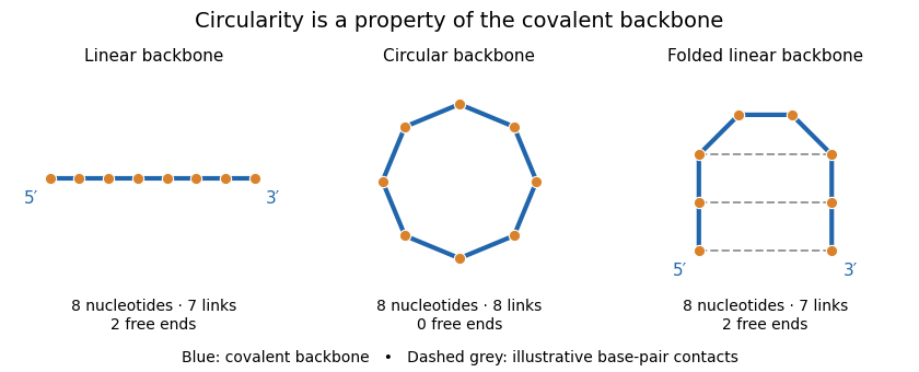
Backbone connectivity distinguishes circular RNA from folded linear RNA
. Original eight-nucleotide schematic, not a molecular structure or folding prediction. Blue lines are covalent backbone links. The circle has eight links and no ends; either linear arrangement has seven links and two ends. Dashed contacts illustrate possible base pairing without joining the ends covalently. No particular sequence or geometric scale is specified. Details.One internal cut in the circle produces a single linear chain. One internal cut in an already linear chain produces two fragments, assuming no other links hold the pieces together. This connectivity difference explains why enzymes requiring a free end can distinguish the molecules, although circularity does not make an RNA immune to every cleavage process.
A 2013 study combined RNA sequencing with and without treatment by RNase R, an enzyme acting from RNA ends. Enrichment of back-splicing junction reads, together with additional validation, supported a substantial population of endogenous circular molecules.[ref]
Circular RNAs are abundant, conserved, and associated with ALU repeats by William R. Jeck and others. Figures1–4, matched treatment comparison and junction validation
Combining chemical resistance with junction evidence is more informative than classifying a molecule solely by its apparent shape or persistence after one treatment.RNA repair restores damaged RNA chemistry or chain continuity. For a strand break, repair can require preparing compatible termini and then using an RNA ligase to form a new phosphodiester bond. The chemical state of an end is therefore part of the substrate: an RNA cyclic phosphate end and a free hydroxyl group need not enter the same joining pathway.[ref]
GTP-dependent RtcB RNA splicing by Anupam K. Chakravarty and Stewart Shuman. Abstract and Figure1, end processing and sealing chemistry
In 2011, purified Escherichia coli RtcB joined a broken transfer RNA-like stem-loop with cyclic-phosphate and hydroxyl termini. Radioactive labelling and denaturing polyacrylamide gel electrophoresis distinguished starting fragments from joined product.[ref]
RtcB is the RNA ligase component of an Escherichia coli RNA repair operon by Naoko Tanaka and Stewart Shuman. Abstract and Figure1, model substrate and product analysis
This established biochemical joining activity and motivated a cellular repair hypothesis. The assay alone did not establish every physiological substrate or prove recovery of all biological functions.For a chain-inventory example, cut an eight-nucleotide RNA into fragments containing three and five nucleotides. Correctly reconnecting their original ends restores eight units in one chain. If one nucleotide has instead been lost, joining the remaining pieces produces seven units; ligation alone does not recreate missing sequence information. Product length, junction chemistry, nucleotide sequence and biological activity consequently provide different tests of what has been restored.
A small nuclear RNA is a member of a class of functional RNAs associated with the cell nucleus, many of which participate in nuclear RNA splicing. Their folded RNA and interactions contribute directly to processing other RNAs, rather than serving as a template for a protein. In a small nuclear ribonucleoprotein, the RNA is assembled with proteins; the RNA molecule and the complete particle are distinct objects.
U1 small nuclear RNA supplies a recognition example. Its complementary pairing with precursor-RNA splice donor sites was tested by introducing altered splice-site and U1 sequences together. A 1986 compensatory base change experiment rescued one tested defect efficiently but another only weakly.[ref]
A compensatory base change in U1 snRNA suppresses a 5′ splice site mutation by Y. Zhuang and A. M. Weiner. Abstract, alternative donor-site perturbations and rescue
This ties sequence recognition to function while showing that restored complementarity alone need not restore every step of splicing.U6 small nuclear RNA illustrates a different role. A 2013 study replaced selected RNA phosphate oxygens with sulfur and tested metal rescue in assembled yeast spliceosomes. The results identified U6 ligands positioning catalytic metals during both splicing reactions.[ref]
RNA catalyses nuclear pre-mRNA splicing by Sebastian M. Fica and others. Page 229, Abstract; Figures 1–3, ligand substitution and metal-rescue comparisons
The evidence concerns RNA-mediated catalysis within a complex machine, not a conclusion that its proteins are dispensable.Thus recognizing a transcript as a small nuclear RNA is a functional classification, not merely observing that it is short and lies in a nucleus. Its molecular partners, target interactions and perturbation response distinguish recognition, assembly and chemical catalysis.
An RNA dimer is a complex containing two associated RNA molecules. The two strands can have identical or different nucleotide sequences, and they need not form one completely paired duplex. Association joins molecular partners; it is different from joining their backbones into one longer covalent RNA strand.
For an original count, two 27-nucleotide strands contain 54 residues altogether. They remain two RNA molecules in a dimer even if much of their base pairing is intermolecular. One continuous 54-residue strand with an intramolecular stem-loop has the same total residue count but a different connectivity. Strand count and RNA secondary structure are separate specifications.
A primary experiment used a 79-nucleotide RNA that gave two bands in native polyacrylamide gel electrophoresis but one band under denaturing conditions. That observation suggested alternative assemblies, but a second method was needed to test their molecular sizes. Sedimentation equilibrium measurements at 20,000 rpm used DNA standards of known size. Samples containing two native-gel bands had an apparent average molecular mass times the monomer mass, compared with for a preparation giving one native band. The combined evidence supported monomer and dimer assignments.[ref]
The study also located an important 21-residue association segment and showed preparation-history effects. Heating and cooling could change the observed assembly, and some dimer states were kinetically trapped.[ref]
Conversion of stable RNA hairpin to a metastable dimer in frozen solution by Xueguang Sun and others. Abstract and Results,21-nucleotide segment and transition reversibility.
Thus two bands are not automatically two sequence lengths, and one predicted low-energy duplex is not sufficient experimental proof of an RNA dimer.For a reversible self-complementary duplex, self-complementary RNA duplex population provides a quantitative two-species model. More complicated contacts, competing folds or RNA melting hysteresis require corresponding experimental and kinetic tests.
RNA integrity describes the preservation of RNA molecules relative to the molecular lengths and structures required by a stated analysis. RNA degradation can leave fragments containing short target sequences even after long transcripts have been broken. Integrity is therefore distinct from total RNA amount, chemical purity, correct nucleotide sequence and preserved biological activity.
An electrophoresis trace measures a size-dependent distribution of material. For a total-RNA preparation, ribosomal RNA contributes prominent features, while smaller fragments can alter the trace as degradation proceeds. The RNA integrity number introduced by Andreas Schroeder and colleagues combines several trace features into an algorithmic score. Its 1-to-10 scale is not a percentage: a score of eight does not mean that of every transcript is intact. A single ribosomal-peak ratio also differs from the multi-feature score.[ref]
The RIN: an RNA integrity number for assigning integrity values to RNA measurements by Andreas Schroeder and others. Abstract, Feature selection, Discussion and Figure8
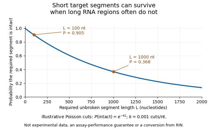
An original cleavage model explains why assay length matters. Suppose cuts form a Poisson process along an RNA strand, with mean density cuts per nucleotide. A required segment of length is unbroken when it contains no cut:At the stipulated density per nucleotide, a 100-nucleotide segment remains intact with probability , while a 1000-nucleotide region survives with probability . Detecting the short sequence consequently does not prove that the full long transcript survived. This is an illustrative independent-cut model, not a fit to a particular RNA sample or a conversion from an integrity score.
A motivating experiment is the 2008 comparison of RNA preservation during laser capture microdissection. Mathieu Clément-Ziza and colleagues measured RNA quality after staining and over the subsequent collection interval. Their tested argon-atmosphere and ethanol-based preparations stabilized the observed RNA quality for up to 90 minutes. Figure1 combines time comparisons with instrumental separation traces.[ref]
Stabilization of RNA during laser capture microdissection by performing experiments under argon atmosphere or using ethanol as a solvent in staining solutions by Mathieu Clément-Ziza and others. Abstract and Figure1: time-dependent measurements, sample scrapes and Bioanalyzer readout
An integrity assessment should state the specimen, handling history, measurement method and downstream requirement. Quantitative PCR directed at one short region and an analysis requiring an intact long transcript need not fail at the same degree of fragmentation. A stable total concentration cannot establish intactness, and a bulk quality score cannot certify every individual molecular species.
RNA degradation is the chemical or enzymatic breakdown of RNA, including cleavage of its phosphodiester bonds into shorter strands. Fragmentation can remove the continuity needed for an RNA molecule's function even while much of its nucleotide sequence remains present. Ribonucleases catalyze RNA cleavage; chemical cleavage also occurs without an enzyme.
A chemical route exploits the hydroxyl group at the position of ribose. Its oxygen can act as a nucleophile toward the adjacent backbone phosphorus, producing an internal transesterification that breaks the chain. In their 1999 primary study, Yingfu Li and Ronald R. Breaker used chimeric DNA/RNA substrates to measure hydroxide-dependent cleavage of RNA phosphodiester bonds. Such a substrate isolates a susceptible linkage within a defined molecular context, allowing chemical instability to be measured separately from a complex sample's overall fragmentation pattern.[ref]
Kinetics of RNA Degradation by Specific Base Catalysis of Transesterification Involving the 2′-Hydroxyl Group by Yingfu Li and Ronald R. Breaker. Abstract and p. 5364: substrates and internal phosphoester-transfer mechanism
An illustrative kinetic model follows the number of molecules that remain intact under constant conditions. If each intact molecule has a constant probability per unit time of undergoing its first destructive cleavage, thenThe last quantity is an RNA half-life in this model. With stipulated , half the starting molecules remain intact after about , and after an hour. The numerical rate is an example, not a universal value for RNA or a measurement from the cited experiment.
A cleavage need not remove material from the tube: breaking one long RNA into two pieces initially preserves its total number of nucleotides. Consequently a concentration measurement can stay almost unchanged while RNA integrity declines. An electropherogram can reveal redistribution toward shorter fragments, while a short target region may remain detectable by reverse transcription and polymerase chain reaction. The relevant question is which molecular continuity the intended assay requires.
In Linear-light RGB, each component is proportional to the amount of its corresponding primary light. Adding or averaging components therefore corresponds to adding or averaging those light contributions, provided the same primaries and reference conditions are used. Nonlinear display encodings such as sRGB must first be decoded for this arithmetic.[ref]
For a nonnegative sRGB channel in , the linear value is when , and otherwise. Thus encoded mid-gray represents approximately0.214 linear light. Averaging encoded black and white gives0.5, while averaging their light gives0.5 linear, encoded as approximately0.735. This explains dark fringes produced by averaging nonlinear channel values directly. The primaries, white point and transfer rule together determine a color space; linearity alone does not specify which colors an RGB triple represents.
An all-trans-retinyl ester is a retinyl ester whose retinyl-chain double bonds have trans geometry. The prefix specifies that part of the molecule; the attached acyl group can still vary. A sample described only by this class name is consequently not a completely specified individual compound.[ref]
All-trans-retinyl ester in ChEBI. CHEBI:63410, Definition and generic structure
Two independent structural choices are involved. Changing the attached acyl group can alter the molecular formula while leaving retinyl-chain geometry unchanged. Changing one retinyl double bond from trans to cis instead changes the geometry without necessarily changing the molecular formula. Esterification and cis-trans isomerism must therefore be distinguished.
The distinction has a concrete role in the visual cycle. A 2005 expression experiment showed that RPE65 converts an all-trans-retinyl ester substrate into 11-cis-retinol, provided the cells also expressed the substrate-supplying lecithin retinol acyltransferase.[ref]
RPE65 is the isomerohydrolase in the retinoid visual cycle by Gennadiy Moiseyev and others. Abstract, supplied ester substrate and 11-cis product
The observed product differs in both ester status and retinyl geometry. Measuring only total retinoid would conceal those chemically important changes.11-cis-retinol is a geometric isomer of retinol with the cis configuration at the double bond identified by position 11 in conventional retinoid numbering. It retains the alcohol group and the formula . The contrast with all-trans-retinol concerns spatial arrangement rather than a change in elemental composition.[ref]
11-cis-retinol in ChEBI. CHEBI:16302, structure and formula
[ref]It is a measured product of RPE65 activity in the visual cycle. In a 2005 study, recombinant expression in cultured cells generated this product when the system also supplied the required all-trans-retinyl ester substrate.[ref]
RPE65 is the isomerohydrolase in the retinoid visual cycle by Gennadiy Moiseyev and others. Abstract, recombinant conversion and substrate requirement
The alcohol must also be distinguished from the aldehyde 11-cis-retinal. Their similar names identify related compounds, not the same oxidation state. Likewise, a mass measurement alone cannot distinguish every retinol geometric isomer: 11-cis-retinol and all-trans-retinol have the same formula. An interpretation of the enzyme experiment needs product identification that resolves the chemical distinction being claimed, rather than simply detecting some vitamin-A-related material.
All-trans-retinol is the form of retinol whose four acyclic double bonds have trans, or , geometry. Its molecular formula is , and it retains the alcohol group characteristic of retinol. The prefix fixes double-bond geometry; it does not specify one frozen conformation about every single bond.[ref]
The distinction from 11-cis-retinol illustrates why elemental composition is incomplete structural information. Both have the same formula, while one specified double bond has different geometry. A mass measurement consistent with that formula cannot on its own identify which isomer is present.
A second distinction concerns the alcohol group. Lecithin retinol acyltransferase can convert retinol into a retinyl ester by transferring an acyl group. Changing ester status and changing cis–trans geometry are separate chemical operations.[ref]
For example, an assay that pools all vitamin-A-related signals could register material before and after esterification without resolving the conversion. An interpretable experiment names the chemical form and the structural distinction being measured, rather than inferring identity solely from a shared retinoid classification.
An ON retinal bipolar cell is a retinal bipolar cell whose central receptive response shows depolarization when illumination increases appropriately. At the input synapse, this reverses the response sign of a photoreceptor, which undergoes hyperpolarization in response to light and releases less glutamate.[ref]
TRPM1 is required for the depolarizing light response in retinal ON-bipolar cells by Catherine W. Morgans and others. Introduction; photoreceptor and ON-bipolar response polarity
The postsynaptic glutamate-sensing receptor mGluR6 controls a current requiring TRPM1. In a 2009 study, TRPM1-deficient mice retained the rod-related electroretinogram a-wave but lacked the b-wave; direct recordings of bipolar responses provided evidence for a postsynaptic signaling defect.[ref]
TRPM1 is required for the depolarizing light response in retinal ON-bipolar cells by Catherine W. Morgans and others. Abstract and Figures3–4; ERG and direct recordings
A local sign model makes the circuit transformation visible. If a small light increment changes glutamate release by and the ON response obeys , with , then . These are illustrative gains, not fitted measurements. The label describes response polarity under specified stimulation, not continuous firing or identical behaviour in every ON subtype.
An OFF retinal bipolar cell is a retinal bipolar cell whose central receptive response shows depolarization for an appropriate decrease in illumination. Its input synapse preserves the sign of a cone photoreceptor's voltage response through excitatory AMPA receptors or kainate receptors, rather than the sign-inverting arrangement of an ON retinal bipolar cell.[ref]
Bipolar cells use kainate and AMPA receptors to filter visual information into separate channels by S. H. DeVries. Introduction; sign-conserving cone-to-OFF transmission
In a 2000 experiment, simultaneous recordings from connected cone cells and OFF retinal bipolar cells, together with receptor-selective manipulations, distinguished receptor mechanisms in different morphological OFF types. Differences in recovery from receptor desensitization filtered the same cone signal into different temporal components.[ref]
Bipolar cells use kainate and AMPA receptors to filter visual information into separate channels by S. H. DeVries. Abstract and Results; paired recordings and receptor pharmacology
Thus two OFF types can agree on whether a light decrement excites them while differing in response timing. Response sign, temporal filtering and neuron identity are related measurements, not interchangeable labels. The result from the studied retina should not be taken as an identical receptor assignment for every OFF subtype in every species.
11-cis-retinal is a geometrical form of retinal with a cis configuration at the carbon-11/carbon-12 double bond. In vertebrate rod rhodopsin, it acts as the bound chromophore that absorbs light. Photoisomerization toward all-trans-retinal triggers a change in the receptor's state, linking molecular geometry to photoreceptor signaling.[ref]
Crystal Structure of Rhodopsin: A G Protein-Coupled Receptor by Krzysztof Palczewski and others. Page 739: bound 11-cis chromophore, photoisomerization and receptor activation.
The name identifies a configuration, not a different number of atoms. 11-cis-retinal and all-trans-retinal have the same molecular composition but differ in the arrangement around a double bond. Replacing a cis arrangement by trans is therefore an example of cis-trans isomerism, rather than cleavage of the retinal molecule into smaller fragments.
The angstrom rhodopsin crystal structure showed the retinal-binding pocket in the dark-state receptor. Bound 11-cis-retinal helps maintain its inactive arrangement; its behavior depends on the surrounding protein, so free retinal and retinal inside a receptor need not have identical optical properties.[ref]
All-trans-retinal is the geometrical form of retinal whose relevant chain double bonds have trans configurations. In rod vision it is formed from the bound 11-cis-retinal chromophore after light absorption by rhodopsin. The change in geometry helps initiate receptor activation, while later processing of retinal participates in the visual cycle.[ref]
Crystal Structure of Rhodopsin: A G Protein-Coupled Receptor by Krzysztof Palczewski and others. Page 739: 11-cis-to-all-trans photoisomerization, receptor change and chromophore dissociation.
“All-trans” describes cis-trans isomerism. It does not mean that the entire molecule is a perfectly straight, rigid line, or that the double bonds have become single bonds. The carbon skeleton and elemental composition are preserved during the idealized isomerization; arrangement changes.
The visual system must subsequently restore a pigment-compatible retinal supply. After bleaching, the retinal-derived material undergoes biochemical processing involving the photoreceptor and retinal pigment epithelium; rebuilding rod rhodopsin requires 11-cis-retinal, not merely any molecule labeled retinal. The RPE65 knockout illustrates the dependence on chemical form: all-trans retinyl esters accumulated while 11-cis retinoids were absent and normal rod responses were lost.[ref]
Rpe65 is necessary for production of 11-cis-vitamin A in the retinal visual cycle by T. M. Redmond and others. Abstract: accumulated all-trans retinyl esters and deficient 11-cis products.
Retinyl esters are distinct chemical species from all-trans-retinal. The experiment demonstrates a disrupted conversion pathway; it should not be paraphrased as though the accumulated ester and the newly photoisomerized aldehyde were the same compound.
Respiratory-system elastance is the local increase in passive elastic pressure per increase in respiratory volume. It is the reciprocal of local respiratory-system compliance:Its units are pressure per volume, such as or . A larger elastance means that the same small volume increment needs a larger elastic pressure increment.
The lung and chest wall share a volume increment while their pressure increments add. Thus their elastances add,whereas their compliances do not. For stipulated and , the elastances are 5 and 10, totaling 15 in reciprocal units. Combined compliance is . The underlying pressure partition is distinct from attributing all measured stiffness to the lung.[ref]
Pulmonary Function Testing by Warren M. Gold and Laura L. Koth. Lung Elasticity, pressure references and compliance
Elastic work in a pressure-volume model gives another meaning to stiffness. Around a passive zero-recoil equilibrium, let added volume be and pressure with constant elastance. The reversible work needed to move from zero to isFor an original SI example, and give final pressure 500 Pa and elastic work 0.125 J. Doubling elastance at the same volume doubles this work; doubling volume at fixed elastance quadruples it. Resistive losses and active muscle work are outside this ideal elastic calculation.
If the pressure-volume curve is nonlinear, local elastance can differ from the interval ratio . Its reciprocal corresponds to the local compliance at that state, not an average across an arbitrary range. A primary ventilator study compared different pressure-volume methods under passive conditions, making this measurement definition operational.[ref]
Static and Dynamic Measurements of Compliance and Driving Pressure: A Pilot Study by Pierre Tawfik and others. Figure 1, methods A–D
Report volume range, passive conditions and whether the value is a derivative or a secant.Plateau airway pressure, , is the airway pressure assessed during an end-inspiratory interval with no flow and sufficiently stable conditions. An end-inspiratory hold can allow the airway and communicating alveolar gas pressures to equilibrate when the respiratory system is passive and the circuit is not leaking.[ref]
Measured values for Ppeak and Pplateau by Hamilton Medical. Definition of airway pressures; Principles of measuring plateau pressures
[ref]The distinction from peak airway pressure follows a simple single-compartment model:During the modeled hold, , leavingHere is respiratory-system compliance, is airway resistance, and total positive end-expiratory pressure supplies the starting reference. The measured difference is the corresponding respiratory driving pressure.
In an original example, total PEEP 8 cm HO, tidal volume 0.40 L and compliance 0.04 L/cm HO give cm HO. If flow immediately beforehand is 0.5 L/s and resistance is 12 cm HO·s/L, the model's airway pressure during that flow is 24 cm HO. The 6 cm HO difference is the modeled resistive contribution, not an extra static distending pressure.
A plateau does not directly measure pleural pressure or isolate the lung from the chest wall. For the same stipulated alveolar pressure 18 cm HO, pleural pressures of 12 and 2 cm HO imply transpulmonary pressures of 6 and 16 cm HO. The airway value alone cannot distinguish those states.
Finally, a stable displayed number is not automatically the same operation as an actual hold. The manufacturer distinguishes measured holds, rapid occlusion and calculated monitoring estimates.[ref]
Measured values for Ppeak and Pplateau by Hamilton Medical. Principles of measuring plateau pressures; Monitoring parameter Pplateau
Reporting the method and its conditions connects plateau airway pressure to the mechanical question it can answer.Positive end-expiratory pressure, PEEP, is airway pressure maintained above atmospheric pressure at the end of exhalation. It specifies an end-expiratory pressure reference, not a tidal volume or a pressure rise measured from the beginning to the end of inspiration. A set external pressure and the total pressure assessed during an appropriate end-expiratory hold need not be identical.[ref]
How to measure driving pressure by Hamilton Medical. Measurement on Hamilton Medical ventilators; End-expiratory hold
One reason is incomplete emptying. In an original passive single-compartment model, let be excess volume above equilibrium with the prescribed outlet pressure. With airway resistance and respiratory-system compliance ,The remaining elastic pressure above the outlet is . Stipulated cm HO·s/L and L/cm HO give a time constant of 1 s. Starting with 0.50 L excess volume, after 0.5 s the model retains about 0.303 L, corresponding to 6.07 cm HO above the outlet pressure. These are constructed mechanics, not ventilator settings for a person.
If the prescribed outlet reference is 5 cm HO, the modeled retained elastic pressure raises the equilibrated total to about 11.07 cm HO. This illustrates intrinsic positive end-expiratory pressure: a pressure contribution that cannot be identified from the external setting alone. The result assumes a communicating passive compartment; active effort, leakage and heterogeneous time constants require further interpretation.
Respiratory driving pressure compares plateau airway pressure with total PEEP, rather than silently subtracting an external setting that may omit retained pressure. The manufacturer's primary measurement account states that distinction and the required passive, leak-free conditions.[ref]
How to measure driving pressure by Hamilton Medical. Driving-pressure definition and measurement conditions
Naming the pressure reference is therefore part of defining the reported mechanical quantity.Respiratory driving pressure, , is the difference between plateau airway pressure and total end-expiratory pressure when measured under suitable passive conditions:The total baseline includes intrinsic positive end-expiratory pressure when present; substituting only the externally imposed baseline can overestimate this tidal pressure difference.[ref]
For the linear static model, respiratory-system compliance is . An original calculation with tidal volume 0.45 L, plateau pressure 23 cm HO and total baseline 8 cm HO gives cm HO and L/cm HO. If the external baseline alone were 5 cm HO, incorrectly subtracting it would give 18 cm HO and a different apparent compliance.
This pressure difference loads both the lung and chest wall. At static endpoints, transpulmonary pressure gives the partitionThus a stipulated airway increment of 12 cm HO corresponds to a lung increment of 4 when the pleural increment is 8, but to a lung increment of 10 when the pleural increment is 2. Equal airway driving pressures do not establish equal lung distension without information about the chest-wall contribution.
The measurement method matters as much as the subtraction. A dynamic estimate during flow can include resistance; an end-inspiratory hold and an end-expiratory occlusion address different endpoints. The primary pilot study explicitly compared static and dynamic ways of obtaining whole-respiratory-system compliance and driving pressure.[ref]
Static and Dynamic Measurements of Compliance and Driving Pressure: A Pilot Study by Pierre Tawfik and others. Introduction, Figure 1 and Study Procedures
These distinctions explain the quantity's mechanical meaning without turning an illustrative number into an individual ventilation recommendation.Peak airway pressure, , is the largest measured airway pressure over the specified breath or inspiratory interval. Its meaning includes the measurement location, reference pressure and temporal response. At the airway opening during positive-pressure inflation, the peak can include the pressure needed for flow through resistance as well as the pressure distending the respiratory system.[ref]
For a passive single-compartment model with constant airway resistance and respiratory-system compliance ,Here is volume above the end-expiratory state and is inspiratory flow. If constant flow persists to the end of inspiration, the largest volume and the resistive pressure occur together, givingWith varying flow, the peak is instead the maximum of the time-dependent sum; separate maxima cannot automatically be added.
An original example takes total end-expiratory pressure 5 cm HO, tidal volume 0.50 L, L/cm HO and L/s. For cm HO·s/L, peak pressure is cm HO. Doubling resistance raises the calculated peak to 25 cm HO while the equilibrated plateau airway pressure stays 15 cm HO. A larger peak can therefore occur without a larger static distending increment.
An end-inspiratory hold removes ongoing airway flow under appropriate conditions and helps separate these contributions. Active respiratory effort, leaks, nonlinear resistance or delayed redistribution complicate that separation. Peak pressure alone neither partitions lung and chest-wall loading nor establishes a unique cause of a pressure change.
Pinned article: Introduction to the OurBigBook Project
Welcome to the OurBigBook Project! Our goal is to create the perfect publishing platform for STEM subjects, and get university-level students to write the best free STEM tutorials ever.
Everyone is welcome to create an account and play with the site: ourbigbook.com/go/register. We belive that students themselves can write amazing tutorials, but teachers are welcome too. You can write about anything you want, it doesn't have to be STEM or even educational. Silly test content is very welcome and you won't be penalized in any way. Just keep it legal!
Intro to OurBigBook
. Source. We have two killer features:
- topics: topics group articles by different users with the same title, e.g. here is the topic for the "Fundamental Theorem of Calculus" ourbigbook.com/go/topic/fundamental-theorem-of-calculusArticles of different users are sorted by upvote within each article page. This feature is a bit like:
- a Wikipedia where each user can have their own version of each article
- a Q&A website like Stack Overflow, where multiple people can give their views on a given topic, and the best ones are sorted by upvote. Except you don't need to wait for someone to ask first, and any topic goes, no matter how narrow or broad
This feature makes it possible for readers to find better explanations of any topic created by other writers. And it allows writers to create an explanation in a place that readers might actually find it.
Figure 1. Screenshot of the "Derivative" topic page. View it live at: ourbigbook.com/go/topic/derivativeVideo 2. OurBigBook Web topics demo. Source. - local editing: you can store all your personal knowledge base content locally in a plaintext markup format that can be edited locally and published either:This way you can be sure that even if OurBigBook.com were to go down one day (which we have no plans to do as it is quite cheap to host!), your content will still be perfectly readable as a static site.
- to OurBigBook.com to get awesome multi-user features like topics and likes
- as HTML files to a static website, which you can host yourself for free on many external providers like GitHub Pages, and remain in full control

Figure 2. You can publish local OurBigBook lightweight markup files to either OurBigBook.com or as a static website.
Figure 3. Visual Studio Code extension installation.
Figure 5. . You can also edit articles on the Web editor without installing anything locally. Video 3. Edit locally and publish demo. Source. This shows editing OurBigBook Markup and publishing it using the Visual Studio Code extension. 
- Infinitely deep tables of contents:




All our software is open source and hosted at: github.com/ourbigbook/ourbigbook
Further documentation can be found at: docs.ourbigbook.com
Feel free to reach our to us for any help or suggestions: docs.ourbigbook.com/#contact