Topics (384k) Articles (398k) Users (410) Discussions (242) Comments (383) Files (13k) New article

Natalia Mast is the first author of a 2011 study explaining how the cholesterol side-chain cleavage enzyme carries out successive reactions converting cholesterol toward pregnenolone. The investigators studied the enzyme bound to the first oxygenated intermediate, combining a crystal structure with solution measurements.[ref]
The active site forms an elongated cavity leading toward the haem group. The intermediate's placement and interactions suggested that movement within this cavity permits successive reactions while the sterol remains associated with the enzyme. Solution spectroscopy tested changes in its position.[ref]
Structural basis for three-step sequential catalysis by the cholesterol side chain cleavage enzyme CYP11A1 by Natalia Mast and others. Abstract and Figure 3: structure and spectral comparisons
The example illustrates why one catalyst can support a multistep transformation without requiring the intermediate to diffuse freely after every step. A bound structure proposes spatial constraints; measurements in solution test whether the arrangement can change, rather than assuming that a crystal captures the only functional position.
Claire Debarnot is the first author of a 2019 structural study of human heparosan-N-sulfate-glucuronate 5-epimerase, an enzyme that modifies heparan sulfate. The reaction changes glucuronic acid residues into iduronic acid residues by epimerization at carbon five.[ref]
The team combined crystal structures with real-time nuclear magnetic resonance measurements. An inactive mutant could bind defined carbohydrate substrates and products, allowing their positions to be examined without rapid conversion obscuring the comparison. The structures showed a bent chain and constrained sugar-ring conformations at the active site.[ref]
Substrate binding mode and catalytic mechanism of human heparan sulfate d-glucuronyl C5 epimerase by Claire Debarnot and others. Abstract; Results, substrate/product complexes; Figures 1A and 2
This approach connects shape and chemical activity. Binding a carbohydrate does not establish that an enzyme catalyses its conversion; the independent activity measurement tested that distinction. Conversely, the bound structures helped explain which local arrangements accompany the reaction.
Carola Hunte is a structural biologist and first author of a 2005 study of NhaA, the sodium/proton antiporter of Escherichia coli. The team determined a crystal structure in a state downregulated by pH, revealing a path from the cytoplasm toward a proposed ion-binding region within the cell membrane.[ref]
The arrangement of short helices and charged groups motivated an alternating access model: changes in the transporter would expose the relevant region toward different sides of the membrane. The paper distinguished the observed structure from the proposed movements and pH-regulatory mechanism.[ref]
That distinction connects two kinds of experiment. Transport measurements count movement of ions; a protein structure supplies candidate routes and interactions. Seeing a possible route in one conformation does not by itself measure transport rate or establish the complete cycle.

A. Malmström coauthored, with L. Åberg, a 1982 assay for the chondroitin-glucuronate 5-epimerase activity involved in dermatan sulfate biosynthesis. The reaction changes the stereochemical configuration of sugar residues already attached to a polysaccharide, rather than synthesizing a free sugar first.[ref]
Biosynthesis of dermatan sulphate. Assay and properties of the uronosyl C-5 epimerase by A. Malmström and L. Åberg. Original byline and Abstract: polymer-level reaction
The experiment used chondroitin carrying tritium at the reacting carbon. Release of the label during incubation was measured with a scintillation counter, converting a hard-to-see molecular exchange into a countable assay signal.[ref]
Biosynthesis of dermatan sulphate. Assay and properties of the uronosyl C-5 epimerase by A. Malmström and L. Åberg. Abstract: labelled substrate, distillation and liquid-scintillation counting
The preparations also distinguished activities associated with dermatan sulfate and heparan sulfate biosynthesis through different requirements for pH and cofactors. Similar overall epimerization chemistry therefore did not establish that the same enzyme performed both biological tasks.[ref]
Biosynthesis of dermatan sulphate. Assay and properties of the uronosyl C-5 epimerase by A. Malmström and L. Åberg. Abstract: two microsomal epimerase activities
L. Åberg coauthored a 1982 investigation of an enzyme involved in making dermatan sulfate. With A. Malmström, L. Åberg used an isotope labelling assay to follow hydrogen exchange during epimerization of residues in chondroitin.[ref]
The label was tritium attached at carbon five. Material released during incubation was distilled and quantified by liquid-scintillation counting. The study also compared conditions supporting the two epimerase activities present in microsome preparations.[ref]
Biosynthesis of dermatan sulphate. Assay and properties of the uronosyl C-5 epimerase by A. Malmström and L. Åberg. Abstract: isotope readout and different cofactor/pH requirements
The assay illustrates a distinction between a chemical event and the signal used to detect it. Radioactive counts report movement of a label under specified conditions; interpreting them as an enzyme activity requires an appropriate labelled substrate and comparisons for background release. Counting the signal alone would not identify which enzyme produced it.
Bert J. C. Janssen is the first author of a 2005 structural study of Complement C3 and its processed fragment C3c. The team determined crystal structures that connected the arrangement of protein domains to activation and regulation in the complement system.[ref]
The structures helped explain how a reactive thioester, needed for later covalent attachment, can remain protected against premature hydrolysis. Comparing the native and processed forms also revealed substantial changes in domain arrangement.[ref]
The comparison is specifically Complement C3 versus C3c. It is not a direct pair of structures showing native Complement C3 and active C3b. Keeping the fragments distinct matters because different stages of processing have different biological roles: an informative structural difference is not automatically the exact movement that creates an active intermediate.
Natallia Strushkevich is the first author of a 2011 structural study of the mitochondrial system that converts cholesterol toward pregnenolone. The team examined the cholesterol side-chain cleavage enzyme together with its electron-transfer partner adrenodoxin.[ref]
An engineered fusion protein helped hold the partners together for crystal structure determination. The structure related the iron-sulfur cluster of adrenodoxin to the haem group of the catalytic protein, while structures with different bound sterols addressed successive reaction stages.[ref]
The engineered connection was an experimental aid, not evidence that the native proteins form one continuous chain. Comparing fusion proteins with different linker lengths also tested how the imposed connection affected reduction of the catalytic partner.[ref]
Structural basis for pregnenolone biosynthesis by the mitochondrial monooxygenase system by Natallia Strushkevich and others. Methods and supporting Figure S8: linker-dependent electron transfer
The example connects a useful structure-stabilizing intervention to an explicit check on its functional consequences.Carissa L. White coauthored a 2002 study identifying a heparosan synthase. The characterized glycosyltransferase combined two sugar-transfer activities in one protein, building a heparosan chain from glucuronic acid and N-acetylglucosamine units. The product was the unsulfated, unepimerized backbone related to heparin, rather than a fully modified heparin molecule.[ref]
Identification and molecular cloning of a heparosan synthase from Pasteurella multocida type D by P. L. DeAngelis and Carissa L. White. Abstract, Journal of Biological Chemistry 277 (2002), pp. 7209–7213; DOI 10.1074/jbc.M112130200.
The study tested which nucleotide sugars supported chain formation and compared the product's susceptibility to different polysaccharide-cleaving enzymes. An illuminating distinction is between building a repeating backbone and modifying that backbone: identifying a heparosan synthase establishes a particular polymerization activity, but does not establish all the later reactions needed to make a differently substituted glycosaminoglycan. The reported one-protein, two-activity mechanism also shows that two alternating chemical additions need not require two separate polypeptides.[ref]
Identification and molecular cloning of a heparosan synthase from Pasteurella multocida type D by P. L. DeAngelis and Carissa L. White. Abstract: dual-action transferase, donor specificity and product digestion.
A. J. Padgett-McCue coauthored a 2000 study identifying a chondroitin synthase and establishing what polysaccharide it made. The work connected an enzyme with the alternating glucuronic acid and N-acetylgalactosamine units of chondroitin. The characterized protein carried both sugar-transfer activities required to extend this repeating chain.[ref]
Identification and molecular cloning of a chondroitin synthase from Pasteurella multocida type F by P. L. DeAngelis and A. J. Padgett-McCue. Abstract, Journal of Biological Chemistry 275 (2000), pp. 24124–24129; DOI 10.1074/jbc.M003385200.
The experimental reasoning combined nucleotide sugar specificity, size-exclusion chromatography and selective enzymatic digestion of the product. This gives a useful example of complementary measurements: a large molecular size establishes that a polymer was made, but does not identify its repeating chemical structure. Agreement between the sugar composition and the response to a chondroitin-cleaving enzyme supplied more specific evidence. The contribution is therefore a chemically tested biosynthetic assignment, rather than a prediction based only on similarity between protein sequences.[ref]
Identification and molecular cloning of a chondroitin synthase from Pasteurella multocida type F by P. L. DeAngelis and A. J. Padgett-McCue. Abstract: donor specificity, gel filtration and contrasting lyase susceptibility.
O. Habuchi coauthored a 1982 study, with N. Miyashita, separating two positional sulfotransferase activities from chick embryo cartilage. The work experimentally distinguished chondroitin 4-sulfotransferase and chondroitin 6-sulfotransferase, which modify different positions in the same kind of carbohydrate backbone.[ref]
Separation and characterization of chondroitin 6-sulfotransferase and chondroitin 4-sulfotransferase from chick embryo cartilage by O. Habuchi and N. Miyashita. original byline and Abstract, positional activities
The separated preparations differed in their biochemical behavior. Under the reported comparison without sulfhydryl compounds, the 4-sulfotransferase activity was very low, whereas the 6-sulfotransferase activity was essentially unaffected.[ref]
This is useful evidence because an assay condition can change the contribution of different activities within a preparation. In a hypothetical mixture with two activities, observing a decline in total signal would not establish that both activities declined equally. Separating them and measuring each under matched conditions supplies more specific information.
The distinction also links reaction chemistry to the meaning of an enzyme assay. Chondroitin modification can vary in the number and location of attached groups, while protein abundance supplies another measurement. The credited study provides a concrete example of using separable properties to investigate how a family of related reactions is implemented.
N. Miyashita coauthored, with O. Habuchi, a 1982 biochemical study distinguishing chondroitin 6-sulfotransferase from chondroitin 4-sulfotransferase in chick embryo cartilage. The activities were separated by size-exclusion chromatography, allowing their properties to be compared in different preparations.[ref]
Separation and characterization of chondroitin 6-sulfotransferase and chondroitin 4-sulfotransferase from chick embryo cartilage by O. Habuchi and N. Miyashita. original byline and Abstract, separation of the two activities
The scientific question concerns position as well as amount. Both enzymes modify chondroitin, but they act at different positions of N-acetylgalactosamine. Equal total group incorporation could therefore conceal different products. Distinguishing the activities gave an experimental way to investigate that structural difference.[ref][ref]
The study also reported removing endogenous acceptors from the enzyme preparations.[ref]
Separation and characterization of chondroitin 6-sulfotransferase and chondroitin 4-sulfotransferase from chick embryo cartilage by O. Habuchi and N. Miyashita. Abstract, acceptor removal
That feature makes the assay logic illuminating: material carried through purification can participate in the measured reaction. Defining the added acceptor is easier when an uncontrolled acceptor pool has been removed. The work thus connects separation of proteins with interpretation of their reaction-specific activity, rather than treating purification as a measurement of protein amount alone.Beom Seok Park is a researcher credited with structural studies of recognition in innate immunity. He was first author of the 2009 study of the Toll-like receptor 4–MD-2 complex with lipopolysaccharide; the original paper records his Department of Chemistry affiliation at KAIST.[ref]
The structural basis of lipopolysaccharide recognition by the TLR4–MD-2 complex by Beom Seok Park and others. Author byline and affiliation; Nature 458, 1191–1195
The crystal structure connected ligand recognition to assembly. Most of the lipopolysaccharide's lipid chains fit inside a pocket in MD-2, while an exposed chain helps make an interface with another Toll-like receptor 4. The assembly contains two receptor-complex units. Thus the ligand helps organize contacts between proteins, rather than merely occupying an isolated binding pocket.[ref]
The structural basis of lipopolysaccharide recognition by the TLR4–MD-2 complex by Beom Seok Park and others. Abstract; Figures 1–3, ligand pocket and dimerization interfaces
A simple comparison shows why this matters. Imagine two ligands that both bind a receptor-associated pocket, but only one presents an exposed surface in the orientation required to join two receptor units. Equal pocket occupancy would not guarantee equal assembly or signaling. This is a conceptual example, not a numerical affinity measurement from the paper.
Park and collaborators' contribution was to provide a molecular account linking the arrangement of lipopolysaccharide, MD-2 and Toll-like receptor 4 to receptor activation. A crystal structure reveals the contacts in the captured complex; claims about response strength or behavior in an intact organism require further experimental measurements.[ref]
Alexander E. Aleshin is a researcher in structural biology credited with studies of the complement system. He was first author of the 2012 determination of the crystal structure of complement C6, with a Sanford-Burnham Medical Research Institute affiliation in the original byline.[ref]
Structure of complement C6 suggests a mechanism for initiation and unidirectional, sequential assembly of membrane attack complex (MAC) by Alexander E. Aleshin and others. Author affiliations; Results, Overall Structure of C6
The biological problem was how soluble proteins assemble a membrane-damaging machine. Complement C6 is recruited during formation of the membrane attack complex. Its structure revealed a closed arrangement of protein domains. Comparing this arrangement with related components suggested how changes in domain orientation could help expose membrane-inserting regions and propagate assembly.[ref]
Structure of complement C6 suggests a mechanism for initiation and unidirectional, sequential assembly of membrane attack complex (MAC) by Alexander E. Aleshin and others. Abstract; Results, C6 Adopts a Default Closed Autoinhibited State; Discussion
A useful mechanical analogy is a folded tool whose working surface is inaccessible until a hinge opens. The analogy motivates two experimentally distinct questions: what configurations exist, and what causes transitions between them? Determining a crystal structure answers the first kind of question for the crystallized state; it does not directly film the assembly sequence.
The distinction is explicit in this work. The complement C6 coordinates and comparisons constrain a proposed model of directional membrane attack complex assembly. Testing the proposed succession of movements requires additional evidence about interacting components and assembly intermediates. Aleshin and colleagues thus connected a measured molecular shape to a testable explanation of a larger biological process.[ref]
Structure of complement C6 suggests a mechanism for initiation and unidirectional, sequential assembly of membrane attack complex (MAC) by Alexander E. Aleshin and others. Discussion, Model for MAC Initiation and Final Steps in MAC Assembly
Brian T. Wimberly is a researcher in RNA structure and ribosome function. The full byline of the 2000 paper on the bacterial small ribosomal subunit establishes the expansion of the credited initials B. T. Wimberly. The paper lists him among six equally contributing authors at the forefront of its structural work.[ref]
Structure of the 30S ribosomal subunit by B. T. Wimberly and others. Publisher author list, affiliations and equal-contribution note
The team determined a 30S ribosomal subunit structure refined to resolution using X-ray crystallography. It located the folded ribosomal RNA and associated ribosomal proteins in a molecular model, providing a framework for interpreting decoding, assembly and biochemical interactions.[ref]
Structure of the 30S ribosomal subunit by B. T. Wimberly and others. Abstract; Figures 1–2, electron-density maps and structure overview
The intellectual advance connected molecular arrangement to a function: how a ribosome reads a messenger RNA sequence. The small ribosomal subunit organizes the interactions of codons with transfer RNA anticodons, so the geometry of its RNA and proteins constrains explanations of decoding.
A constructed comparison clarifies what a structural measurement contributes. Two models could contain the same list of molecules yet place a functional contact apart in one and apart in the other. Composition alone cannot distinguish them; spatial evidence can. This is not a distance reported in the paper, and nominal spatial resolution should not be interpreted as identical coordinate precision for every atom.
Wimberly's institutional biography also describes using complementary structural methods to relate RNA organization to biological function.[ref]

Brian T. Wimberly
. Portrait identified by the New York Structural Biology Center profile: nysbc.org/people/brian-wimberly .Marina Serna is a researcher in structural biology whose work includes cryo-electron microscopy of large protein complexes. She was first author of the 2016 study determining the architecture of the complement system's membrane attack complex.[ref]
Structural basis of complement membrane attack complex formation by Marina Serna and others. Original author list; Results, Cryo-EM structure of the MAC
The experiment linked function to structure. The researchers assembled complexes on liposomes, checked release of a fluorescent marker to establish functional pores, then isolated the complexes for frozen-hydrated imaging. The reconstruction identified eighteen copies of complement C9 in the analyzed completed pore.[ref]
Structural basis of complement membrane attack complex formation by Marina Serna and others. Results, Cryo-EM structure of the MAC and Molecular architecture; Figure 1
A constructed fluorescence example explains why assay controls matter. Suppose a no-release control gives signal , a sample gives , and a complete-release reference gives , all in compatible instrument units. A normalized release readout would beThis is an invented linear-calibration example, not the paper's measured values. The raw sample signal is forty units, but the estimated fraction is thirty percent because the background and reference determine the denominator.
The functional check and structural reconstruction answer different questions. Marker release tests whether a preparation makes membranes permeable; the reconstructed density constrains the architecture of selected complexes. Eighteen C9 copies in the studied end product do not establish that eighteen are a universal minimum for membrane damage.[ref]
Structural basis of complement membrane attack complex formation by Marina Serna and others. Results, completed-pore stoichiometry distinguished from lysis by fewer C9 molecules

Marina Serna
. Portrait by Esther Sánchez / CNIO, explicitly identified in the institutional research profile: www.cnio.es/en/news/cnio-researcher-marina-serna-awarded-for-her-contribution-to-solving-a-crucial-biological-problem-2/ .D. R. Davies is the published author name credited on a structural study of Tn5 transposase. The original 2000 paper lists a University of Wisconsin biochemistry affiliation. Its initials are retained here because the scientific byline establishes this contribution without establishing a full personal identity.[ref]
Three-dimensional structure of the Tn5 synaptic complex transposition intermediate by D. R. Davies and others. Published byline, affiliation and Abstract; Science 289, 77–85
Davies and collaborators determined a protein–DNA assembly at ångströms crystallographic resolution. The protein dimer held two DNA ends, with each end contacted by both subunits. The arrangement explained how one subunit could bind an end while the other supplied the active site for cleavage. It supplied a structural framework for understanding coordinated DNA transposition.[ref]
Three-dimensional structure of the Tn5 synaptic complex transposition intermediate by D. R. Davies and others. Abstract: dimeric complex, shared DNA contacts and cleavage in trans
The geometry is more revealing than a count of components. Imagine two subunits A and B and two DNA ends L and R. Knowing merely that the assembly contains two subunits and two ends leaves open an arrangement in which A independently processes L and B independently processes R. Contacts that span the two subunits instead couple recognition of an end to the partner's catalytic machinery. Such an arrangement provides a concrete reason why an isolated, correctly bound DNA end need not behave like the complete transpososome.
The quoted distance is nanometres, since one ångström is nanometres. Crystallographic resolution describes the spatial detail supported by the diffraction data; it is not a frame interval in a movie and does not mean that every atomic coordinate has an uncertainty of precisely nanometres. A static structure constrains possible reaction mechanisms, while the sequence and rates of chemical steps require additional evidence. The distinction matters whenever an attractive structural model is used to explain a dynamic enzyme reaction.
Sk Tofajjen Hossain is the published author name credited with biochemical studies of how RNase R processes structured RNA. The original 2016 paper gives this name and a University of Miami biochemistry affiliation; the initial element Sk is retained without expansion.[ref]
How RNase R Degrades Structured RNA: Role of the Helicase Activity and the S1 Domain by Sk Tofajjen Hossain and others. Published byline and affiliation
Hossain and collaborators compared the enzyme's RNA-unwinding and nuclease activities using designed substrates and changes in its S1 domain. They also used intrinsic tryptophan fluorescence to investigate changes associated with RNA binding. Measuring these different observables helped distinguish RNA recognition, structural change and degradation rather than treating all of them as one event.[ref]
A generic binding model makes one important distinction clear. If a single binding site has dissociation constant and free ligand concentration , its ideal equilibrium occupancy isAt , half the sites are occupied; at , the occupancy is , not twice as large. These are mathematical examples, not fitted values from the paper. They show why an increase in measured binding need not be proportional to ligand concentration, and why an equilibrium binding result alone does not measure chemical turnover.
In particular, detecting ATP bound to an enzyme is different from demonstrating ATP hydrolysis. A binding signal does not, by itself, identify how energy is used during RNA unwinding. Likewise, an altered fluorescence signal can report a changed local environment without directly counting phosphodiester bonds cleaved. The strength of the study lies in relating separate measurements to a model of how RNA secondary structure is handled, rather than inferring the entire mechanism from one signal.
William N. Beavers is a researcher whose work combines analytical chemistry with the study of chemical changes to proteins. His institutional profile identifies post-translational modification as a research interest and directly lists his scientific publications.[ref]
William N. Beavers research profile at Louisiana State University. Named faculty profile, Research Interest and Select Publications
In a 2017 study, Beavers and collaborators investigated proteins modified by endogenously generated lipid electrophiles. Chemical labelling and selective capture enabled a broad analysis of modified targets. The results highlighted mitochondria as both a source associated with electrophile generation and a prominent location of modified proteins in that experimental system.[ref]
A useful quantitative distinction is the amount of a modified protein versus the fraction of that protein that is modified. For an original ideal measurement model, suppose response-corrected signals correspond to modified and unmodified molecules in sample A, and modified and unmodified molecules in sample B. The modified amount doubled, but the modified fraction is in both samples. These are invented counts, not results from the paper.
The response correction is essential. A chemically modified molecule may be captured or detected differently from an unmodified one, so raw signal intensities cannot automatically be substituted for molecule counts. Nor does more captured material alone establish a higher probability of modification if total protein abundance also changed. An informative interpretation therefore distinguishes chemical selectivity, total abundance and the denominator used for a reported fraction.
Beavers is also credited as a coauthor on a separate 2020 microbial-community study, illustrating the range of scientific contexts in which chemical measurements and biological comparisons meet.[ref]

M. Bycroft is the first-named author of a 1997 structural study of the S1 RNA-binding domain. The paper examined this protein domain from Escherichia coli polynucleotide phosphorylase using nuclear magnetic resonance. Its initial-only byline is retained here.[ref]
The resulting protein structure contained a five-stranded antiparallel beta barrel. Conserved amino acid residues and neighboring loops suggested a possible RNA-binding face. Comparing this fold with other proteins connected a small structural module with a broader family of nucleic-acid interactions.[ref]
The solution structure of the S1 RNA binding domain: a member of an ancient nucleic acid-binding fold by M. Bycroft and others. Abstract: solution structure, putative RNA-binding face and related proteins
The distinction between an observed fold and a proposed binding face is scientifically useful. Finding several conserved amino acid residues on the same exposed surface suggests an experiment: alter selected residues, then compare RNA binding while independently checking whether the protein still folds. Weaker binding after a change that also unfolds the protein would not establish a direct contact at that residue. This is an experimental interpretation of the structural hypothesis, not a report that every such test was performed in the 1997 paper.
For a simple architectural example, two proteins can each contain one S1 RNA-binding domain while having different additional protein domains. Sharing that module alone does not imply identical enzyme activity, the same RNA preference, or an equal number of active sites.
Jeong Ho Chang is a researcher in structural biology and a coauthor of a 2011 study of Xrn1, an exoribonuclease that degrades RNA from its end toward its end. The paper's contribution statement explicitly credits Chang with mutagenesis, nuclease assays and thermal shift assays, as well as shared crystallographic work.[ref]
The team combined X-ray crystallography with biochemical measurements. A defined protein construct yielded structures at resolution. Additional protein domains lay away from the active site, yet alterations in these regions affected activity. Comparisons of RNA binding, degradation and thermal stability helped separate possible explanations for a defective enzyme.[ref]
Structural and biochemical studies of the 5′→3′ exoribonuclease Xrn1 by Jeong Ho Chang and others. Results, Structure determination and Domain D1 may stabilize the N-terminal segment; Figures 5–6
One important experimental distinction is that the crystallized E178Q construct was catalytically defective; its detailed protein structure was not itself a demonstration of normal Xrn1 activity. A structural model and a functional assay supply complementary evidence.[ref]
Structural and biochemical studies of the 5′→3′ exoribonuclease Xrn1 by Jeong Ho Chang and others. Structure determination and Figure 5b: E178Q structure and essentially absent nuclease activity
For an original measurement example, suppose arbitrary units of intact RNA remain in a control and after an assay. The disappearance fraction is . That observation alone does not identify the degradation products, the processivity, or whether binding became weaker. Each requires an additional readout; the invented numbers illustrate why several assays are useful.

Jeong Ho Chang research profile at Kyungpook National University. Named profile, linked portrait and explicit listing of the 2011 Xrn1 paper
Pinned article: Introduction to the OurBigBook Project
Welcome to the OurBigBook Project! Our goal is to create the perfect publishing platform for STEM subjects, and get university-level students to write the best free STEM tutorials ever.
Everyone is welcome to create an account and play with the site: ourbigbook.com/go/register. We belive that students themselves can write amazing tutorials, but teachers are welcome too. You can write about anything you want, it doesn't have to be STEM or even educational. Silly test content is very welcome and you won't be penalized in any way. Just keep it legal!
Intro to OurBigBook
. Source. We have two killer features:
- topics: topics group articles by different users with the same title, e.g. here is the topic for the "Fundamental Theorem of Calculus" ourbigbook.com/go/topic/fundamental-theorem-of-calculusArticles of different users are sorted by upvote within each article page. This feature is a bit like:
- a Wikipedia where each user can have their own version of each article
- a Q&A website like Stack Overflow, where multiple people can give their views on a given topic, and the best ones are sorted by upvote. Except you don't need to wait for someone to ask first, and any topic goes, no matter how narrow or broad
This feature makes it possible for readers to find better explanations of any topic created by other writers. And it allows writers to create an explanation in a place that readers might actually find it.
Figure 1. Screenshot of the "Derivative" topic page. View it live at: ourbigbook.com/go/topic/derivativeVideo 2. OurBigBook Web topics demo. Source. - local editing: you can store all your personal knowledge base content locally in a plaintext markup format that can be edited locally and published either:This way you can be sure that even if OurBigBook.com were to go down one day (which we have no plans to do as it is quite cheap to host!), your content will still be perfectly readable as a static site.
- to OurBigBook.com to get awesome multi-user features like topics and likes
- as HTML files to a static website, which you can host yourself for free on many external providers like GitHub Pages, and remain in full control

Figure 2. You can publish local OurBigBook lightweight markup files to either OurBigBook.com or as a static website.
Figure 3. Visual Studio Code extension installation.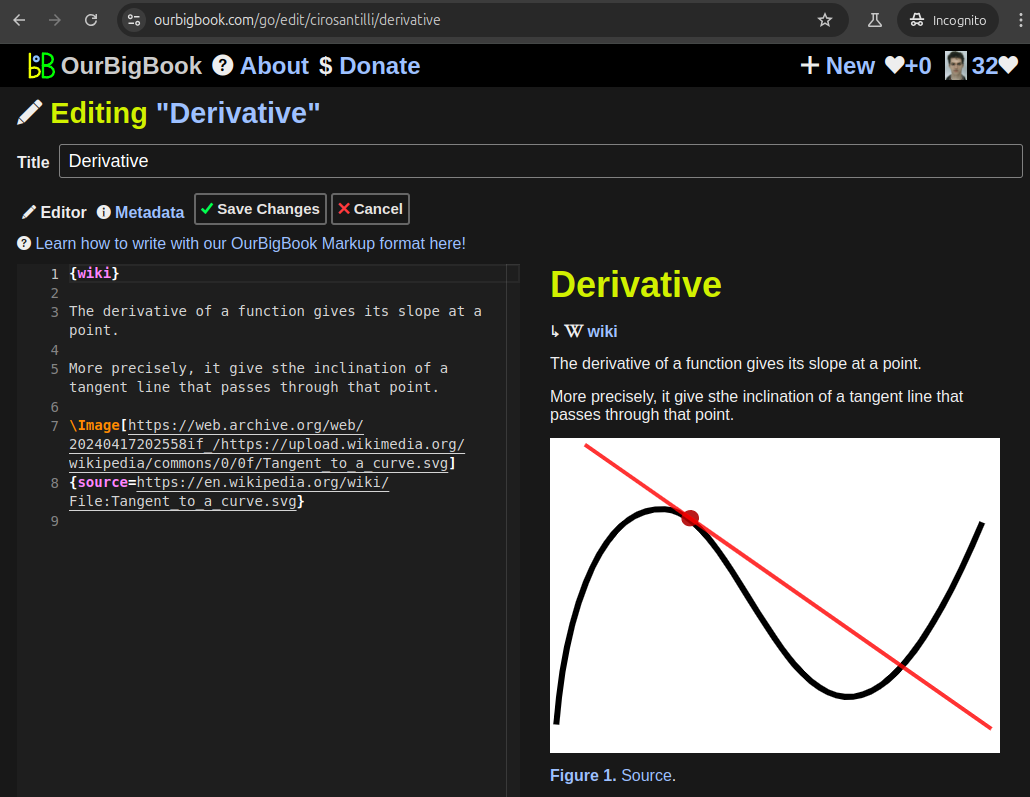
Figure 5. . You can also edit articles on the Web editor without installing anything locally. Video 3. Edit locally and publish demo. Source. This shows editing OurBigBook Markup and publishing it using the Visual Studio Code extension. 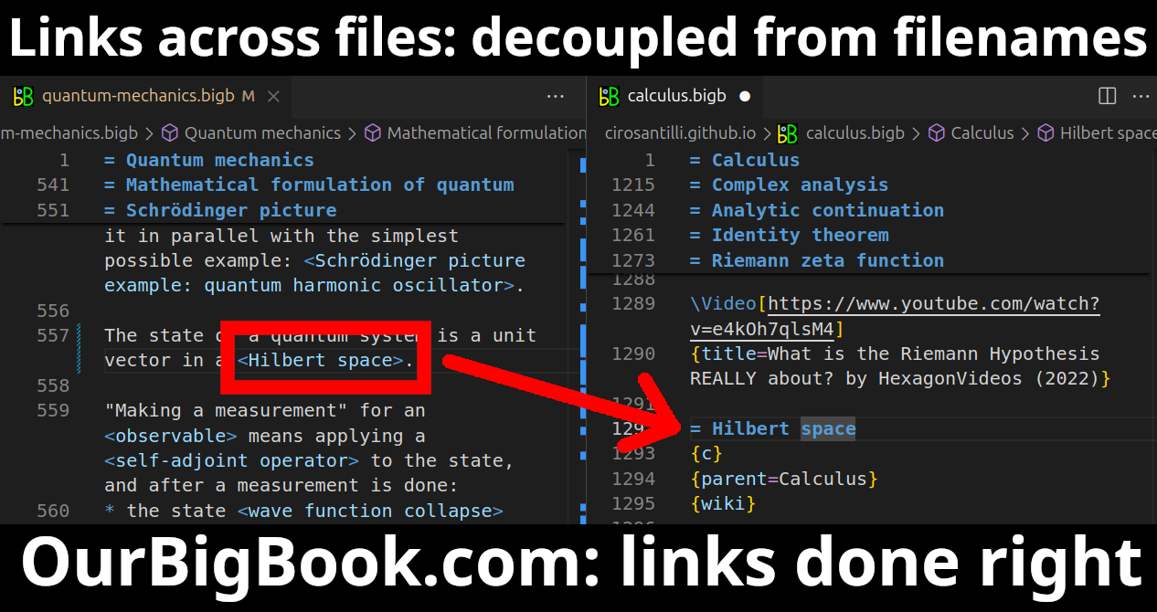
- Infinitely deep tables of contents:
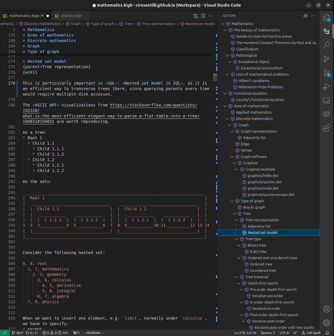

All our software is open source and hosted at: github.com/ourbigbook/ourbigbook
Further documentation can be found at: docs.ourbigbook.com
Feel free to reach our to us for any help or suggestions: docs.ourbigbook.com/#contact