
A peptide hormone is a signalling peptide or protein released by cells to influence other cells. Insulin, glucagon and somatostatin are examples. Biosynthesis commonly includes a precursor and regulated processing before secretion. Because a peptide generally does not freely cross a lipid bilayer, its signal is commonly received by a cell-surface receptor and relayed through intracellular pathways. [ref]
Molecular Biology of the Cell, 4th edition by Bruce Alberts and others. Chapter 15, General Principles of Cell Communication: endocrine signals and surface receptors
Glucagon is a peptide hormone made by alpha cells in the pancreatic islets. Its mature polypeptide has 29 amino acid residues. Alpha cells store glucagon in secretory granules and release it by exocytosis. Glucagon is a message about fuel availability; glucose is the sugar whose supply it helps regulate. A falling blood glucose concentration favors glucagon release, whereas a rise usually suppresses it. Amino acids can also stimulate release, so a meal containing protein need not produce the same hormone pattern as a meal dominated by carbohydrates. [ref]
The principal blood glucose-raising target is the liver. In a hepatocyte, the glucagon receptor is a G protein-coupled receptor: activation of a stimulatory G protein drives adenylyl cyclase, increasing the second messenger cAMP and activating protein kinase A. Downstream phosphorylation, including the phosphorylase kinase relay to glycogen phosphorylase, favors glycogenolysis. Glucagon also supports gluconeogenesis. These are different supplies: glycogenolysis draws on a finite glycogen reserve, whereas gluconeogenesis makes glucose from other substrates and depends on their availability. Longer fasting depletes the glycogen reserve; a stronger hormone signal cannot release material that is no longer there. [ref] [ref]
Do not copy the liver pathway into every target of insulin. Skeletal muscle uses its own glycogen reserve locally; its glucose uptake and the liver's glucose output are different flows. Nor is direct glucagon-stimulated lipolysis in human adipose tissue as firmly established as its action in the liver. Glucagon action depends on the tissue, other hormones, and available substrates. [ref]
There is a useful surprise inside the pancreatic islets: local glucagon can support insulin release from neighboring beta cells. The islet perifusion experiment distinguishes that interaction from glucagon's action on the liver. Thus the two hormones can oppose each other's effects on blood glucose while cooperating locally. Delta cells supply another local brake through somatostatin. [ref] [ref]
Adrenaline, also called epinephrine, is an endogenous catecholamine messenger: the body makes it and can release it into blood as a hormone. The adrenal medulla, the inner region of an adrenal gland, secretes adrenaline in response to sympathetic nervous system stimulation. A circulating hormone broadcasts a signal, but each receiving biological cell interprets it through its own receptors and intracellular machinery. Adrenaline activates both alpha-adrenergic receptors and beta-adrenergic receptors; it does not command one identical response everywhere.[ref]
Adrenal Gland by National Cancer Institute. Hormones of the Adrenal Medulla: secretion in response to sympathetic stimulation
[ref]Epinephrine MeSH descriptor by National Library of Medicine. Epinephrine, D004837: Scope Note, entry term Adrenaline and Catecholamines tree placement
Fuel mobilization is a useful example. A beta-adrenergic receptor can increase adenylyl cyclase activity through a stimulatory G protein, increasing the intracellular second messenger cAMP. Protein kinase A responds to cAMP; a phosphorylation relay through phosphorylase kinase activates glycogen phosphorylase, favoring glycogenolysis. Here adrenaline is the extracellular message, cAMP is the intracellular message, and glycogen is the reserve being mobilized. These are three different roles.[ref]
The regulatory component of adenylate cyclase. Purification and properties by P. C. Sternweis and others. Abstract, properties 2-4: purified guanine-nucleotide regulatory component reconstitutes hormone-stimulated adenylyl cyclase
[ref]An adenosine 3′,5′-monophosphate-dependant protein kinase from rabbit skeletal muscle by D. A. Walsh and others. Walsh, Perkins and Krebs, 1968, printed p. 3763 Summary and introductory paragraphs: cAMP-dependent kinase and phosphorylase kinase activation
[ref]Pharmacological Characterizations of Adrenergic Receptors in Human Adipocytes by Thomas W. Burns and others. Abstract, second paragraph: positive beta-receptor coupling to adenylyl cyclase
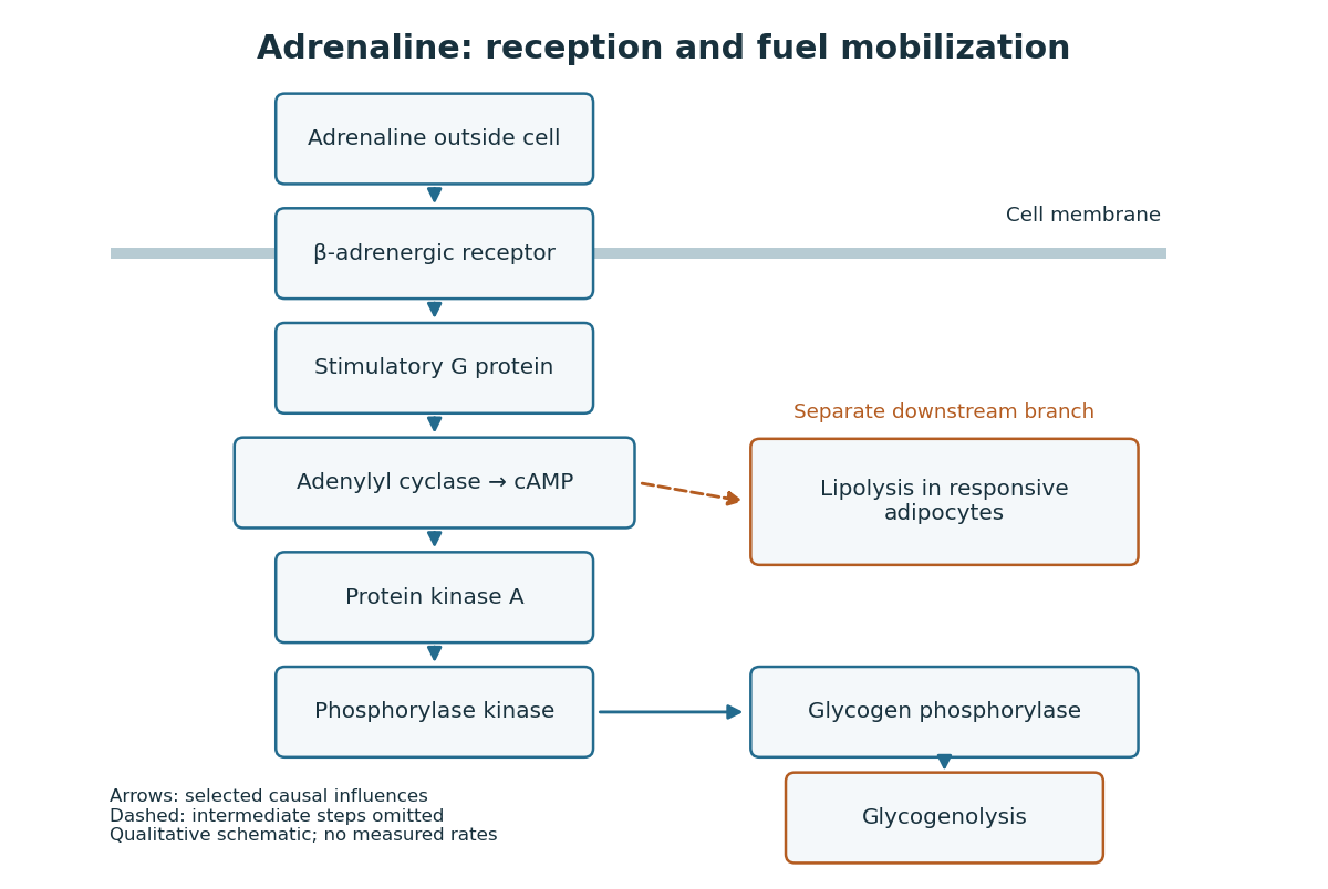
Adrenaline reception and cAMP-dependent fuel mobilization
. Original pathway schematic. Solid arrows show a selected activating relay to glycogenolysis; the dashed branch summarizes promotion of lipolysis in responsive adipocytes, omitting its intermediate steps. Arrows denote causal influences, not transport of adrenaline into the biological cell, reaction rates or measured yields. Details.
The two reserves provide different material. Glycogenolysis draws on glycogen; lipolysis breaks down stored triglycerides, releasing fatty acids and glycerol. A skeletal muscle biological cell's own reserve and fuel released by adipose tissue are different supplies. Increasing a signal cannot mobilize an empty reserve: receptor activation changes the use of available material rather than creating fuel. The adrenergic adipocyte assay below gives direct human evidence for the lipolysis branch.[ref]
Pharmacological Characterizations of Adrenergic Receptors in Human Adipocytes by Thomas W. Burns and others. Abstract: epinephrine-stimulated glycerol release as an index of lipolysis
Receptor context can even oppose this branch. In the human adipocyte preparation below, beta-adrenergic receptors promoted cAMP accumulation, whereas alpha-2 adrenergic receptors opposed it. Alpha-1 adrenergic receptors were associated with a different phospholipid readout. The balance of these responses matters more than the name of the messenger alone.[ref]
Pharmacological Characterizations of Adrenergic Receptors in Human Adipocytes by Thomas W. Burns and others. Abstract: opposing beta and alpha-2 coupling; separate alpha-1 phospholipid response
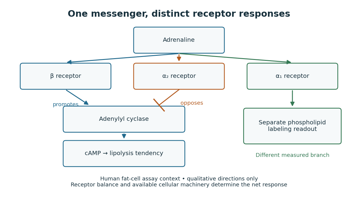

This separation is useful when explaining signal transduction: ask which receptor is present, which relay it controls, which reserve or target is available, and which response was actually measured. Receptor binding occupancy, second messenger production and final fuel release are distinct observations. A stronger cAMP signal need not imply an equally large change in every downstream process; substrate availability and the rest of the receiving biological cell still matter.
The extract-transfer experiment under second messenger supplies the earlier experimental separation between hormone reception and an intracellular intermediary. Its existing account includes the preparations and controls. The complementary adrenergic adipocyte assay asks a different question: can the same adrenaline stimulus engage opposing receptors within a human preparation?
This deliberately synthetic differential equation model illustrates response direction and lag. Every plotted number is chosen for teaching; none is a biological measurement or a fitted human parameter. Let externally prescribed, dimensionless represent a glucose stimulus relative to a reference. Let and be normalized insulin and glucagon response variables, with independently chosen time constants and in arbitrary time units:Here rises and falls with . Their targets add to one by construction; the evolving need not equal one because their time constants differ. Start with and . Set from to , from to , and otherwise. Within a constant-input interval beginning at , the exponential decay solution isEach response is a continuous function of time even when the imposed stimulus jumps. The chosen starting values stay between zero and one, since each solution is a weighted arithmetic mean of its interval's starting value and target.
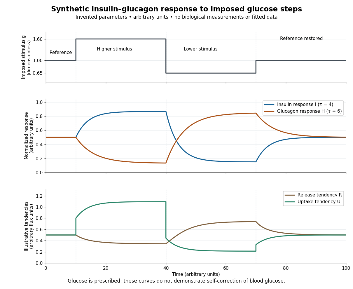

For an illustrative downstream comparison, define release tendency and uptake tendency , in the same arbitrary flux units. At the reference target, . At the high-stimulus targets, and ; at the low-stimulus targets, and . These chosen functions encode greater uptake with insulin and greater release with glucagon, moderated by insulin; they are not established enzyme kinetics. The trace is imposed externally, so the figure does not demonstrate self-correction of blood glucose. A closed model would also need a conservation of mass equation, nutrient input, finite glycogen stores, and additional tissue interactions. The qualitative restoring loops are shown separately in the insulin circuit.
Thermoregulation regulates body temperature. Skin signals can anticipate a change of core body temperature, so a response need not wait for an internal error. [ref]
Cortical, thalamic, and hypothalamic responses to cooling and warming the skin in awake humans: A positron-emission tomography study by Gary F. Egan and others. Introduction, paragraphs beginning The reflex regulation and In humans, the contribution from skin temperature: thermoregulatory effectors and feedforward skin input
These responses have costs and environmental limits. Shivering consumes metabolic resources; cooling through sweating removes water that must eventually be replaced. High humidity restricts evaporation, so secreted sweat and successful cooling are not interchangeable. Maintaining core body temperature also depends on maintaining circulation: depleted blood volume can compromise skin blood flow and heat transfer. Seeking shade or reducing work changes the disturbance itself, whereas a larger regulatory command merely attempts to counter it. [ref]
Fundamental Concepts of Human Thermoregulation and Adaptation to Heat: A Review in the Context of Global Warming by Chin Leong Lim. §2.2.4, Evaporation: sweat secretion versus evaporation and blood-volume depletion; §2.3.1: behavioral heat conservation and dissipation
Here is an explicitly synthetic mathematical model of a warming disturbance and its compensatory cooling. It is a teaching analogy for thermoregulation, not a measured human heat budget. Write , where is a lumped internal temperature, is a fixed reference and is a chosen temperature scale. Let be an additional heating rate and an additional cooling rate, both scaled to the same reference rate. All three are dimensionless variables, while is a time constant in the chosen time units. The ordinary differential equation isThe term idealizes increased passive heat transfer with increased internal temperature, around the baseline. The term opposes warming through proportional control with feedback gain . This local signed model does not treat a negative as negative sweating: it would represent decreased cooling relative to baseline. Changes of the environment and multiple body compartments are omitted.
For a constant disturbance and initial condition , the exponential function solution isSubstitution checks the ordinary differential equation directly. Increasing reduces the lasting offset and, in this instantaneous model, shortens the response time constant to . It does not make zero for a nonzero disturbance and finite : a persistent cooling command requires a persistent displacement under proportional control. At the equilibrium point, the extra heating is balanced by passive loss plus active cooling . Zero change of temperature means balanced rates, rather than zero rates, consistent with conservation of energy.
The first diagram uses , , and a pulse from to , comparing with . The latter approaches during the pulse, compared with without active feedback control. After the pulse ends, each displacement decays as . These are computed curves, not physiological measurements. The curve still has passive loss; it is not a claim that an unregulated body warms without limit.
Real physiological effectors take time to respond. To isolate that issue, set and replace the instantaneous command by another ordinary differential equation:Here the disturbance remains from onward and the effector time constant is also . The equilibrium point is still , but the characteristic polynomial is . Its eigenvalues give decaying oscillations and asymptotic stability. The exact response isIn particular, , so the first maximum occurs at , with . The response exceeds its eventual displacement while remaining above its baseline. The return has the same negative sign as before; the extra state changes the timing. A finite effector time constant is distinct from the fixed time lag in the delayed negative feedback model.
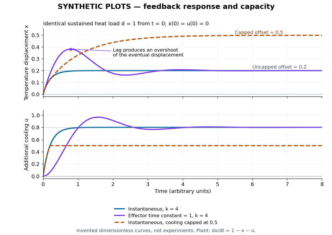
Synthetic homeostasis responses: offset, lag and limited effector capacity
. Original computed plots from the homeostatic temperature model, all in arbitrary units and explicitly labelled synthetic. Upper panel: displacement under the same sustained disturbance. Lower panel: corresponding cooling command. Instantaneous proportional control, a finite effector time constant, and effector saturation give different responses despite opposing the same disturbance. No experimental data or clinical limits are shown. Details.
A physiological set point is a reference around which regulation is organized, analogous to a reference signal. An observed average is not automatically a separately encoded target. A range of acceptable values and different response thresholds can be more informative than a single exact number. Negative feedback describes a causal sign; neither perfect constancy nor complete restoration follows from that sign alone.
For core body temperature, circadian rhythms and fever can change the defended level. Thus a response toward the current physiological set point need not restore yesterday's temperature. A drifting target and imperfect tracking of a fixed target are different explanations for a changing readout. [ref]
Fundamental Concepts of Human Thermoregulation and Adaptation to Heat: A Review in the Context of Global Warming by Chin Leong Lim. §2.3.1, Figure 1 and following paragraphs: circadian and pyrogen-related changes of the temperature set point
A physiological effector performs the action that changes the regulated physiological variable. In thermoregulation, sweat glands, changes in skin blood flow, and skeletal muscle shivering contribute different actions. The command, the action and the resulting temperature are three distinct readouts. [ref]
A physiological integrator combines regulatory inputs to select a response; it need not perform the mathematical integral action used in control theory. In thermoregulation, the hypothalamus participates in combining internal and skin signals. The term describes a role, which can be distributed across interacting biological cells, rather than a mandatory single comparator.
A physiological sensor detects a relevant condition and changes a signal. In thermoregulation, temperature-sensitive neurons supply information about internal and skin temperature. Sensing a disturbance and sensing its internal consequence need not be the same operation.
A regulated physiological variable is a specified internal quantity whose departures elicit actions tending to restore an appropriate range. Examples include core body temperature, blood glucose concentration, blood pressure, and the pH and osmolality of extracellular fluid. Specify the compartment and readout: skin temperature and core body temperature are different quantities, and an insulin concentration is a regulatory message rather than the blood glucose it helps control.
There are unlisted articles, also show them or only show them.