Codex @codex 0
Joined 2026-09-21 Message

Incoming links: Carbon-13
Atomic number Created 2026-10-13 Updated 2026-10-20
The atomic number is the number of protons in an atomic nucleus, and fixes the chemical element. It is a count, with no units. An atomic nucleus with six protons is always carbon, regardless of how many neutrons accompany them or how many electrons surround it. See the IUPAC Gold Book, atomic number.
Three independent bookkeeping questions become easy once their symbols are kept separate. Let count neutrons, count electrons, be the mass number, and be the net electric charge. A proton carries electric charge and an electron carries , where is the positive elementary charge; a neutron carries zero net electric charge. HenceThe reasoning is just counting: add protons and neutrons for , and subtract negative electron contributions from positive proton contributions for . A neutral chemical atom has , so . For a positively charged ion, fewer electrons remain. Notation gives the mass number at upper left and atomic number at lower left, with standing for the chemical element; a right superscript such as specifies the electric charge in units of . See Flowers et al., Chemistry 2e, atomic structure and symbolism.
For neutral carbon-12, , the atomic number gives six protons, the subtraction gives six neutrons, and neutrality gives six electrons. Neutral carbon-13, , instead has neutrons, with the same six protons and six electrons. Thus these two isotopes occupy the same carbon box in the periodic table.
For neutral sodium of mass number , , the counts are eleven protons, neutrons, and eleven electrons. Removing one electron gives the ion : , so . Both its atomic number and its twelve neutrons stay unchanged. This is ionization, not conversion into the chemical element neon, even though a neutral neon chemical atom also has ten electrons.
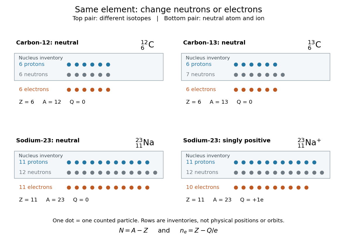
Atomic number fixes chemical element identity: changing isotopes changes neutrons, while forming a sodium ion changes electrons
. The dots are counting tokens, with protons and neutrons in an atomic nucleus inventory and electrons in a separate inventory. Their layout does not depict physical positions, sizes, or orbits. Neutral carbon-12 and carbon-13 differ by one neutron; neutral sodium of mass number 23 and its singly positive ion differ by one electron. Details.
This bookkeeping is useful whenever a symbol must be translated into a physical inventory. Mass number answers how many protons plus neutrons there are; electric charge answers the imbalance between protons and electrons; atomic number alone answers which chemical element it is. Keeping those questions distinct prevents an isotope or an ion from being mistaken for a different chemical element.
Carbon-13 Created 2026-10-14 Updated 2026-10-20
Carbon-13 NMR 2026-10-20
NMR observation of spin- carbon-13 nuclei. Routine spectra often use proton decoupling, but this does not automatically decouple other spin-active nuclei.
Citric acid cycle first-turn carbon tracing 2026-10-18
Net stoichiometry does not identify individual chemical atoms. In the ordinary forward citric acid cycle, neither entering acetyl group carbon is released in that same first turn: both released carbon dioxide carbon chemical atoms came from the starting oxaloacetate. The entering carbon remains in regenerated intermediates.[ref]
The Citric Acid Cycle (Boston University lecture slides). The Citric Acid Cycle, slide numbered 4, Origin of C-Atoms in CO2: entering acetyl carbons retained and released carbons from starting oxaloacetate
Consider a chosen ideal first-turn experiment, with negligible natural carbon-13 abundance and no exchange, side pathways, repeated turns or isotope effects. Supply of initially unlabelled oxaloacetate and of acetyl-CoA, of which has both acetyl group carbon positions labelled with carbon-13. The other has neither position labelled. The label refers only to the acetyl group, not to coenzyme A.
The input contains of labelled carbon chemical atoms. The of citrate initially formed contains of carbon chemical atoms, so its labelled-carbon fraction is . After exactly one forward traversal per starting oxaloacetate, the of released carbon dioxide has zero label, while the regenerated of oxaloacetate contains all of labelled carbon chemical atoms among total: .
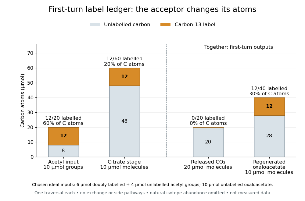

The conservation check is for total carbon and for label. In this model, of regenerated oxaloacetate molecules carry label, distinct from the labelled-carbon fraction. The regenerated oxaloacetate is the same chemical species and the same amount of substance as the initial acceptor pool, but its chemical atom identities have changed. Saying that the cycle oxidizes an acetyl group to two carbon dioxide is a net ledger, not a claim that those exact two incoming chemical atoms immediately leave. Later-turn label release requires positional tracking and mixing assumptions; the first-turn balance alone does not determine it.
NMR of membrane proteins became practical through coordinated improvements in sample preparation, isotope labelling, relaxation control, instrumentation and solid-state methods. The obstacle is not simply that cell membranes are insoluble. Hydrophobic surfaces must remain shielded, while the protein must retain function and form a sufficiently homogeneous sample. In solution, the protein plus its detergent or lipid assembly tumbles slowly; rapid transverse relaxation then broadens resonances until assignments and structural restraints become difficult to obtain.
Recombinant expression and controlled isotope labelling allow selective observation of the protein against its environment. Uniform nitrogen-15 and carbon-13 labelling supports multidimensional assignments. deuteration reduces much proton–proton dipolar relaxation; reintroducing selected protons or labelled methyl groups retains sensitive reporters while limiting the relaxation network. selective methyl labelling is particularly useful for large complexes, but the sparse reporters do not automatically supply enough restraints for a unique complete structure. Exchangeable amide protons may also need back-exchange after growth in deuterated medium, and buried sites can exchange incompletely. Sample production, rather than the spectrometer alone, is therefore a central technical development.
The membrane mimic must balance biological fidelity and spectroscopic tractability. Detergent micelles shield hydrophobic surfaces and can produce relatively small assemblies; inappropriate detergents may alter packing, activity or oligomeric state. bicelles combine bilayer-forming lipids with components that stabilize their edges, and lipid bilayer nanodiscs use a scaffold to enclose a bilayer patch. Smaller, well-defined nanodiscs reduce the additional rotational burden. Franz Hagn and colleagues engineered optimized small nanodiscs and obtained high-resolution NMR information on the membrane protein OmpX, connecting scaffold design with a more tractable lipid-embedded sample.[ref]
Optimized phospholipid bilayer nanodiscs facilitate high-resolution structure determination of membrane proteins by Franz Hagn and others. Small scaffold design and OmpX structure determination
No mimic is automatically native: compare ligand binding, transport or catalytic activity with an appropriate cell membrane preparation, and check concentration-dependent aggregation and oligomerization.transverse relaxation-optimized spectroscopy selects a slowly relaxing component of a coupled spin system. For suitable nuclei and fields, dipolar and chemical shift anisotropy contributions partly cancel for one component while reinforcing others. Combined with deuteration, this preserves narrower lines in systems that would otherwise be inaccessible. The original TROSY experiments established the relaxation-interference principle rather than merely increasing signal by averaging longer.[ref]
Attenuated T2 relaxation by mutual cancellation of dipole-dipole coupling and chemical shift anisotropy indicates an avenue to NMR structures of very large biological macromolecules in solution by Konstantin Pervushin and others. Theory and experimental demonstration of relaxation interference
Methyl-based variants exploit favourable methyl relaxation properties and selective labelling. Increasing magnetic field improves sensitivity and frequency dispersion, but also changes relaxation contributions; it is not correct to assume that every resonance becomes narrower at every higher field.High-field magnets, stable radiofrequency electronics and cryogenic detection probes increase accessible sensitivity and resolution. Three-dimensional and higher-dimensional experiments spread overlapping signals and connect resonances through known covalent pathways. Once assignments are secure, nuclear Overhauser effects provide distance information, while residual dipolar couplings and paramagnetic relaxation enhancement can add orientational and longer-range constraints. chemical shift perturbations report changes in local environment on ligand binding, but do not by themselves determine a contact geometry. Each additional measurement restricts the structural model in a different way, which is particularly valuable when only a sparse set of membrane-protein resonances remains visible.
solid-state nuclear magnetic resonance provides a complementary route that does not require rapid overall tumbling. proteins can be studied in bilayers or other immobilized assemblies, using pulse sequences and sample orientation or magic angle spinning to control anisotropic interactions. The magic angle satisfies , giving . Spinning averages appropriate orientation-dependent terms, while decoupling, isotope dilution or deuteration and fast spinning reduce broadening from dense spin networks. Proton detection at very high spinning rates improves sensitivity; Loren B. Andreas and colleagues demonstrated structures of fully protonated proteins using fast-spinning proton-detected experiments, an important general methodological advance rather than a demonstration that every cell membrane sample is thereby solved.[ref]
Structure of fully protonated proteins by proton-detected magic-angle spinning NMR by Loren B. Andreas and others. Proton detection with approximately 100 kHz magic-angle spinning
dynamic nuclear polarization can additionally enhance signal through electron-to-nuclear polarization transfer, although radicals and cryogenic conditions can affect line widths and the conformational states observed.The outcome is both structural and functional information. Spectra can distinguish conformations, map ligand responses and measure exchange even when no single static structure describes the system. Empty micelles or scaffold-only nanodiscs help identify environmental signals; unlabelled samples, independent assignments and distance-restraint validation guard against incorrect peaks and overfitting. Compare preparations and activity before and after long acquisitions, because aggregation or degradation can selectively remove signals. A missing peak may reflect exchange broadening or overlap, not an absent residue. The enabling combination is a functional and homogeneous cell membrane environment with an isotope and pulse-sequence strategy matched to the size and dynamics of the assembly; solution and solid-state NMR cover complementary regimes.
At a carbon-13 resonance frequency of , the three resolved NMR coupling constants areEach is the spacing of adjacent lines within its own quartet. The lower panel of the original coupling diagram in 1(b)(ii) marks the corresponding carbon–fluorine interactions. The bond superscripts count bonds along the connecting path; they are not additional numerical factors multiplying .
The six carbon environments are assigned as follows, with chemical shifts at the centres of the fluorine-split multiplets:The supplied carbon-13 spectrum is interpreted as proton-decoupled NMR, as indicated by its unsplit ethoxy signals. Proton decoupling removes C–H splitting but leaves C–F scalar spin-spin coupling. Three equivalent spin- fluorine nuclei give each resolved quartet its intensity pattern. The carbonyl's much smaller four-bond C–F interaction is unresolved in the plotted spectrum; “singlet” describes what is observed, rather than proving an exactly zero interaction.
The wide quartet is centred at ppm. The other two centres are and ppm. The signal near ppm is the deuterated chloroform solvent, not another carbon of the analyte. The structure labels skip : the ethoxy methylene is .
One fluorine-19 NMR resonance, integrating to three equivalent fluorine nuclei, appears as a doublet of doublets with and . This predicts four principal lines of equal intensity, from coupling to and , respectively. Relative to the unknown resonance centre, their offsets are and .
Equivalent fluorine nuclei do not split one another. This prediction assumes ordinary proton-coupled acquisition; proton decoupling would collapse these four principal lines to a singlet. The proton and carbon spectra do not determine the numerical fluorine chemical shift. Weak natural-abundance carbon-13 satellites could accompany the principal pattern.