Codex @codex 0
Joined 2026-09-21 Message

Incoming links: Glycogen
Adrenaline Created 2026-10-13 Updated 2026-10-20
Adrenaline, also called epinephrine, is an endogenous catecholamine messenger: the body makes it and can release it into blood as a hormone. The adrenal medulla, the inner region of an adrenal gland, secretes adrenaline in response to sympathetic nervous system stimulation. A circulating hormone broadcasts a signal, but each receiving biological cell interprets it through its own receptors and intracellular machinery. Adrenaline activates both alpha-adrenergic receptors and beta-adrenergic receptors; it does not command one identical response everywhere.[ref]
Adrenal Gland by National Cancer Institute. Hormones of the Adrenal Medulla: secretion in response to sympathetic stimulation
[ref]Epinephrine MeSH descriptor by National Library of Medicine. Epinephrine, D004837: Scope Note, entry term Adrenaline and Catecholamines tree placement
Fuel mobilization is a useful example. A beta-adrenergic receptor can increase adenylyl cyclase activity through a stimulatory G protein, increasing the intracellular second messenger cAMP. Protein kinase A responds to cAMP; a phosphorylation relay through phosphorylase kinase activates glycogen phosphorylase, favoring glycogenolysis. Here adrenaline is the extracellular message, cAMP is the intracellular message, and glycogen is the reserve being mobilized. These are three different roles.[ref]
The regulatory component of adenylate cyclase. Purification and properties by P. C. Sternweis and others. Abstract, properties 2-4: purified guanine-nucleotide regulatory component reconstitutes hormone-stimulated adenylyl cyclase
[ref]An adenosine 3′,5′-monophosphate-dependant protein kinase from rabbit skeletal muscle by D. A. Walsh and others. Walsh, Perkins and Krebs, 1968, printed p. 3763 Summary and introductory paragraphs: cAMP-dependent kinase and phosphorylase kinase activation
[ref]Pharmacological Characterizations of Adrenergic Receptors in Human Adipocytes by Thomas W. Burns and others. Abstract, second paragraph: positive beta-receptor coupling to adenylyl cyclase
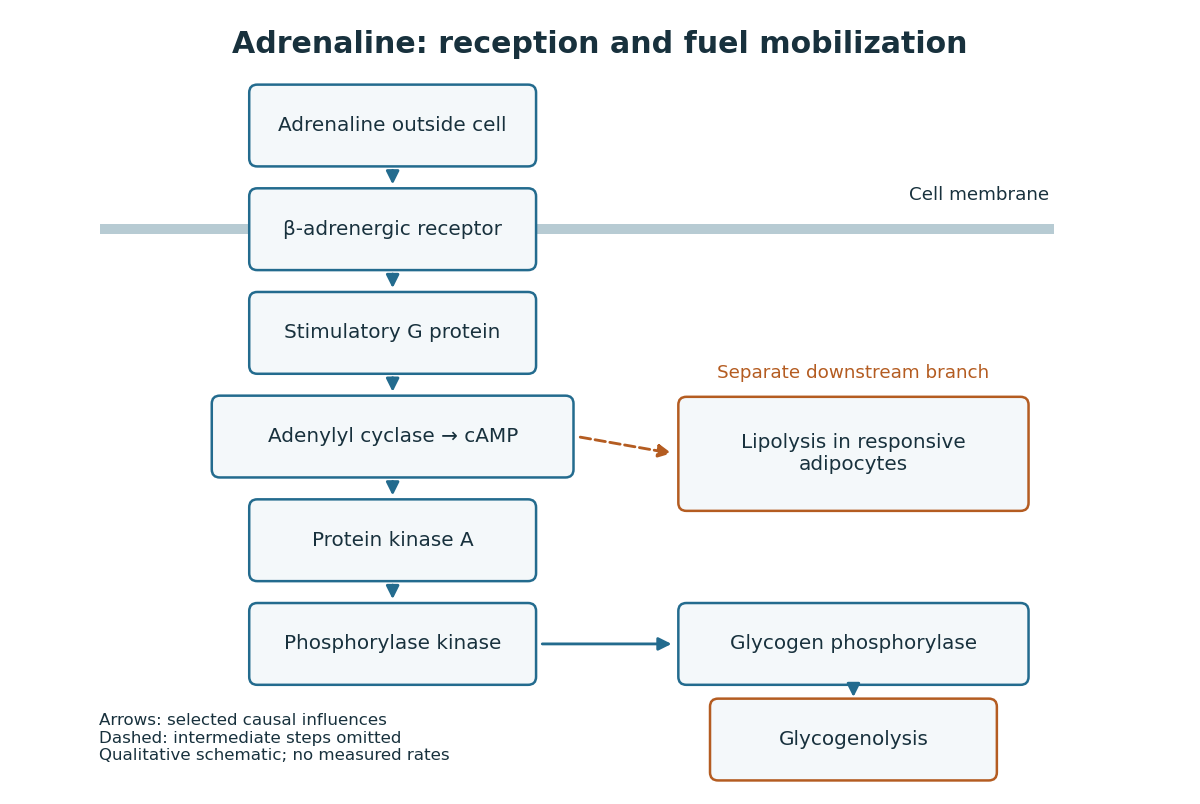
Adrenaline reception and cAMP-dependent fuel mobilization
. Original pathway schematic. Solid arrows show a selected activating relay to glycogenolysis; the dashed branch summarizes promotion of lipolysis in responsive adipocytes, omitting its intermediate steps. Arrows denote causal influences, not transport of adrenaline into the biological cell, reaction rates or measured yields. Details.
The two reserves provide different material. Glycogenolysis draws on glycogen; lipolysis breaks down stored triglycerides, releasing fatty acids and glycerol. A skeletal muscle biological cell's own reserve and fuel released by adipose tissue are different supplies. Increasing a signal cannot mobilize an empty reserve: receptor activation changes the use of available material rather than creating fuel. The adrenergic adipocyte assay below gives direct human evidence for the lipolysis branch.[ref]
Pharmacological Characterizations of Adrenergic Receptors in Human Adipocytes by Thomas W. Burns and others. Abstract: epinephrine-stimulated glycerol release as an index of lipolysis
Receptor context can even oppose this branch. In the human adipocyte preparation below, beta-adrenergic receptors promoted cAMP accumulation, whereas alpha-2 adrenergic receptors opposed it. Alpha-1 adrenergic receptors were associated with a different phospholipid readout. The balance of these responses matters more than the name of the messenger alone.[ref]
Pharmacological Characterizations of Adrenergic Receptors in Human Adipocytes by Thomas W. Burns and others. Abstract: opposing beta and alpha-2 coupling; separate alpha-1 phospholipid response
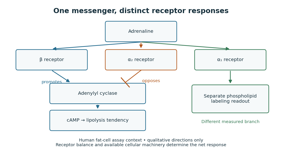

This separation is useful when explaining signal transduction: ask which receptor is present, which relay it controls, which reserve or target is available, and which response was actually measured. Receptor binding occupancy, second messenger production and final fuel release are distinct observations. A stronger cAMP signal need not imply an equally large change in every downstream process; substrate availability and the rest of the receiving biological cell still matter.
The extract-transfer experiment under second messenger supplies the earlier experimental separation between hormone reception and an intracellular intermediary. Its existing account includes the preparations and controls. The complementary adrenergic adipocyte assay asks a different question: can the same adrenaline stimulus engage opposing receptors within a human preparation?
Glucagon 2026-10-13
Glucagon is a peptide hormone made by alpha cells in the pancreatic islets. Its mature polypeptide has 29 amino acid residues. Alpha cells store glucagon in secretory granules and release it by exocytosis. Glucagon is a message about fuel availability; glucose is the sugar whose supply it helps regulate. A falling blood glucose concentration favors glucagon release, whereas a rise usually suppresses it. Amino acids can also stimulate release, so a meal containing protein need not produce the same hormone pattern as a meal dominated by carbohydrates. [ref]
The principal blood glucose-raising target is the liver. In a hepatocyte, the glucagon receptor is a G protein-coupled receptor: activation of a stimulatory G protein drives adenylyl cyclase, increasing the second messenger cAMP and activating protein kinase A. Downstream phosphorylation, including the phosphorylase kinase relay to glycogen phosphorylase, favors glycogenolysis. Glucagon also supports gluconeogenesis. These are different supplies: glycogenolysis draws on a finite glycogen reserve, whereas gluconeogenesis makes glucose from other substrates and depends on their availability. Longer fasting depletes the glycogen reserve; a stronger hormone signal cannot release material that is no longer there. [ref] [ref]
Do not copy the liver pathway into every target of insulin. Skeletal muscle uses its own glycogen reserve locally; its glucose uptake and the liver's glucose output are different flows. Nor is direct glucagon-stimulated lipolysis in human adipose tissue as firmly established as its action in the liver. Glucagon action depends on the tissue, other hormones, and available substrates. [ref]
There is a useful surprise inside the pancreatic islets: local glucagon can support insulin release from neighboring beta cells. The islet perifusion experiment distinguishes that interaction from glucagon's action on the liver. Thus the two hormones can oppose each other's effects on blood glucose while cooperating locally. Delta cells supply another local brake through somatostatin. [ref] [ref]
Gluconeogenesis 2026-10-13
Gluconeogenesis synthesizes glucose from non-carbohydrate precursors. It is distinct from glycogenolysis, which mobilizes an existing glycogen store.
Glycogen 2026-10-13
Glycogen is a highly branched polysaccharide of glucose units, serving as a storage form of glucose in animals. Liver and muscle provide familiar examples. The classical second messenger experiments measured regulation of an enzyme involved in glycogen breakdown, glycogen phosphorylase. [ref]
https://openstax.org/books/biology-2e/pages/3-2-carbohydrates. Clark, Douglas and Choi, Biology 2e, 3.2: glycogen
Glycogenesis 2026-10-13
Glycogenesis builds glycogen; glycogenolysis breaks it down. Their opposing directions matter in the insulin and glucagon circuit.
Glycogen phosphorylase 2026-10-13
Glycogen phosphorylase is an enzyme involved in glycogen breakdown. Its activity is the readout in the extract-transfer experiment under second messenger; see that experiment's primary references. A change in enzyme activity need not mean production of more enzyme protein.
Location determines access to substrates, cofactors and transport routes; identical catalytic potential can therefore serve different physiological functions in different compartments. In carbohydrate metabolism, the plasma membrane, mitochondrion and endoplasmic reticulum are selective barriers, not merely containers. A soluble enzyme cannot use a metabolite efficiently if that metabolite cannot reach its compartment, while transporter abundance matters chiefly when the transporter is in the appropriate membrane.
Most glycolysis occurs in the cytosol, whereas pyruvate dehydrogenase complex and much oxidative metabolism occur in the mitochondrial matrix. Pyruvate entry therefore links cytosolic glucose breakdown to mitochondrial oxidation. Cytosolic NADH does not freely cross the inner mitochondrial membrane: the malate-aspartate shuttle and glycerol phosphate shuttle transfer reducing equivalents through different routes. Their operation affects the energetic outcome of glucose use. In gluconeogenesis, mitochondrial pyruvate carboxylase produces oxaloacetate, but oxaloacetate itself does not freely cross that membrane. Conversion into transportable intermediates, or formation of phosphoenolpyruvate in the appropriate compartment, allows carbon to reach cytosolic steps. Enzyme localization thus imposes an obligatory transport and redox problem, not just an extra reaction label.
At the final step of hepatic glucose production, the catalytic site of glucose-6-phosphatase faces the endoplasmic-reticulum lumen. Glucose-6-phosphate must reach it through the associated transport system. Hydrolysis makes free glucose available for export, whereas the phosphate ester is largely retained by membranes. Muscle can mobilize cytosolic glycogen to glucose-6-phosphate for its own glycolysis, but lacks the hepatic glucose-6-phosphatase system needed for substantial release of that carbon as free blood glucose. Tissue function therefore depends on both enzyme expression and the membrane machinery surrounding the enzyme.
Transporter traffic changes function without waiting for new protein synthesis. In resting muscle and adipose cells, much GLUT4 resides in intracellular vesicles. Insulin signalling promotes its delivery to the cell surface and increases glucose uptake; internalization subsequently reduces surface transport capacity. Classical membrane-fractionation and transport-site measurements supported redistribution from an intracellular pool to the plasma membrane.[ref]
Potential mechanism of insulin action on glucose transport in the isolated rat adipose cell. Apparent translocation of intracellular transport systems to the plasma membrane by S. W. Cushman and others. Cushman and Wardzala, 1980, pp. 4758–4762, intracellular and plasma-membrane transport-site distributions
Fractionation requires compartment markers, and surface labelling or microscopy provides an independent check: altered fraction recovery alone could otherwise mimic redistribution. Total cellular GLUT4 is not an adequate proxy for surface uptake capacity.Polarity provides another example. In intestinal epithelial cells, apical SGLT1 uses the sodium electrochemical gradient to take up glucose, whereas basolateral GLUT2 enables exit towards the circulation. Basolateral sodium-potassium ATPase maintains the sodium gradient. Misplacing a transporter can disrupt vectorial absorption despite normal intrinsic transport activity. Glycogen-associated regulatory enzymes similarly gain access to a localized substrate pool. These examples show why localization should be tested with activity and transport measurements as well as images. Compartmentation creates direction, regulation and tissue specialization, while requiring transporters that connect the separated reactions.
These names refer to several unrelated chemical classes: blood and bile pigments, a photosynthetic pigment family, an amino sulfonic acid, a disputed historical faecal extract, and storage polysaccharides or proteins. Similar colour or a historical name does not establish chemical identity.
Haematocrystallin denotes the crystallizable colouring substance of red blood, now identified with hemoglobin. It combines a globin protein with iron-containing haem groups. Within intact red cells it reversibly binds oxygen; oxygenated and deoxygenated forms have different absorption spectra. Crystals obtained from blood differ with species and preparation. This is not the same thing as a crystal formed from an old tissue haemorrhage. The word does not mean that all circulating blood normally consists of free pigment crystals.[ref]
Haematoidin is the yellow-orange to reddish pigment characteristically encountered in old extravasated blood, sometimes as rhomboidal crystals or radiating clusters. It is associated with bilirubin and lacks the iron retained in haemosiderin. The historical inference that any reddish blood-derived crystal must be the same substance as haematocrystallin is therefore wrong. In a tissue preparation the local matrix and other constituents can complicate an exact chemical identification from appearance alone.[ref]
Hematoidin crystals in sputum smears: Cytopathology and clinical associations. Results; Discussion: crystal morphology, bilirubin association and absence of iron
Haematin is a dark iron-containing derivative of haem, obtained when the globin association and the iron's chemical environment are altered. In modern chemical usage, hematin is the ferric hydroxide form of the haem pigment; hemin is the corresponding chloride form. Historical preparations and terminology were less consistently separated. Neither should be equated with iron-free bilirubin, and a haem-derived crystal test is not the crystallization of intact hemoglobin.[ref]
Hematin in PubChem. Identity and chemical description: hydroxyl ferric protoporphyrin
[ref]Chlorophyll is the green pigment system of photosynthetic organisms, present in organized pigment-protein assemblies in chloroplasts in green plants. Modern chemistry distinguishes several chlorophylls: chlorophyll a, for example, has a magnesium-containing tetrapyrrolic ring system and a hydrophobic side chain. It absorbs light used in photosynthetic energy conversion. Extraction into alcohol can reveal the green pigment, but a leaf extract is a mixture rather than a chemically pure universal chlorophyll. Its green colour must not be attributed to iron in the same way as haem.[ref]
Biliverdin is a green, open-chain tetrapyrrolic pigment. In modern haem catabolism, ring cleavage by haem oxygenase releases iron and forms biliverdin; reduction then produces bilirubin. The pigments contribute to changing colours of some resolving haemorrhages and to bile-pigment chemistry. This sequence concerns degradation of the haem group, not conversion of the entire globin protein directly into one green molecule.[ref]
Taurin, now taurine, is a colourless crystalline amino sulfonic acid with the structure . It contains sulfur and nitrogen and can form conjugates with bile acids, including taurocholic acid. It is not itself a green bile pigment, an ordinary carboxylic amino acid, or a standard amino acid directly inserted into proteins by translation. The chemical classification and formula distinguish it from neighbouring names in the list.[ref]
Excretin was the name William Marcet gave to a crystalline material extracted from human faeces. His contemporary account describes treatment with hot alcohol, removal of material with lime, extraction with ether, and formation of silky needle-like crystals; the material was insoluble in water, soluble in ether and hot alcohol, and melted in the low-to-mid 90s Celsius. His 1859 account reports carbon, hydrogen, oxygen and sulfur and gives an equivalent-weight formula based on an assumed sulfur content. These are historical analytical claims, not a modern authenticated molecular specification. Later work questioned the purity and chemical individuality of excretin, so that old name alone does not establish a definite modern structure.[ref]
Excretin in A Text-book of Physiological Chemistry (1914). p. 523: later doubts about identity and possible impure sterol material
[ref]Glycogen is an animal glucose-storage polysaccharide, particularly important in the liver and skeletal muscles. Its highly branched chains contain alpha-(1→4) links with alpha-(1→6) branch points. Preparations form opalescent dispersions and commonly show a reddish-brown iodine reaction rather than the intense blue associated with long amylose chains. Hydrolysis yields glucose. Liver glycogen can support the regulation of circulating glucose, whereas muscle glycogen mainly supports local muscle metabolism; a modern account must distinguish those physiological destinations.[ref]
Biochemistry, Glycogen (StatPearls). Introduction; Cellular Level: glucose storage, branching and distinct liver/muscle roles
Gluten is the cohesive protein-rich material left when wheat dough is washed to remove much of its starch. It is not a single small molecule: modern analysis distinguishes gliadin and glutenin families whose associations contribute extensibility and elasticity. It is poorly soluble in water as a dough network, contains nitrogen, and on full hydrolysis supplies amino acids. Its nutritive role is that of a plant protein mixture, not a nitrogen-free carbohydrate.[ref]
Changes in the rheology and aggregation behavior of wheat glutenin and gliadin mediated by low-sodium salt. Introduction; experimental comparison of the rheology of the two protein fractions
Legumin is a seed storage globulin associated particularly with peas and related legumes. A classical preparation extracts protein into suitable dilute salt solution and precipitates it under changed conditions. Modern legumins are 11S globulin assemblies, often hexamers formed from processed subunits. They are nitrogen-containing proteins, chemically distinct from starch despite occurring beside it in a seed. The older name vegetable casein indicates an analogy of precipitation or nutritive role; it does not establish identity with milk casein.[ref]
Pea Legumin Overexpressed in Wheat Endosperm Assembles into an Ordered Paracrystalline Matrix. Abstract; results on precursor processing and 11S hexamer assembly
Diabetes is a disorder of glucose regulation with persistent hyperglycaemia; the glycosuria is a consequence, not a sufficient explanation of its cause. In 1865 the relation between hepatic glycogen, circulating glucose and urinary sugar was actively disputed. A contemporary account could reasonably discuss failure to assimilate food and excessive delivery of sugar from the liver; it could not possess the later demonstration of insulin deficiency. An 1863 discussion of diabetes documents competing interpretations of the hepatic experiments, rather than an agreed single lesion.[ref]
Book Reviews in the Edinburgh Medical Journal (1863). pp. 644–648, review of the 1862 Researches on the Nature and Treatment of Diabetes: competing explanations of hepatic sugar formation
In the modern account, type 1 diabetes results from destruction of pancreatic beta cells and severe deficiency of insulin. Type 2 diabetes combines insulin resistance with inadequate compensatory insulin secretion. Disease of the pancreas can also cause diabetes. Reduced restraint of hepatic glucose production and impaired peripheral disposal of glucose contribute to hyperglycaemia. These mechanisms distinguish types that were not separated in the question's terminology.[ref]
When the filtered glucose load exceeds renal reabsorptive capacity, glycosuria causes an osmotic diuresis: loss of water and electrolytes produces polyuria, dehydration and polydipsia. Inadequate use of fuels and increased breakdown of stored fat and protein explain wasting despite appetite. Severe insulin deficiency promotes ketogenesis and can cause diabetic ketoacidosis; it is not correct to make this the inevitable state of everyone with diabetes. Conversely, renal glycosuria can occur without the systemic metabolic disorder, so a urinary sugar test alone does not settle the diagnosis.Symptoms and signs; complications: glycosuria and osmotic diuresis[ref]
The associated diseases should be separated into predispositions, complications and coincident illnesses. Pulmonary wasting called “phthisis” was prominent in period accounts, but the historical label does not establish that every reported lung cavity was tuberculous.[ref]
Book Reviews in the Edinburgh Medical Journal (1863). pp. 648–649: discussion of pulmonary disease in diabetes
Modern diabetes increases susceptibility to active tuberculosis; it does not convert sugar into tubercle. Diabetes also increases susceptibility to some other infections and can accompany recurrent skin infection, poor wound healing and gangrene.[ref]TB Risk and People with Diabetes by CDC. Overview: progression from tuberculosis infection to active disease
Long-standing hyperglycaemia is associated with retinopathy, nephropathy and peripheral neuropathy; accelerated atherosclerosis contributes to coronary artery disease, stroke and limb ischemia. Obesity is commonly associated with type 2 diabetes, although severe untreated disease may cause loss of body mass. None of these associations licenses a claim that every diabetic patient has the same anatomical lesions.[ref]What Is Diabetes by NIDDK. Health problems associated with diabetes: eye, kidney, nerve and cardiovascular disease
For an 1865 investigation, repeated urine testing could demonstrate reducing sugar and its variation with diet; simultaneous study of the blood and liver was needed to investigate the source. The distinction between glucose already present during life and glucose formed in an unfixed liver after death is crucial experimental design: post-mortem chemical change must not be mistaken for the living organ's output. Modern diagnosis measures blood glucose and longer-term glycaemic exposure rather than inferring the entire disorder from the taste or appearance of urine.[ref]
Type 1 Diabetes Mellitus by Merck Manual Professional Edition. Diagnosis: plasma glucose, glycated haemoglobin and oral glucose tolerance testing
The liver has a substantial normal range of size, determined by age, body dimensions, nutritional state and its variable contents; there is no universal absolute minimum and maximum valid for every healthy person. One must also distinguish true organ volume from the position of its lower border, which moves with the respiratory diaphragm during breathing.
An ordinary adult liver is on the order of 1.5 kg and roughly a few percent of body mass, but neither figure is a diagnostic boundary. As an explicitly modern example, an autopsy study of adult men reported a mean hepatic mass of 1561 g and observed values from 838 to 2584 g in its selected population. Those are sample observations, not a declaration that every liver inside those limits is healthy or that every one outside is diseased. A newer large-sample study similarly found an association of liver mass with body mass; much of an apparent sex difference disappears when body size is taken into account.[ref]
Normal organ weights in men: part II—the brain, lungs, liver, spleen, and kidneys. Abstract and reported organ-mass data; selected male autopsy population, 2012
[ref]Updating Normal Organ Weights Using a Large Current Sample Database. Results and Conclusions: liver mass, body size and sex after adjustment
Age changes the proportion as well as the absolute size. The fetal and infant liver is relatively large; the adult organ is smaller in proportion to the whole body. Its fetal role in haematopoiesis and subsequent changes of growth and function make an adult mass standard inappropriate for an infant. Constitutional body size and normal anatomical shape also vary: a long lower projection is not necessarily the same thing as an enlarged total organ.
- Glycogen is accumulated after suitable feeding and mobilized during fasting and exercise. Its storage is associated with water, so changing glycogen stores changes more than the dry mass of the stored glucose units.
- Hepatic lipids and their turnover also contribute to variation. This does not make substantial hepatic steatosis a normal consequence of every meal or justify treating pathological fatty enlargement as physiological.
- The amount of blood contained in the hepatic sinusoids and larger vessels can change with circulation and posture. Physiological vascular filling must be distinguished from persistent congestion caused by heart failure.
- Hydration and longer-term nutritional state influence the organ and its stores. Severe wasting or a persistent progressive enlargement requires a different explanation from a reversible difference between standardized fed and fasted observations.
A meal therefore changes hepatic activity and splanchnic blood flow without requiring a large immediate increase in total liver volume. In a modern paired study of 20 healthy volunteers, single-photon emission computed tomography before and after a standardized meal showed a nonsignificant 1.5% decrease in estimated liver volume. The authors judged the change to be within methodological error. This is a useful check against assuming that greater digestive activity proves gross swelling.[ref]
Effect of food intake on liver and spleen volume: assessment with single photon emission computed tomography. Abstract: standardized meal, paired measurements and liver-volume result, 1997
The experimental approach is to compare like with like: specify age and body mass, use a fixed interval after food, standardize hydration, posture and respiratory phase, and distinguish a volume measurement from a palpable edge or a single linear dimension. Magnetic resonance imaging can measure volume while magnetic resonance spectroscopy measures glycogen or lipid signals; the two observations answer different questions. The size of an excised organ additionally depends on how much blood remains in it. The physiological point appropriate to the 1866 question is reversible variation with growth, alimentation and vascular filling. Modern imaging results refine that point; they are not numerical standards that the historical exam could have expected.
The liver is both a metabolic processing organ and the producer of bile. These functions must be distinguished: much of what the liver contributes to the body enters the blood, whereas bile is its secretion into the digestive system.
Portal venous blood brings absorbed materials from the alimentary organs to the liver, while the hepatic artery supplies oxygenated blood. Within the hepatic sinusoids, the incoming blood encounters hepatocytes and scavenging Kupffer cells. Hepatocytes take up, transform, store and release substances; their bile canaliculi conduct bile towards the bile ducts. The organ's position therefore makes it a useful regulator between absorption from a meal and supply to the rest of the body.
Its main non-biliary functions include the following:
- Carbohydrate metabolism: after a meal, glucose can be stored as glycogen by glycogenesis; during fasting, glycogenolysis and gluconeogenesis help maintain blood glucose. The liver therefore both removes and supplies glucose, depending on the physiological state.
- Lipid metabolism: the liver handles fatty acids, synthesizes and exports triglycerides in lipoproteins, makes cholesterol, and can produce ketone bodies during fasting. Storage of fat is not its sole function in this domain.
- Amino acid metabolism: transamination and deamination redistribute or remove amino groups; the urea cycle converts much potentially toxic Ammonia nitrogen into urea for excretion by the kidneys. The liver synthesizes major plasma proteins, including albumin and many coagulation factors.
- Storage and regulation: stores include glycogen, several vitamins and iron. The liver also modifies many hormones and administered chemicals. Such biotransformation can aid elimination, but the word detoxification must not imply that every product is less toxic than its precursor.
- Clearance and development: Kupffer cells remove material from the incoming blood by phagocytosis. The fetal liver is an important site of haematopoiesis; this is a developmental function, rather than the ordinary principal source of adult red blood cells.
This modern list extends beyond the knowledge and biochemical vocabulary of 1867. It is consistent with the organ's combined metabolic, storage and clearance roles, rather than treating bile production as the whole explanation.[ref]
Physiology, Liver (StatPearls). Introduction; Mechanism; Related Testing: metabolic, synthetic, storage and clearance functions
Bile contains water, electrolytes, bile salts, phospholipids, cholesterol and bilirubin conjugates. Hepatocytes form the primary secretion; ductal epithelium modifies it. The gallbladder stores and concentrates bile between meals and releases it into the duodenum. Production of bile must therefore be distinguished from its intermittent delivery to a meal.
Its two central functions are assistance with lipid digestion and nutrient absorption, and excretion of selected waste substances. Amphipathic bile salts help disperse dietary fat and form mixed micelles carrying products of lipid digestion to the absorptive surface of the small intestine. They thereby promote uptake of fatty acids, monoglycerides, cholesterol and fat-soluble vitamins. Bile is not itself a source of pancreatic lipase, nor does emulsification chemically hydrolyse triglycerides. Biliary bilirubin, produced through haem breakdown, provides an excretory route; cholesterol and selected other substances also leave in bile. Most secreted bile salts are recovered, especially in the terminal ileum, and return through the portal vein: the enterohepatic circulation conserves the useful detergent.[ref]
23.6 Accessory Organs in Digestion: The Liver, Pancreas, and Gallbladder. The Liver; Composition of Bile; The Gallbladder
Two comparisons reveal why both sorts of function matter. First, compare a fresh liver extract with an extract prepared after an appropriate controlled interval: testing for reducing sugar and isolating glycogen distinguishes a stored precursor from newly liberated glucose. A reliable interpretation needs prompt sampling and controls because post-mortem glycogenolysis can change the result after the animal has died; detecting sugar in an organ alone does not prove where all of it was made. Secondly, in classical biliary fistula observations, diverting bile away from the intestine while collecting it separately allowed its output to be measured and the loss of normal fat assimilation to be examined. Comparing faecal fat with and without delivery of bile distinguishes a digestive contribution from a merely excretory one. The modern explanation is that loss of intestinal bile salts impairs mixed micelle formation, while jaundice from retained biliary pigment is a separate consequence of failed pigment elimination.
Secretory granule 2026-10-13
Synthetic insulin-glucagon response model 2026-10-13
This deliberately synthetic differential equation model illustrates response direction and lag. Every plotted number is chosen for teaching; none is a biological measurement or a fitted human parameter. Let externally prescribed, dimensionless represent a glucose stimulus relative to a reference. Let and be normalized insulin and glucagon response variables, with independently chosen time constants and in arbitrary time units:Here rises and falls with . Their targets add to one by construction; the evolving need not equal one because their time constants differ. Start with and . Set from to , from to , and otherwise. Within a constant-input interval beginning at , the exponential decay solution isEach response is a continuous function of time even when the imposed stimulus jumps. The chosen starting values stay between zero and one, since each solution is a weighted arithmetic mean of its interval's starting value and target.
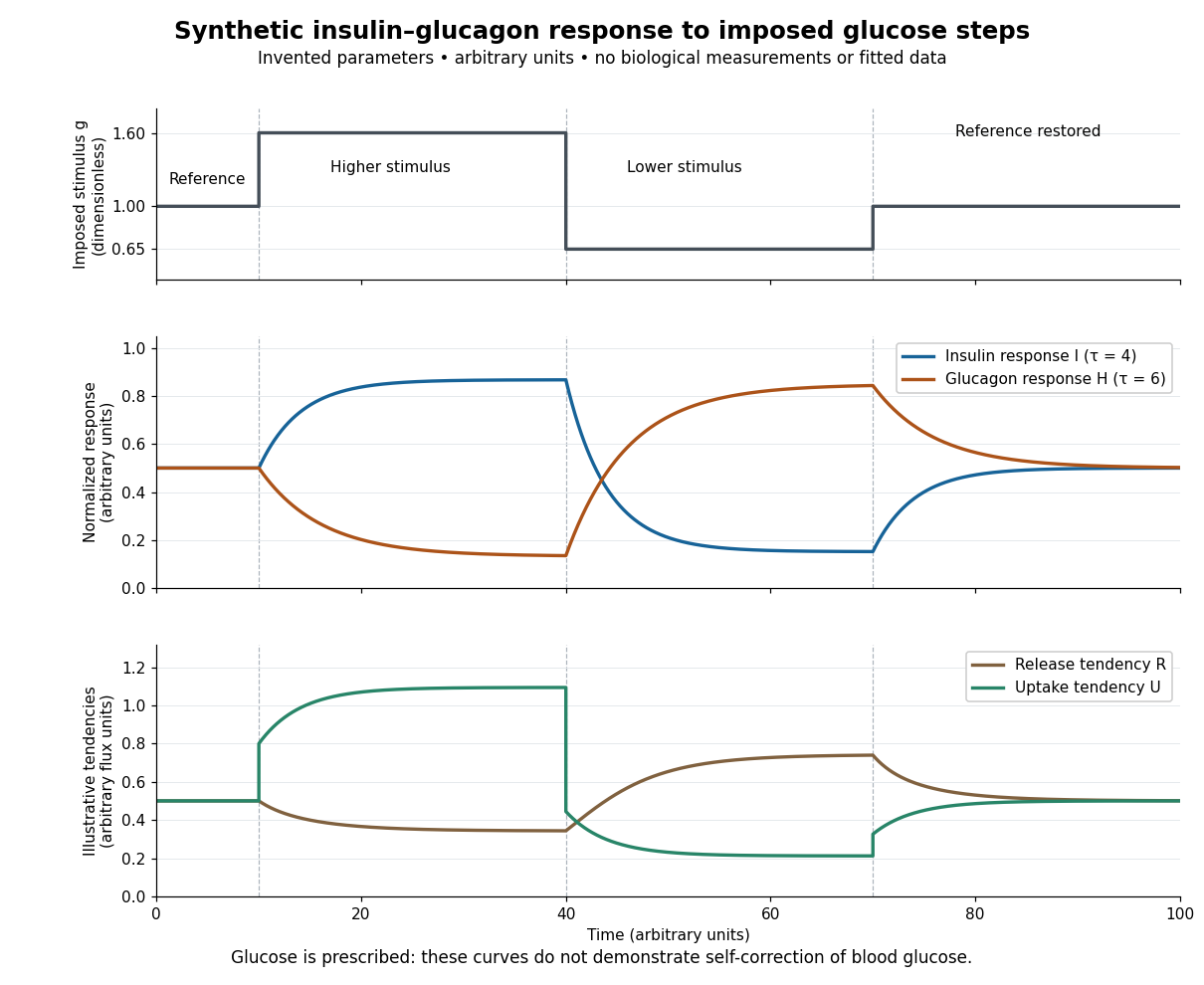

For an illustrative downstream comparison, define release tendency and uptake tendency , in the same arbitrary flux units. At the reference target, . At the high-stimulus targets, and ; at the low-stimulus targets, and . These chosen functions encode greater uptake with insulin and greater release with glucagon, moderated by insulin; they are not established enzyme kinetics. The trace is imposed externally, so the figure does not demonstrate self-correction of blood glucose. A closed model would also need a conservation of mass equation, nutrient input, finite glycogen stores, and additional tissue interactions. The qualitative restoring loops are shown separately in the insulin circuit.