Topics (384k) Articles (398k) Users (410) Discussions (242) Comments (383) Files (13k) New article

A cardiac short-axis stack is a series of images across a cardiac chamber at successive positions approximately transverse to its cardiac long axis. In ventricular volumetry, the series must cover the intended cavity from its basal boundary toward the cardiac apex. A primary cardiovascular magnetic resonance imaging protocol specifies linked long-axis planning and short-axis coverage.[ref]
Standardized cardiovascular magnetic resonance imaging (CMR) protocols: 2020 update by Christopher M. Kramer and others. General techniques, Left ventricular structure and function; Figures 1–2
The stack supplies a spatial sum rather than a single diameter. Let be the segmented cavity area in slice at phase , and let be the represented distance along the stack normal. A basic approximation isAll areas in a given sum must refer to a compatible phase of the cardiac cycle. Image segmentation, slice coverage and phase matching are separate inputs to the volume estimate.
An original example uses four representative areas of 2, 6, 10 and 6 cm, each assigned 1 cm of axial extent. Their represented volume is 24 cm, or 24 mL. Omitting the basal 6 cm contribution reduces the result to 18 mL without changing the tracing quality of the other slices. If slice thickness is 0.8 cm but center spacing is 1 cm, the numerical integration convention must also account for the intervening extent; treating thickness and represented spacing as interchangeable changes the estimate.
Phase mixing causes a different error. An area from one slice near end-filling and another near end-ejection do not generally describe a cavity that existed at one time. A stack can therefore be complete spatially yet inconsistent temporally. With correctly matched phases, the derived end-diastolic volume and end-systolic volume can be used for stroke volume and ejection fraction; those formulas do not repair missing anatomy or mismatched acquisition phases.
The Fick principle infers blood flow from the rate at which a transported substance is added to or removed from blood and the corresponding concentration difference. For whole-body oxygen consumption in a steady state, conservation of mass giveswhere is cardiac output, is arterial oxygen content and is mixed venous oxygen content. The arteriovenous oxygen content difference must match the tissue bed whose oxygen consumption is measured. The equation follows by subtracting outgoing oxygen transport from incoming transport ; it assumes no continuing accumulation of oxygen within that system.
For a constructed example, oxygen consumption of mL/min and an arteriovenous oxygen content difference of mL/dL imply dL/min, or L/min. Keeping the decilitre in the denominator prevents a factor-of-ten unit error. Using only a difference of oxygen saturation would omit the hemoglobin concentration needed to obtain oxygen content.
Direct implementation measures oxygen consumption, together with arterial and appropriately mixed venous blood samples. An estimated implementation substitutes a model for oxygen consumption. These are different measurement procedures even though they use the same balance equation. A 2017 comparison explicitly studied thermodilution against estimated-oxygen-uptake output, rather than treating estimated uptake as an exact measured input.[ref]
At fixed measured content difference, a error in assumed oxygen consumption produces a error in inferred cardiac output. More generally, differentiating the Fick principle gives . Thus a small content difference magnifies a fixed absolute sampling error.
Pressure-flow distinction in cardiovascular assessment is the separation of blood pressure, a mechanical loading quantity, from blood flow, a volume transported per unit time. Cardiac output is a flow rate. A pressure reading alone does not specify the volume of blood delivered by the heart.
In a simplified steady model,where is the pressure difference across the represented vascular region, is its blood flow and its effective vascular resistance. The difference between upstream and downstream blood pressure matters, rather than one pressure considered in isolation. The pressure-gradient and resistance account is developed in the primary physiology text.[ref]
Anatomy and Physiology 2e by J. Gordon Betts and others. §20.2, Blood Flow, Blood Pressure, and Resistance
An original counterexample holds the pressure difference fixed at 90 mmHg. With mmHg/(L/min), flow is 5 L/min; with mmHg/(L/min), flow is 3 L/min. The same driving pressure can accompany different delivery rates. Conversely, at fixed flow a higher measured pressure difference can represent a higher effective resistance.
The quantities also have different dimensions. The product has units of energy per time, describing a hydraulic rate of mechanical energy transfer in this model. A pressure alone has neither flow units nor power units.
A primary coronary thermodilution apparatus combined a pressure/temperature-sensing guidewire with a controlled infusion catheter; its flow estimate used infusion rate and temperature measurements.[ref]
Direct volumetric blood flow measurement in coronary arteries by thermodilution by Wilbert Aarnoudse and others. Abstract, Methods
The availability of pressure and thermal signals in one instrument does not make them the same observable. Finally, pulsatile circulation includes storage and changing vessel geometry, so the steady resistance relation is an explanatory approximation. Matching time averages, anatomical sites and model assumptions is part of interpreting pressure and flow together.Cardiac index is cardiac output divided by body surface area:When is in L/min and area in m, its units are L/(min m). Despite the word index, it is not dimensionless. A primary comparison of thermodilution and estimated Fick principle measurements explicitly indexed output to body surface area for its analyses.[ref]
An original comparison shows how normalization can change ranking. Output 5 L/min with area 2 m gives 2.5 L/(min m). Output 4 L/min with area 1.5 m gives about 2.67 L/(min m). The second output is lower in absolute terms but higher after this area normalization. Neither statement contradicts the other.
The denominator is often estimated rather than directly measured. If the Mosteller formula is chosen, with numerical height in centimetres and body mass in kilograms, thenThe unit-specific area equation is documented in the official VA technical manual.[ref]
Formulas for Body Surface Area Calculator in the VA Clinical Reminders Manager's Manual. Printed p.36
Choosing an area model is consequently part of defining the reported index.For small input errors, . An overestimated area can lower the index without changing the underlying output measurement. Area normalization also does not by itself account for every difference in metabolism or body composition. The quantity is a standardized way of expressing flow relative to one body-size measure, not an additional independent observation of how much blood the heart pumps.
Cardiac image foreshortening occurs when a cardiac view represents a shorter apparent base-to-apex extent than the intended anatomical chamber length, often because the imaging plane misses the true cardiac apex. It can create an apparently thick false apex and affect derived ejection fraction and deformation measurements.
A primary echocardiography study acquired optimal and deliberately foreshortened views in 72 participants using a GE Vivid E9 system. The altered acquisition window was one rib-space above the optimal window; two analysis systems were used to compare derived quantities.[ref]
Impact of apical foreshortening on deformation measurements: a report from the EACVI-ASE Strain Standardization Task Force by Serkan Ünlü and others. Abstract, Aims and Methods and results
This tests acquisition geometry while keeping the participant the same, rather than interpreting every shorter-looking chamber as a biological change.An original ellipsoid model distinguishes a missed apex from a simple projected length. Letwith the true cardiac long axis along . The central long-axis plane includes both axial tips and has full length . A parallel offset plane instead intersects an ellipse whose apparent axial extent isAt , . A 10 cm true extent therefore appears as 8 cm in this model even though the slice was translated rather than rotated. Its apparent tips are not the original chamber tips.
This model is a geometrical explanation, not a clinical correction formula. A shorter outline can alter measured volume differently at end-diastolic volume and end-systolic volume; the ejection fraction error need not follow the length error by one fixed percentage. Consistent anatomical landmarks and independent views help distinguish actual chamber shape from the chosen section. Improving the repeatability of an outline inside an incorrect plane does not recover anatomy that was never acquired.
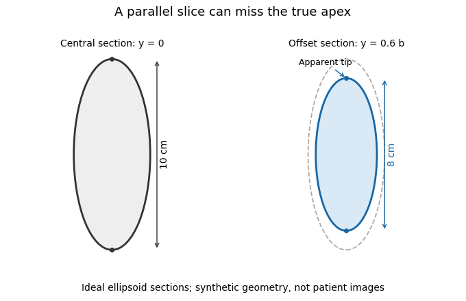
A parallel cardiac slice can miss the true apex
. Original sections of an ideal ellipsoid with axial semiaxis 5 cm and transverse semiaxis 2 cm. The central plane has full length 10 cm; a parallel plane offset by 0.6 times the transverse semiaxis has length 8 cm. Dashed outline shows the central section for comparison. Synthetic geometry, not patient data. Details.Nicotinic acid, , is pyridine-3-carboxylic acid: a pyridine ring bearing a carboxyl group at position 3. It is one form included under vitamin B3, and can contribute to production of the coenzymes nicotinamide adenine dinucleotide and nicotinamide adenine dinucleotide phosphate.[ref]
PubChem Compound database by National Center for Biotechnology Information. Nicotinic acid, CID 938, formula and pyridine-3-carboxylic-acid structure
[ref]Niacin Fact Sheet for Health Professionals by the Office of Dietary Supplements. Introduction, niacin forms and NAD/NADP production
The related nicotinamide has a carboxamide group in place of the acid group. Their related names reflect related structures, while the different functional groups make them distinct chemical compounds. “Niacin” can be used broadly for the vitamin family, so an experiment reporting a particular chemical form should specify which molecule was used.
A carboxyl group is the functional group : one carbon has a double bond to oxygen and a single bond to a hydroxyl group. It gives a carboxylic acid its characteristic acid functionality. In acetic acid, the group is attached to a methyl group, giving .[ref]
Loss of the acidic hydrogen produces a carboxylate, the conjugate base:The pH influences the balance between these forms. A structural drawing of the neutral acid therefore need not show the predominant charge state in every solution. The carboxyl group is a part of the molecule, while acetic acid names the entire compound.
An alpha-hydroxy acid is a carboxylic acid with a hydroxyl group on the carbon immediately adjacent to the carboxyl group. That neighboring carbon is the alpha carbon; in simple systematic numbering it is carbon 2 because the carboxyl carbon is carbon 1. The hydroxyl oxygen within the carboxyl group itself is not the additional hydroxyl that defines this class.[ref]
Organic Chemistry by John McMurry. Chapter 20, carboxylic-acid structure and nomenclature
For a common saturated monocarboxylic example, the local structure can be writtenwith representing hydrogen or carbon-containing substituents. Glycolic acid, , has two hydrogens at that carbon. Lactic acid, , has hydrogen and methyl groups there and is also called 2-hydroxypropanoic acid.[ref]
Glycolic acid in the NIST Chemistry WebBook. Identity and structural record
[ref]Lactic acid in the NIST Chemistry WebBook. Identity and Other Names
Position distinguishes the class. The structure has its additional hydroxyl group at carbon 3 and is a beta-hydroxy acid, even though it has the same molecular formula as lactic acid. A molecular formula therefore does not determine whether a compound is an alpha-hydroxy acid; the connectivity is essential.
The alpha carbon can also be a stereogenic centre. In lactic acid it is bonded to four different groups: hydrogen, hydroxyl, methyl and carboxyl. Its two enantiomers share the alpha-hydroxy classification but differ in three-dimensional arrangement. Glycolic acid does not have that stereogenic centre because two substituents are identical hydrogens.
Both functional groups participate in the chemistry. Loss of the carboxyl proton gives a carboxylate, such as lactate from lactic acid, while the additional hydroxyl remains part of the structure. Distinguishing group identity, position, protonation and stereochemistry makes the class useful in organic and biological chemistry without reducing it to a list of commercial uses.
A beta-hydroxy acid is a carboxylic acid with a hydroxyl group on the beta carbon: the carbon two carbon–carbon bonds away from the carboxyl group carbon. For a simple chain numbered from the carboxyl carbon as carbon 1, the beta carbon is carbon 3. The hydroxyl within the carboxyl group itself does not establish the additional hydroxy substitution.
The small example 3-hydroxypropionic acid has structureStarting at the carboxyl carbon, the adjacent is alpha and the terminal is beta. The primary ChEBI record gives the systematic name 3-hydroxypropanoic acid, formula and its classification as a 3-hydroxy monocarboxylic acid.[ref]
An alpha-hydroxy acid instead places the additional hydroxyl group one carbon closer to the carboxyl group. Thus lactic acid, , and 3-hydroxypropionic acid, , have the same molecular formula but different atom connectivity. They are constitutional isomers; counting oxygen atoms or measuring molecular mass alone cannot distinguish them. The distinction is structural, not a statement about acidity or biological activity.[ref]
The two functional groups also describe different possible transformations. Removing the carboxyl proton forms a carboxylate while preserving the beta-hydroxy carbon skeleton. Converting the hydroxyl group to a carbonyl changes the hydroxy-acid functionality. Which transformation occurs depends on reagents and conditions; the class name alone supplies no reaction rate or treatment claim. Tracking positions relative to the carboxyl group makes such structural changes easier to compare.
Malonic acid is propanedioic acid, a dicarboxylic acid in which two carboxyl groups are separated by one methylene group:Its neutral molecular formula is and its average molar mass is approximately .[ref]
Two of the four hydrogen atoms belong to carboxyl OH groups; the other two belong to the central carbon. Complete neutralization of both carboxyl groups therefore requires two equivalents of a suitable base, not four merely because the formula contains four hydrogens. An original stoichiometric model isFor acid, the two-equivalent amount of hydroxide solution is . The pH at an experimental endpoint also depends on acid-base equilibria and the measurement method; the equivalent count alone does not determine it. Malonate names the corresponding deprotonated system, whose protonation state should be specified.
Heating can promote decarboxylation, producing acetic acid and carbon dioxide. Malonic-acid derivatives underlie the carbon-chain construction described in the malonic ester synthesis.[ref]
Organic Chemistry by John McMurry. Section22.7, Alkylation of Enolate Ions, malonic ester hydrolysis and decarboxylation
For the unsubstituted acid, the stoichiometric balance isAn original ideal mass balance for converted by only this reaction gives about acetic acid and carbon dioxide from starting material. Following evolved gas and residual material would test conversion, whereas a change in sample mass alone would not distinguish this reaction from evaporation or another decomposition pathway.Terephthalic acid is benzene-1,4-dicarboxylic acid, an aromatic dicarboxylic acid with two carboxyl groups attached at opposite positions of a benzene ring. Its neutral molecular formula is . The positions of the groups are part of its identity, not information recoverable from the elemental formula alone.[ref]
The two carboxyl groups make it a useful difunctional building block. Reaction with ethylene glycol, which has two alcohol groups, forms ester connections in polyethylene terephthalate, a polyester. This is step-growth polymerization: growing molecules react through their remaining functional groups.[ref]
Organic Chemistry by John McMurry. Section31.4, Step-Growth Polymers, terephthalic acid and ethylene glycol example
An original repeat-unit bookkeeping calculation isUsing rounded masses, per repeat-unit contribution. The subtraction represents two esterification connections per repeat in the long-chain description; it is not an elementary reaction equation for one isolated pair of monomers forming a complete polymer.
For a finite linear chain assembled from acid molecules and glycol molecules, with no ring closure or side reaction, there are connections. The chain therefore loses water molecules, rather than . End groups supply the difference: its mass is approximately under the same rounded-mass convention.
The molar mass and end-group balance consequently matter alongside conversion. Measuring consumption of acid groups and formation of ester connections tests the reaction model; identifying an aromatic diacid does not determine the final chain length or material properties.
A dicarboxylic acid is a carboxylic acid containing two carboxyl groups. The groups may be attached to an aliphatic framework, as in malonic acid, or an aromatic framework, as in terephthalic acid.[ref] The definition counts functional groups rather than specifying chain length, ring substitution or acid strength.
A dicarboxylic acid can supply two acidic protons through its carboxyl groups. In the simplest diprotic acid model, write the successive equilibriaThe two acid dissociation constants and characterize different equilibria. Two chemically similar groups do not justify treating the two proton-removal steps as identical. Other ionizable groups, if present, require additional equilibria.
For an original ideal dilute-solution example, use concentration-form constants , and hydrogen-ion concentration . Mass balance and the two equilibrium relations give species fractionsHere . The average charge is per acid-derived species even though only of the molecules are monoanions: equal neutral and dianion populations balance around that mean. The constants are stipulated for illustration, not assigned to a particular compound. Precise thermodynamic calculations use activities rather than equating them with concentrations.
A titration that fully removes both carboxyl protons consumes two moles of hydroxide per mole of initially neutral dicarboxylic acid, in the absence of other reactions. For example, 0.50 mmol requires 1.00 mmol hydroxide by stoichiometry. Two stoichiometric equivalents do not guarantee two separately resolved features on a titration curve; the acid dissociation constants and measurement conditions determine their separation.
Two carboxyl groups also provide two possible connections in esterification. Terephthalic acid and a diol can therefore build a polyester, illustrating how the same functional-group count matters both to acid-base measurements and to polymer connectivity.[ref]
Organic Chemistry by John McMurry. Section 31.4, Step-Growth Polymers, terephthalic acid and ethylene glycol
A carbon sink takes up more carbon than it releases over a specified interval. It is a statement about a net transfer, whereas carbon storage describes an amount already held. If a reservoir's carbon stock is , its balance isA positive balance makes it a carbon sink relative to the stated boundary. A large, nearly unchanged stock can provide substantial carbon storage without acting as a strong current carbon sink.
Forests acquire carbon through photosynthesis and lose it through cellular respiration, decay, fire and removal of biomass. Forest inventories and long-term ecosystem measurements therefore have to account for both accumulation and losses: the carbon sink in growing forests is not automatically the net effect of all changes in forested land.[ref]
A large and persistent carbon sink in the world's forests by Yude Pan and others. Science 333, pp. 988–993; separate forest uptake, regrowth and land-use-change emissions for 1990–2007
Pyrolytic carbon is a form of deposited carbon produced by thermal decomposition of a carbon-containing precursor. The name specifies a preparation route, not one uniquely fixed crystal structure or degree of ordering.
The deposited material can contain differently oriented graphitic domains. Texture therefore matters: properties measured parallel and perpendicular to preferred layers can differ strongly. Highly ordered pyrolytic graphite is a particular well-oriented material, whereas the broader term pyrolytic carbon also covers less ordered deposits. Preparation conditions, porosity and microstructure are needed to interpret a measured mechanical or thermal property.
The capillary electrophoresis heat-dissipation tradeoff is the balance between rapid electrically driven separation and the temperature rise caused by Joule heating. Narrow capillaries offer a large wall area per liquid volume, while high electric fields increase migration speed and electrical heat production. The 1981 primary experiment demonstrated efficient separations in a 75-micrometre-bore, one-metre glass capillary at 30 kV; it does not imply that arbitrarily high fields are thermally harmless.[ref]
Free-zone electrophoresis in glass capillaries by J. W. Jorgenson and K. D. Lukacs. Abstract, capillary dimensions, applied potential and fluorescent separations
A simple model makes the competing scalings explicit. For a cylindrical liquid column of radius , uniform electrical conductivity and axial field , current and heat production per unit length areAt fixed conductivity and bore, doubling the field doubles drift speed for constant electrophoretic mobility but quadruples generated heat. Halving the bore radius reduces heat per length by four at fixed field.
For steady radial heat conduction, let the liquid's thermal conductivity be and impose a fixed wall temperature . With uniform volumetric heating , the cylindrical heat equation givesRegularity at the axis and the wall condition fix the integration constants. The center-to-wall rise is . The symbol here denotes thermal conductivity, not an optical decay constant.
For stipulated , , and , the model gives W/m and center-to-wall rise 0.02344 K. Doubling the radius or field multiplies that rise by four. This ideal calculation holds the wall temperature fixed; it does not include heating through the capillary wall, instrument housing or cooling system.
Temperature gradients can change liquid viscosity, migration and band dispersion. Conversely, reducing bore size also reduces sample volume and optical path through the sample. A useful instrument experiment therefore records current and separation behavior while controlling cooling conditions, rather than judging success only by reduced migration time. The thermal model predicts what to test; it is not a measured temperature profile from the cited experiment.
Pinned article: Introduction to the OurBigBook Project
Welcome to the OurBigBook Project! Our goal is to create the perfect publishing platform for STEM subjects, and get university-level students to write the best free STEM tutorials ever.
Everyone is welcome to create an account and play with the site: ourbigbook.com/go/register. We belive that students themselves can write amazing tutorials, but teachers are welcome too. You can write about anything you want, it doesn't have to be STEM or even educational. Silly test content is very welcome and you won't be penalized in any way. Just keep it legal!
Intro to OurBigBook
. Source. We have two killer features:
- topics: topics group articles by different users with the same title, e.g. here is the topic for the "Fundamental Theorem of Calculus" ourbigbook.com/go/topic/fundamental-theorem-of-calculusArticles of different users are sorted by upvote within each article page. This feature is a bit like:
- a Wikipedia where each user can have their own version of each article
- a Q&A website like Stack Overflow, where multiple people can give their views on a given topic, and the best ones are sorted by upvote. Except you don't need to wait for someone to ask first, and any topic goes, no matter how narrow or broad
This feature makes it possible for readers to find better explanations of any topic created by other writers. And it allows writers to create an explanation in a place that readers might actually find it.
Figure 1. Screenshot of the "Derivative" topic page. View it live at: ourbigbook.com/go/topic/derivativeVideo 2. OurBigBook Web topics demo. Source. - local editing: you can store all your personal knowledge base content locally in a plaintext markup format that can be edited locally and published either:This way you can be sure that even if OurBigBook.com were to go down one day (which we have no plans to do as it is quite cheap to host!), your content will still be perfectly readable as a static site.
- to OurBigBook.com to get awesome multi-user features like topics and likes
- as HTML files to a static website, which you can host yourself for free on many external providers like GitHub Pages, and remain in full control

Figure 2. You can publish local OurBigBook lightweight markup files to either OurBigBook.com or as a static website.
Figure 3. Visual Studio Code extension installation.
Figure 5. . You can also edit articles on the Web editor without installing anything locally. Video 3. Edit locally and publish demo. Source. This shows editing OurBigBook Markup and publishing it using the Visual Studio Code extension. 
- Infinitely deep tables of contents:



All our software is open source and hosted at: github.com/ourbigbook/ourbigbook
Further documentation can be found at: docs.ourbigbook.com
Feel free to reach our to us for any help or suggestions: docs.ourbigbook.com/#contact