
An action potential is a brief regenerative excursion of membrane potential, produced by changing electrical resistance and conductance through voltage-gated ion channels. In a typical fast neuronal action potential, sodium entry raises membrane potential, activating further sodium channels. This feedback produces the rapid rising phase. Inactivation of sodium channels and increased conductance to potassium then lower membrane potential again; persisting potassium conductance can produce an undershoot below the resting level. See Purves and colleagues: the ionic basis of action potentials.
For an illustrative electrical calculation, write inward sodium electric current as . At , taking and gives inward. A potassium current with and gives , outward. These illustrative values expose opposing driving forces; a waveform requires the conductances to evolve with membrane potential and time, as in the Hodgkin-Huxley model.
An action potential propagates along an axon because local electric currents bring neighboring regions of cell membrane into the regime where their voltage-gated ion channels regenerate the signal. It is a traveling pattern of excitation, not a packet of sodium ions traversing the entire axon. Regeneration allows an action potential to travel without the passive attenuation of a small local voltage change. See Ion Channels and the Electrical Properties of Membranes.
The neuronal refractory period constrains how soon another action potential can follow. Under comparable conditions, successive action potentials have roughly stereotyped shapes, making event count and timing useful for neural coding; waveform details can nevertheless change with channel state and conditions. The leaky integrate-and-fire model abstracts each action potential to a threshold event and reset. Its reset jump is a mathematical rule, not the actual regenerative waveform produced by sodium and potassium currents.
A synapse is a signaling junction between a neuron and another biological cell. The sending side is presynaptic and the receiving side postsynaptic. At a chemical synapse, released neurotransmitter acts on a receptor in the receiving cell membrane. At an electrical synapse, gap junctions provide a direct path for electric current between cells. See 1.1 Elements of Neuronal Systems.
In a common sequence at a chemical synapse, an arriving action potential opens voltage-gated ion channels selective for calcium. Calcium entry triggers fusion of synaptic vesicles with the presynaptic cell membrane, releasing neurotransmitter into the synaptic cleft. Binding to a receptor changes postsynaptic electrical resistance and conductance, directly through an ion channel or through an intracellular signaling pathway. The resulting change in membrane potential is a synaptic potential. See Ion Channels and the Electrical Properties of Membranes.
A useful conductance approximation writes the inward-positive synaptic electric current aswhere describes the activated electrical resistance and conductance, is the postsynaptic membrane potential, and is the reversal potential, the voltage at which this current vanishes. Opening these channels tends to pull toward ; it does not prescribe a current of fixed sign independently of .
For a worked example, let and . A reversal potential of gives , an inward electric current that raises membrane potential. A reversal potential of instead gives , which lowers membrane potential. These are illustrative calculations, not measurements. A reversal potential of can raise from yet oppose reaching a threshold of : a depolarizing input need not be excitatory.
An excitatory synapse favors a subsequent action potential, whereas an inhibitory synapse opposes it. The effect depends on the receptor, the permeating ions, their gradients and the reversal potential; a neurotransmitter does not have one fixed excitatory or inhibitory sign in every context. Even with , an added conductance can reduce responses to other inputs by lowering effective electrical resistance, an effect called shunting inhibition. See Purves and colleagues: excitatory and inhibitory postsynaptic potentials. This dependence matters when translating a biological synapse into a signed weight or an imposed electric current in computational neuroscience.
Changes in responsiveness during sustained or repeated input. Adaptation makes activity depend on recent stimulation and can invalidate a model whose rate depends only on the present stimulus.
The soma is a neuron's cell body, containing its cell nucleus and much of the machinery for translation and metabolism. Dendrites and the axon extend from, or are connected to, this central compartment. The soma also receives synapses and participates electrically in synaptic integration.[ref]
A voltage recorded at the soma is a local measurement. In an extended neuron, cable properties and active ion channels can make the simultaneous membrane potential in a distant dendrite different.
A pyramidal cell is a multipolar neuron named for its approximately triangular soma, with an apical dendrite extending toward the cortical surface and basal dendrites spreading from the cell base. Pyramidal cells are prominent excitatory projection neurons in the cerebral cortex and hippocampus; their detailed branching and targets vary.
Their separated compartments make pyramidal cells useful for testing how local dendrite events affect whole-cell output. Simultaneous recordings from the soma and a dendrite or axon showed active back-propagation of somatically recorded action potentials, connecting anatomy to measurable signal propagation.[ref]
A pseudounipolar neuron has one main process leaving its soma and dividing into peripheral and central branches. Many primary sensory neurons in dorsal root ganglia have this shape. The peripheral branch receives sensory input and the central branch conducts toward the spinal cord; the soma lies to the side of this principal conducting route.[ref]
This arrangement explains why the familiar drawing of input entering the soma before reaching an axon is not a universal circuit diagram for a neuron.
A bipolar neuron has two main processes extending from its soma, conventionally an axon and a dendrite. Retinal bipolar cells provide a sensory example connecting photoreceptors to downstream retinal circuitry. The anatomical label does not mean that the biological cell has two axons, and it does not imply that every signal must be an action potential.[ref]
12.2 Nervous Tissue. Classifying Neurons: structural classification
A multipolar neuron has one axon and more than one dendrite extending from its soma. Many motor neurons and pyramidal neurons have this organization. Multiple branching dendrites provide spatially distributed sites for synaptic integration; the label specifies overall shape rather than one unique neurotransmitter or firing pattern.[ref]
12.2 Nervous Tissue. Classifying Neurons: structural classification
Synaptic integration is the conversion of inputs at a neuron's synapses into a changing membrane potential and, in a spiking neuron, the timing or absence of action potentials. Spatial summation concerns inputs at different locations; temporal summation concerns inputs at different times. A useful question is whether individually ineffective inputs can act together, and whether an inhibitory postsynaptic potential changes that cooperation. The answer depends on location, timing and the receiving neuron's state.
A synaptic potential is a voltage response, whereas synaptic conductance describes activated ion channels. With inward-positive electric current, the contribution of input isHere is its synaptic conductance, its reversal potential and the local membrane potential. The driving force is an electric potential difference. An EPSP generally brings the membrane potential toward a more positive reversal potential, but its driving force shrinks during depolarization. An IPSP need not produce hyperpolarization: a reversal potential above rest but below the neuronal firing threshold can produce depolarization while opposing a larger excursion. At , the added synaptic conductance initially carries zero electric current yet can cause shunting inhibition of another input.[ref]
Two distinct nonlinearities matter. Even a passive cell membrane can show sublinear synaptic integration when added synaptic conductances reduce driving force and effective electrical resistance. Active voltage-gated ion channels and NMDA receptors can instead amplify sufficiently concentrated inputs and produce a dendritic spike. Neither summing prescribed electric currents nor adding separate measured EPSPs is an exact universal rule for a branching neuron.[ref]
1.4 Limitations of the Leaky Integrate-and-Fire Model. §1.4.2 and §1.4.4: shunting, saturation and active dendritic interactions
Neuronal polarity is the structural and molecular differentiation of a neuron into compartments such as an axon and dendrites. Different distributions of proteins, transport machinery and synapses support different roles in receiving and sending signals. This organization does not require every electrical event to travel in only one direction: action potentials can propagate back into dendrites.
In 1988, Carlos G. Dotti, Christopher A. Sullivan and Gary A. Banker followed individual embryonic rat hippocampus neurons in culture using repeated photography and time-lapse imaging. Several initially similar neurites preceded rapid growth of one future axon; staining for microtubule-associated protein 2 helped distinguish later dendrites. Following the same biological cells over time directly tested the developmental sequence, which a collection of mature shapes could only suggest.[ref]
The establishment of polarity by hippocampal neurons in culture by Carlos G. Dotti and others. Abstract; developmental observations, pp. 1454–1468
Neuroimaging measures structural or functional properties of the nervous system. Different methods record different physical signals, so anatomical images and activity-sensitive measurements support different inferences.
For example, electroencephalography records electrical potential differences, while many functional vascular measurements depend on neurovascular coupling. Spatial overlap in an image is not by itself proof that one location implements a complete psychological process.
Reproductive success is an organism's contribution of offspring, or later descendants, under a stated measurement convention. Counting eggs laid, offspring surviving to maturity and descendants that themselves reproduce gives different outcomes. The relevant interval must therefore be explicit.
Biological fitness often compares expected reproductive contributions among inherited types in a specified environment. A realized count from one individual also includes chance: two individuals with the same underlying expected contribution can leave different numbers of descendants. Survival is one contributor to reproductive success, rather than a complete substitute for it.
Consider an explicitly idealized haploid population with two inherited types , one allele per individual at the tracked genetic locus, and discrete, nonoverlapping generations. Assume faithful type transmission, no mutation or gene flow, a constant environment, and reproductive contributions independent of type frequency. Let the expected values of offspring surviving to the next census be , with relative genotype fitness .
For frequency , expected offspring contributions are proportional to and . Normalizing gives the deterministic large-population mathematical modelThe denominator is mean relative genotype fitness. For and , . Equivalently, 200 and 800 parents with contribute expected counts 240 and 800. Fractional per-parent expected values are averages, not literal fractional offspring. The ratio of these expectations is a deterministic approximation, not generally the expectation of a random ratio.
The odds multiply particularly simply:Thus : repeated modest advantage can substantially change composition. For and , . The deterministic boundary remains unchanged: natural selection needs existing variation.
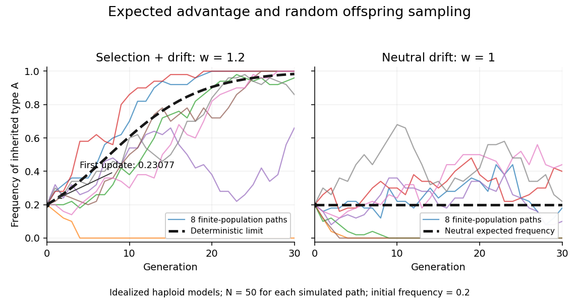

A distinct stochastic extension fixes offspring and samples their types independently using a binomial distribution: , where , then . Its conditional expected value is and conditional variance is . With , the conditional expected value remains , yet realized frequencies wander through genetic drift. With , genetic drift can still lower or lose entirely. Over several generations, the deterministic curve need not equal the mean stochastic trajectory, because the recurrence is nonlinear.
There are unlisted articles, also show them or only show them.