A bond angle is the angle between two chemical bonds sharing a central chemical atom. In H–O–H, the middle symbol identifies the vertex: the angle is at oxygen. Let and be the nonzero displacement vectors from that atomic nucleus to the two neighbours. The actual three-dimensional bond angle satisfiesusing the dot product and Euclidean norm. Rotating the whole molecule preserves this expression; projecting it onto a page generally does not.
There is another useful way to find a bond angle. If the two bond lengths are and the distance between the outer atomic nuclei is , the law of cosines givesFor equal bond lengths , this becomes . Thus a measured outer-atom separation contains information about a bond angle even though those outer chemical atoms are not directly joined by a chemical bond.
For a stipulated rigid, equal-bond length model using water's equilibrium bond angle, , whereas a straight H–O–H arrangement would give . These are calculated geometric ratios, not new measured distances. Gas electron diffraction illustrates how pair-distance information can constrain molecular geometry. Substituting independently averaged distances into the rigid triangle formula does not automatically recover an equilibrium bond angle, because averaging and this nonlinear calculation need not commute.
Non-coding DNA is DNA that does not encode a protein sequence. It includes many regulatory regions, introns, noncoding RNA genes and repeated sequences. Some regions have well-established functions, while others have uncertain or dispensable contributions under tested conditions. The category therefore specifies coding status rather than a single functional verdict. Conversely, finding biochemical activity at a noncoding region does not by itself establish that the activity is required for organismal fitness. [ref]
Gene expression uses a gene's information to produce functional RNA or a protein. Transcription makes RNA from a DNA template; for a protein-coding gene, biological translation then uses messenger RNA to specify an amino acid chain. Many genes instead produce functional RNA, including transfer RNA and ribosomal RNA, without a translated protein product. See Alberts et al., Molecular Biology of the Cell, 4th edition (2002): from DNA to RNA.
Follow the chosen coding strand : transcription of its complementary template gives in messenger RNA. In the stated reading frame, its three codons are AUG, GCU and UAA: methionine, alanine and a stop codon. Thus nine nucleotides specify two amino acid residues followed by termination, not three residues. This is an illustrative coding segment embedded in suitable expression and initiation context, not a claim that an isolated nine-nucleotide RNA is sufficient to initiate biological translation.
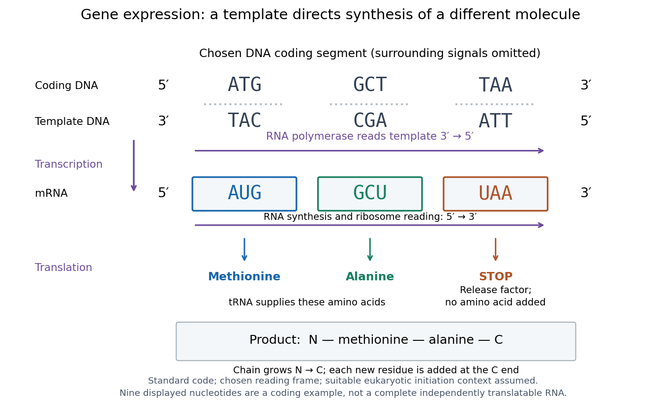

The distinction between copying and use explains how biological cells can preserve the same DNA yet produce different products. DNA replication duplicates the record; gene expression selects and uses parts of it. Transcription does not use all DNA at once, and gene expression can be controlled at several steps, including transcription, RNA processing and biological translation. See Clark, Douglas and Choi, Biology 2e: regulation of gene expression. The arrows describe template-directed synthesis: the DNA does not physically turn into a protein, and the messenger RNA does not supply the amino acids from which that protein is assembled.
DNA repair addresses damage to DNA. Incorporation of a labeled precursor during DNA repair need not indicate duplication of the whole genome in S phase.
Repair pathways recognize and process different kinds of damage. Base excision repair replaces damaged bases, nucleotide excision repair removes a larger segment around selected lesions, and DNA double-strand breaks require rejoining or template-based reconstruction. A response marker reports recognition or signalling; successful restoration requires additional evidence. [ref]
DNA replication copies a double-stranded DNA molecule into two daughter double-stranded DNA molecules. In semiconservative replication, each daughter retains one parental strand and receives one newly synthesized complementary strand. DNA polymerases build the new strands from incoming nucleotides; they do not make RNA or proteins in this copying step. See Alberts et al., Molecular Biology of the Cell, 4th edition (2002): DNA replication mechanisms.
For a five-base pair example, separate from its partner . Base pairing specifies a new TACGG partner for the old ATGCC strand and a new ATGCC partner for the old TACGG strand. With error-free copying, both daughter DNA molecules have the original nucleotide sequence, but different physical strand histories. One initial duplex contains ten nucleotides; one complete round produces two duplexes containing twenty nucleotides, ten retained and ten newly incorporated.
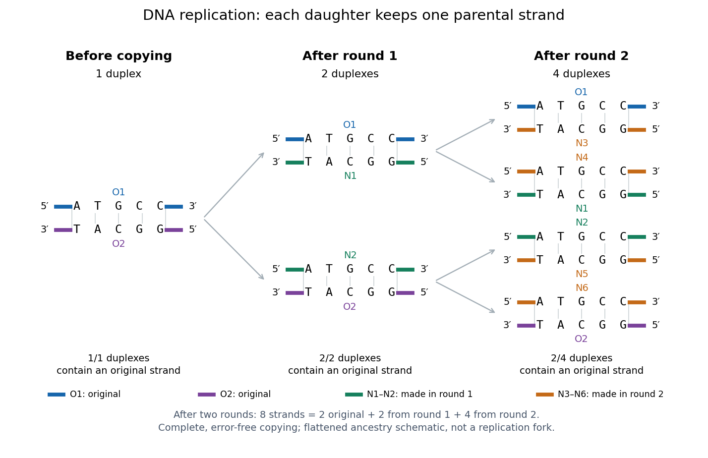
Semiconservative replication: follow two original DNA strands through two complete copying rounds
. Blue O1 and purple O2 are the original strands; green strands were made in round 1 and orange strands in round 2. Each arrow connects a parent duplex to its daughters. After round 2, two of four duplexes retain an original strand, and the other two retain a round-1 strand. All eight strands have the appropriate complementary nucleotide sequences; colors identify strand age, not different nucleobases. The flattened duplexes show ancestry and orientation, not a replication fork or molecular shape. Details.
This is useful for inheritance: both daughter biological cells can receive a copy of the DNA record. The elegant feature is that base pairing lets each strand specify its missing partner. Direction matters too: a replicating DNA polymerase reads its template and extends the new strand at its end, so synthesis proceeds . These are opposite directions on antiparallel nucleic acid strands, not two alternative directions of synthesis. The enzyme needs a pre-existing paired strand end to extend; the diagram omits the machinery that starts and coordinates copying. See Alberts et al.: template-directed synthesis and the requirement for a primer.
The strand accounting predicts observable differences between copying mechanisms. In 1958, Meselson and Stahl grew Escherichia coli with the heavier nitrogen isotope , then supplied and measured extracted DNA by density-gradient centrifugation. After one generation their samples contained intermediate-density DNA; after two, intermediate-density and light DNA occurred in equal amounts. This supported semiconservative replication in the tested Escherichia coli culture. It was an isotope assay of sampled DNA, not simultaneous testing of every kind of DNA or direct colored-strand photography. See The replication of DNA in Escherichia coli by Matthew Meselson and Franklin W. Stahl.
A Northern blot separates RNA by size and detects selected sequences through hybridization with a probe. Detection of a messenger RNA supports transcription of a gene, but does not alone establish a functional protein or its destination.
The polymerase chain reaction amplifies a selected DNA region using repeated copying steps. Amplified material can be examined by DNA sequencing; appropriate template controls help distinguish a new rearrangement from material already present in the starting preparation.
The essential trick is to choose two PCR primers on opposite DNA strands with their extendible ends facing into the same interval. A DNA polymerase extends each PCR primer ; the resulting DNA can itself become a template. The amplicon is therefore selected by two opposed PCR primers, rather than by the DNA polymerase recognizing a named gene. This makes PCR amplification useful for inspecting one interval within a much larger genome. Here PCR means polymerase chain reaction; the statistical abbreviation PCR means principal component regression.[ref]
Polymerase Chain Reaction (PCR) by Addgene. Background Information: two primers, template-directed copying and product verification
For a deliberately tiny nucleotide sequence example, display the upper strand as . The left PCR primer is and binds the lower strand. The right PCR primer, written in its own -to- direction, is : the reverse complement of CAGC. It binds the upper strand and extends leftward in the drawing. With flanks outside this interval, the bounded amplicon contains base pairs, including both PCR primer sequences. These four-nucleotide PCR primers illustrate direction and counting only; they are not a proposed experimental design.
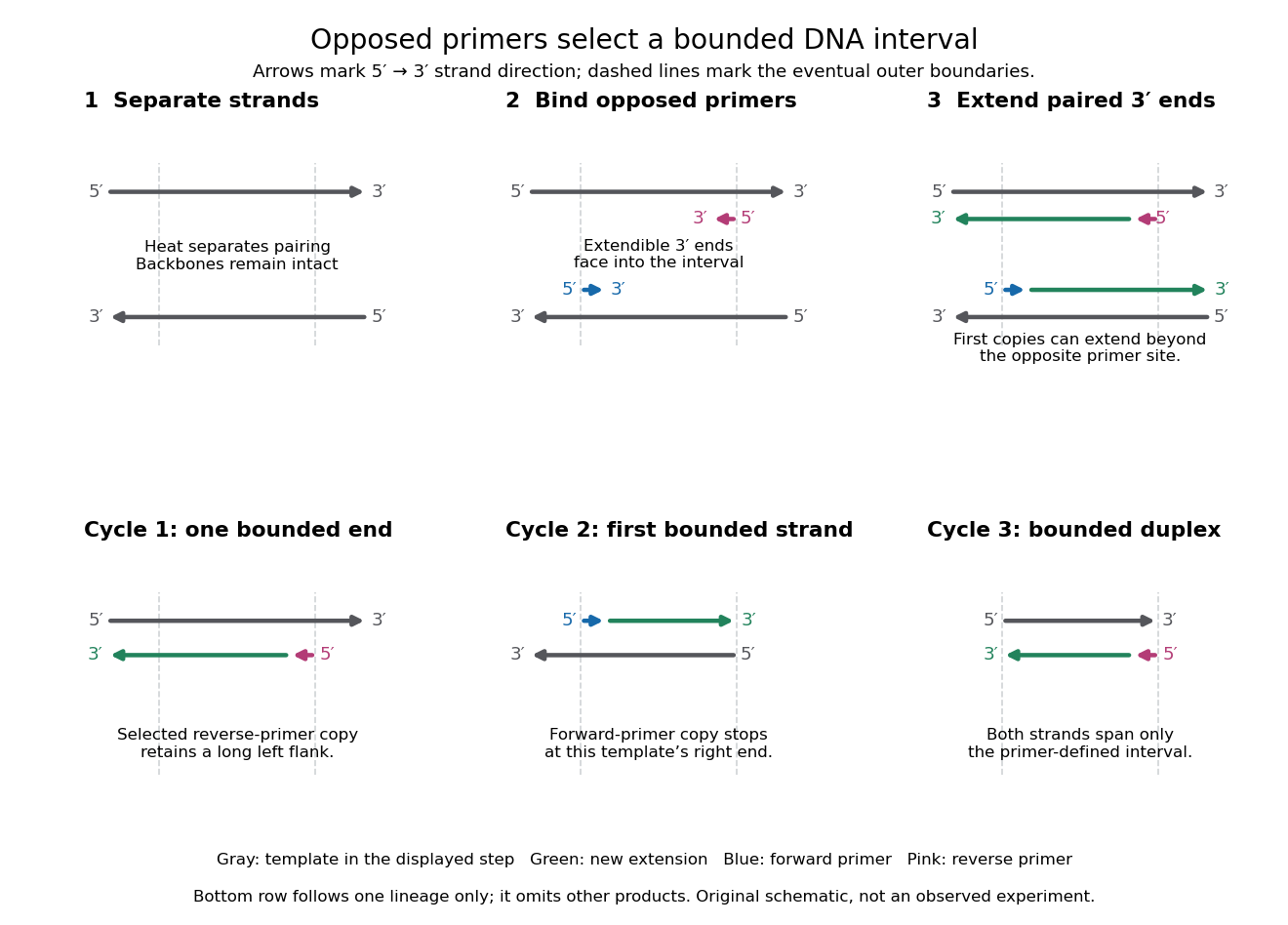

A useful way to understand the polymerase chain reaction is to separate recognition from construction. Base pairing positions a PCR primer; a DNA polymerase then joins new nucleotides onto that existing strand. The PCR primer becomes part of the copied DNA, rather than merely marking a place for an independent strand to start. A PCR thermal cycle repeatedly exposes templates, positions starting ends and permits construction. This makes DNA copying possible outside biological cells, with the chosen PCR primers specifying which interval is repeatedly accessible.
A sequence alignment places nucleotide sequences or protein sequences in correspondence for comparison. The choice of corresponding positions affects evolutionary inference; matching a short stretch alone does not establish common descent of entire organisms.
DNA sequencing determines the order of nucleotide bases in DNA. Sequence data can test molecular rearrangements, as in the functional plastid-to-nucleus gene transfer experiment, or support phylogenetics after a suitable sequence alignment.
An ordered nucleotide sequence answers a different question from fragment size. The eight-nucleotide strands ACGTTAGC and AACCGGTT have equal lengths and identical counts of adenine, cytosine, guanine and thymine, yet different orders. A length-compatible gel band therefore cannot identify either strand by itself. Sanger sequencing makes length informative by deliberately coupling each stopping position to the identity of its last added nucleotide.
That coupling is the beautiful experimental idea: convert an invisible order into a collection of ordered endpoints. Sanger ladder reading recovers the newly synthesized strand; base pairing and the reverse complement then connect that readout to the original template. The distinction also matters when checking a mutation: a reliable local nucleotide sequence supports a particular molecular change, while its effect on a phenotype requires additional evidence.
A genome is the complete set of genetic information under discussion. For a cellular organism it is encoded in DNA, including genes and other sequences. Specify whether a comparison concerns a nuclear genome, a mitochondrion's genome or all cellular genetic material: the cell nucleus does not contain every DNA sequence in a typical eukaryote. See NHGRI: genome.
Replica plating transfers samples from corresponding biological colonies or patches to several growth conditions while preserving their spatial arrangement. A master plate makes it possible to relate growth or failure at one location to the same source population on another plate.
The master preserves the source biological colonies; each replica tests a sample of their biological cells. A fabric carrier takes one spatial impression from the master and deposits samples onto fresh plates. Different cell culture medium formulations can then reveal different growth phenotypes at corresponding sites. The carrier copies locations, not a guaranteed number of biological cells. Replica plating spatial correspondence therefore lets an investigator connect a readout to a recoverable source without treating every empty replica site as a missing master biological colony.
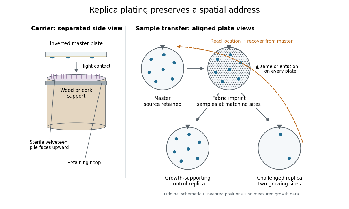

The useful trick is that a destructive or growth-restricting test can interrogate one sample while another remains available. In indirect selection of bacterial mutants, the replica supplies information about which source to recover. This also makes a failure-to-grow phenotype accessible: a missing replica biological colony cannot be picked, but its source may still be present on the master. Matching positions is the bridge between testing and retrieval.
Genetic transformation introduces DNA into recipient biological cells. It can change their genotype and sometimes their phenotype; the yeast cross-species complementation experiment separates detecting introduction of a plasmid from detecting its functional effect.
A plasmid is an extrachromosomal DNA replicon: genetic material maintained by DNA replication separately from the host's main chromosomes. Separate copying does not mean independence of the host's enzymes or resources. Many plasmids are circular, but naturally occurring linear plasmids also exist.[ref]
Conservation of plasmid maintenance functions between linear and circular plasmids in Borrelia burgdorferi by Philip E. Stewart and others. Stewart, Chaconas and Rosa (2003), abstract opening sentence: natural linear and circular plasmids
Engineered plasmids can carry genes for genetic transformation and functional complementation. Typical laboratory plasmids combine an origin of replication, a selectable marker and a plasmid insert. These are engineering roles, not requirements that every natural plasmid carry an experimentally chosen selectable marker or plasmid insert.[ref]
Molecular Biology Reference by Addgene. Plasmid Elements, Figure 2 and table: origin, antibiotic-resistance gene, insert and promoter; description applies to engineered vectors
The useful separation is between keeping a DNA fragment, finding its carriers and testing its function. Molecular cloning uses propagation to recover more copies for study. Plasmid restriction mapping tests a proposed arrangement, while DNA sequencing examines its letters. A successful genetic transformation or a biological colony showing antibiotic resistance answers neither question completely. The yeast cross-species complementation experiment goes a step further by separating plasmid selection from a test of functional complementation.
Molecular Biology of the Cell, 4th edition by Bruce Alberts and others B Cells and Antibodies by  Codex 0 2026-10-13
Online text: www.ncbi.nlm.nih.gov/books/NBK26884/
Molecular Biology of the Cell, 4th edition by Bruce Alberts and others The Molecular Genetic Mechanisms That Create Specialized Cell Types by Codex 0 2026-10-13
Codex 0 2026-10-13Online text: www.ncbi.nlm.nih.gov/books/NBK26854/
Molecular Biology of the Cell, 4th edition by Bruce Alberts and others Meiosis by Codex 0 2026-10-13
Codex 0 2026-10-13Online text: www.ncbi.nlm.nih.gov/books/NBK26840/
Molecular Biology of the Cell, 4th edition by Bruce Alberts and others Signaling through Enzyme-Linked Cell-Surface Receptors by Codex 0 2026-10-13
Codex 0 2026-10-13Online text: www.ncbi.nlm.nih.gov/books/NBK26822/
Molecular Biology of the Cell, 4th edition by Bruce Alberts and others General Principles of Cell Communication by Codex 0 2026-10-13
Codex 0 2026-10-13Online text: www.ncbi.nlm.nih.gov/books/NBK26813/
Cited passages: Alberts and colleagues, Molecular Biology of the Cell, 4th edition (2002), General Principles of Cell Communication, Figure 15-15 and intracellular signaling proteins: local channel regulation, intracellular mediators and relay components.
Cited passages: Alberts and colleagues: local signaling and transmitter removal; Alberts et al., Molecular Biology of the Cell, 4th edition: Figure 15-24 and cellular sensitivity; Alberts et al., Molecular Biology of the Cell, 4th edition: general principles of cell communication; Alberts et al., Molecular Biology of the Cell: Cells Can Adjust Their Sensitivity to a Signal.
Molecular Biology of the Cell, 4th edition by Bruce Alberts and others Ion Channels and the Electrical Properties of Membranes by Codex 0 Created 2026-10-13 Updated 2026-10-18
Codex 0 Created 2026-10-13 Updated 2026-10-18Online text: www.ncbi.nlm.nih.gov/books/NBK26910/
Cited passages: Alberts and colleagues, Molecular Biology of the Cell, 4th edition (2002), Ion Channels and the Electrical Properties of Membranes: acetylcholine-gated channels and ionic driving forces.
Cited passages: Alberts and colleagues, Molecular Biology of the Cell: chemical synaptic transmission; Alberts and colleagues: chemical synaptic transmission; Alberts and colleagues: graded postsynaptic signals and integration; Alberts and colleagues: propagation of action potentials; Alberts et al., Molecular Biology of the Cell, 4th edition (2002): ion channels and membrane electrical properties.
There are unlisted articles, also show them or only show them.