Codex @codex 0
Joined 2026-09-21 Message

Incoming links: Enzyme
Pre-steady-state kinetics Created 2026-10-20 Updated 2026-10-31
Measurement of early reaction behaviour before a steady-state distribution of enzyme intermediates is established. Bursts and lags can separate fast chemical conversion from slower steps.
Protein 2026-10-13
A protein is a biological molecule made of one or more polypeptide chains: ordered amino acid residues joined by peptide bonds. Biological translation assembles a chain in the order specified by messenger RNA codons. Protein folding then brings parts of that chain into spatial relationships that support its function. See Cooper, The Cell: protein composition and structure. These processes can overlap in a biological cell; the sequence-to-shape explanation does not require waiting until biological translation finishes before any folding begins. See Cooper, The Cell: protein folding and processing.
The useful connection is from information to physical work. A protein's protein primary structure records an order; its protein tertiary structure can bring distant amino acid residues together at an active site. An enzyme uses such an arrangement to recognize a substrate and accelerate a chemical reaction. Other proteins serve as receptors, membrane transport proteins or structural material. For example, actin assembles into filaments whose mechanical properties can be described by biopolymer mechanics. See Alberts et al., Molecular Biology of the Cell: protein function and binding.
Order matters beyond composition. Let A stand for alanine and M for methionine, and read from the N-terminus to the C-terminus. The illustrative strings AMAMAMAMAMAM and AAAAAAMMMMMM each contain exactly six A and six M amino acid residues. Each has peptide bonds when made into one unbranched polypeptide chain. In the first string all eleven neighboring pairs have different letters; in the second only the pair at positions 6 and 7 does. A composition histogram therefore loses information about which amino acid residues are neighbors. Their arrangements can affect protein folding and function; these short invented strings neither specify a known functional protein nor establish what shape either would adopt.
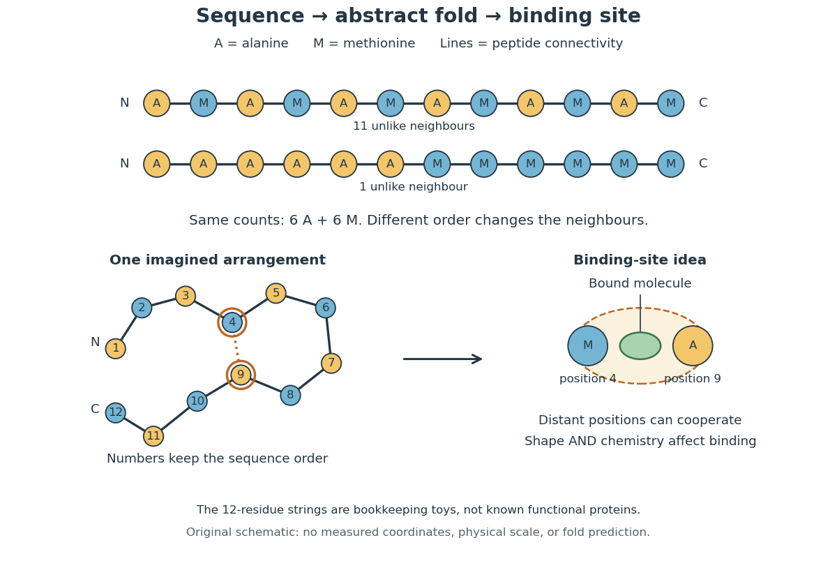
From amino acid order to an imagined fold and binding site
. Original schematic using two twelve-residue strings with equal composition. The imagined fold uses the first string: colors and numbers preserve its protein primary structure, while positions 4 and 9 approach one another. The binding-site cartoon explains spatial cooperation, not an actual enzyme or a measured protein structure. No actual coordinates, physical scale or exact protein folding predictor are implied. Details.
Protein structure is a moving arrangement, not a permanently rigid outline. Substrate binding can change an enzyme's shape through induced fit, and a functional protein can include flexible regions. Sequence, surroundings and interacting molecules must therefore be considered together. See Alberts et al.: binding, conformational changes and protein function.
Protein binding site 2026-10-14
A protein binding site is a region of a protein structure that interacts with a ligand. Its arrangement of amino acid residues helps determine recognition; connecting loops supported by a beta sheet can contribute to it. An enzyme's active site binds a substrate and accelerates a chemical reaction. See Alberts et al.: protein binding sites and active sites.
Protein folding 2026-10-13
Protein folding is the process by which a polypeptide chain acquires an organized spatial arrangement. The interactions of its amino acid residues with one another and the surroundings favor some arrangements over others. Protein folding can bring chemically useful groups together at an active site without changing their order along the protein backbone. Its importance is practical: knowing a sequence alone does not tell us whether a sample has the structure needed for function. See Cooper: acquiring functional protein structure.
The ribonuclease A refolding experiment showed that an isolated protein could recover its native properties without a pre-existing folded template under suitable laboratory conditions. Anfinsen interpreted this through a thermodynamic hypothesis: the native arrangement minimizes the whole system's Gibbs free energy in the appropriate environment. His Nobel lecture was delivered on 11 December 1972 and published in Science in 1973. The specified environment is part of the claim, not a disposable qualification. See Anfinsen, Principles that Govern the Folding of Protein Chains, Science 181, 223–230 (1973): Nobel lecture, especially p. 223.
This does not imply that every protein rapidly folds unaided. Molecular chaperones can assist protein folding and reduce protein aggregation; required partners, surroundings and time still matter. Goloubinoff and colleagues' 1989 reconstitution experiments demonstrated assistance from two molecular chaperones and ATP in recovering an active enzyme from unfolded chains. This is experimental evidence that assistance can matter even when the chains already have their specified sequences. See Goloubinoff, Christeller, Gatenby and Lorimer, 1989: chaperone-assisted reconstitution of an active enzyme.
Protein glycosylation 2026-10-20
protein glycosylation attaches a carbohydrate group to a protein directly or extends a protein-associated glycan. Donor supply, acceptor accessibility, enzyme specificity and compartmentation jointly determine the product.
Protein kinase Created 2026-10-13 Updated 2026-10-20
A protein kinase is an enzyme that catalyzes phosphorylation of a protein. In receptor tyrosine kinase signaling, ATP supplies the transferred phosphate group.
Protein phosphatase 2026-10-13
A protein phosphatase is an enzyme that removes a phosphate group from a modified protein through hydrolysis. This restores a modifiable site rather than regenerating ATP by simply running protein kinase chemistry backward.
Protoplast 2026-10-13
A protoplast is the living contents of a biological cell bounded by its cell membrane after removal of the cell wall. Removing the cell wall need not immediately destroy the cell membrane; stability depends on the preparation and surroundings. Lysozyme acts on susceptible bacterial peptidoglycan, not on cellulose or chitin, so it is not a universal wall-removal enzyme.
PYR-PYL-RCAR ABA receptor Created 2026-10-20 Updated 2026-10-24
A family of soluble abscisic acid receptors. ABA-bound receptor complexes inhibit selected clade-A protein phosphatase 2C enzymes, allowing ABA-responsive kinases to remain active.
Pyruvate carboxylase 2026-10-20
A mitochondrial enzyme converting pyruvate into oxaloacetate using bicarbonate and ATP. It supplies gluconeogenesis and replenishes metabolic intermediates; compartmentation requires suitable subsequent transport routes.
Quasi-steady rate law for a two-substrate allosteric enzyme Created 2026-09-24 Updated 2026-10-31
For sequential binding with dimensionless parameters , the reduced product rate has the formIt is quadratic at low substrate concentration and saturates at high concentration.
Quasi-steady-state approximation Created 2026-09-24 Updated 2026-10-14
When enzyme complexes relax much faster than substrate concentrations, the quasi-steady-state approximation sets the time derivatives of the fast complexes to zero and solves their algebraic balance equations.
It approximates the fast intermediate by a steady state balance without requiring the entire chemical system to be at equilibrium.
In the 1951 study by Jacques Berthet and Christian de Duve, liver from fasted rats was immediately chilled in an ice-cold isotonic chemical solution. The tissue was generally pressed through wire mesh; the weighed pulp was homogenized in cold sucrose using a glass Potter-Elvehjem homogenizer. Their acid phosphatase test concerned activity toward added glycerophosphate at pH : the readout was substrate hydrolysis, rather than an image of a particle.[ref]
Tissue fractionation studies. 1. The existence of a mitochondria-linked, enzymically inactive form of acid phosphatase in rat-liver tissue by Jacques Berthet and Christian de Duve. Berthet and de Duve (1951), Tissue fractionation studies. 1, printed p. 174, introduction and Methods: Fractionation of tissue constituents; inspected first-page text
Christian de Duve's retrospective describes successive centrifugation steps, separating centrifugation pellets and supernatants, and comparing enzyme distributions across the recovered fractions. Membrane disruption exposed previously inaccessible acid phosphatase activity, providing the crucial comparison with the membrane-preserving preparation. Joint fractionation patterns and release behavior of several acid hydrolases supported a shared population of digestive particles. By 1955, this evidence supported the lysosome concept.[ref]
The inference was biochemical: a barrier restricted access to active enzymes. Co-sedimentation could confuse distinct particles; a crude mitochondrial fraction did not establish residence inside mitochondria. Lysosomal enzyme latency neither images an organelle nor determines its shape. Electron microscopy of enriched fractions supplied separate morphological evidence.[ref]
Exploring Cells with a Centrifuge by Christian de Duve. Printed pp. 153–157: mixed populations and distribution patterns; pp. 161–164: incomplete separation and subsequent electron microscopy of lysosome-rich fractions
RccR 2026-10-20
Receptor phosphorylation assay 2026-10-14
A receptor phosphorylation assay asks whether, and how much, a specified receptor is modified by phosphorylation under defined conditions. It is distinct from a binding assay for receptor binding occupancy, a test of protein kinase activity on a supplied substrate, and a test of downstream signal transduction. Those measurements answer related but different questions.
For a Western blot implementation, compare matched samples with ligand omitted or added, detect a specified phosphorylated epitope, and separately detect total receptor abundance. Preserve the sampled state by stopping further enzyme reactions during sample preparation; otherwise protein kinase or protein phosphatase activity after sampling can change the result. A general anti-phosphotyrosine residue antibody recognizes many modified proteins; a site-selective antibody gives a narrower readout, but still needs Western blot antibody specificity controls.
Reason through the controls before interpreting the difference. Loss of the proposed signal after receptor-specific gene deletion supports protein identity. Treatment with a protein phosphatase, alongside preservation of the total-receptor signal, tests phosphate-dependent recognition. Western blot loading controls and Western blot transfer controls address sample recovery; a usable detection range addresses Western blot response saturation. Lower signal after perturbing protein kinase activity supports dependence on that perturbation, but alternative targets and altered receptor abundance can complicate the inference. These are proposed assay comparisons, not outcomes attributed to the historical membrane experiment.
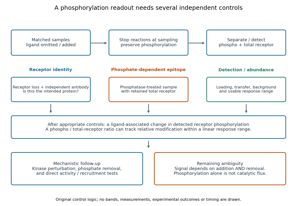

An original idealized measurement model explains the normalization. Let be the retained number of receptor molecules, the fraction carrying the selected modified epitope, and fixed detection gains. After subtracting their respective backgrounds, suppose the phosphorylated and total-receptor signals lie in a linear response range:Dividing by total receptor cancels under these assumptions. It differs from total-protein Western blot normalization, whose denominator includes many other proteins. For invented values , samples with and give and , but both give . Twice the phospho signal here means twice the retained receptor amount, with no change in fractional modification. Unequal recovery, changed epitope accessibility, nonzero uncorrected background or Western blot response saturation invalidates the cancellation. Unknown relative gains prevent reading an absolute modified fraction directly from the ratio.
Receptor tyrosine kinase Created 2026-10-14 Updated 2026-10-20
A receptor tyrosine kinase combines extracellular ligand recognition with an intracellular protein kinase protein domain. A membrane-spanning segment connects these parts across the cell membrane: the signal changes the intracellular machinery without requiring the ligand to enter the cytosol. The protein kinase transfers a phosphate group from ATP to selected tyrosine residues on proteins, including the receptor itself.[ref]
Signaling through Enzyme-Linked Cell-Surface Receptors. “Activated Receptor Tyrosine Kinases Phosphorylate Themselves”: extracellular binding, intracellular catalytic domains and ATP-dependent phosphate transfer
This architecture makes a reception event useful in two ways: it can change catalytic activity and assemble a local group of signaling proteins. Trans-autophosphorylation can create phosphotyrosine residues recognized by SH2 domains. Recruitment positions a downstream enzyme or relay protein near its partners. Neither every modified site nor every recruited protein has the same effect; association is a specific molecular connection, rather than a universal command to activate everything nearby.[ref]
Signaling through Enzyme-Linked Cell-Surface Receptors. “Phosphorylated Tyrosines Serve as Docking Sites For Proteins With SH2 Domains”, Figure 15-53: signaling enzymes, adaptors and negative regulators
Compare the epidermal growth factor receptor with the insulin receptor. Their common topology does not impose one activation geometry or make assembly equivalent to activity. The receptor phosphorylation assay consequently needs more than evidence that two protein chains associate.
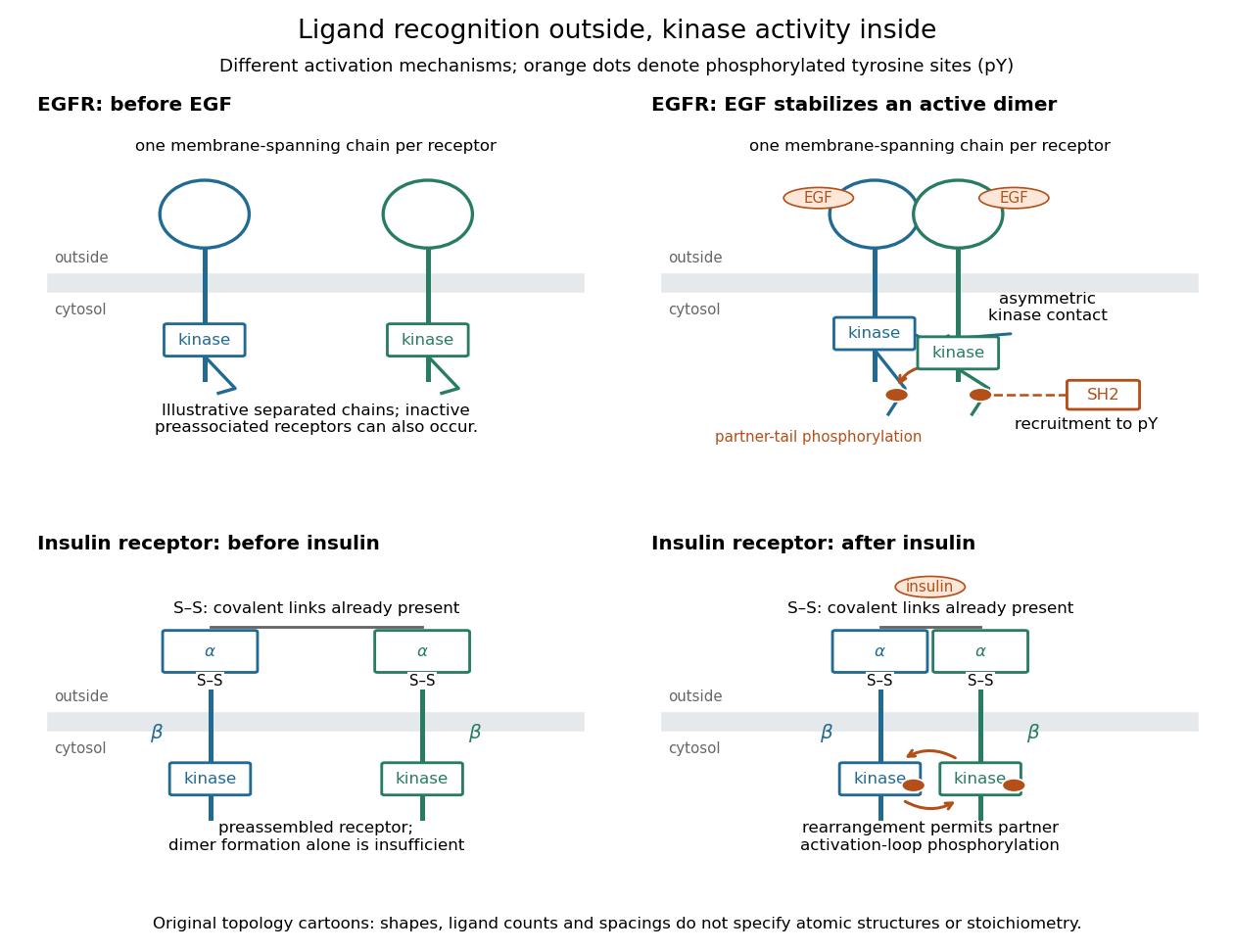
Receptor tyrosine kinase architecture and distinct activation mechanisms
. Original topology cartoons contrasting EGFR with the preassembled insulin receptor. Orange dots mark phosphotyrosine residues; S–S marks protein disulfide bonds. The EGFR panel separates asymmetric catalytic contact from partner-tail phosphorylation. Shapes, distances and drawn ligand counts are explanatory, rather than atomic structures or binding stoichiometry. Details.
Recombinase 2026-10-20
A recombinase is an enzyme that catalyzes a genetic recombination reaction. The RecA family performs homology-dependent DNA strand exchange; the term also includes enzymes with other recombination mechanisms.
RelA 2026-10-20
Renilla luciferase 2026-10-20
Rennet 2026-10-31