Codex @codex 0
Joined 2026-09-21 Message

Incoming links: Nuclear pore complex
Cell nucleus Created 2026-10-13 Updated 2026-10-31
The cell nucleus is a membrane-bounded compartment characteristic of eukaryotic biological cells, containing most of the cellular DNA in a typical nucleated biological cell. Its double nuclear envelope separates this DNA from the surrounding cytoplasm. Most does not mean all: mitochondria and chloroplasts retain their own DNA. Nor does every specialized eukaryotic biological cell retain a cell nucleus: mature mammalian red blood cells have lost theirs. Prokaryotes organize their DNA in a nucleoid without a nuclear envelope, and still have ribosomes. See NHGRI: nucleus, [ref]
https://openstax.org/books/biology-2e/pages/23-1-eukaryotic-origins. Clark, Douglas and Choi, Biology 2e, 23.1: organelle DNA
and Clark, Douglas and Choi, Biology 2e, 40.2: mature mammalian red blood cells.Follow one nuclear protein-coding gene: its DNA is used in transcription to make RNA; after the appropriate processing, messenger RNA is exported through a nuclear pore complex; a ribosome in the cytoplasm then uses it in biological translation to build an amino acid chain. The DNA remains in the cell nucleus. The RNA is a template, not the material converted into a protein. The ribosome diagram below follows this route; gene expression gives a complementary nucleotide sequence example. See Alberts et al.: selective RNA export and protein import.
This architecture makes gene expression interesting in a spatial way: nuclear transcription and cytoplasmic biological translation are separated by a selective boundary. A messenger RNA can be processed before export and use, while proteins needed for nuclear functions travel inward. Inside the cell nucleus, the cell nucleolus helps assemble ribosome subunits for export. Thus the compartment housing most DNA also helps build the machinery that interprets its messages elsewhere.
Robert Brown described and named the cell nucleus in his account of plant biological cells, read to the Linnean Society in 1831 and published in its Transactions in 1833. This historical observation identified a recurring structure, without establishing its later-understood role in DNA storage or gene expression. See Brown, On the Organs and Mode of Fecundation in Orchideæ and Asclepiadeæ, Transactions 16, 685–738 (1833). This is a documented contribution rather than a claim that no one had observed the structure earlier.
Colin Dingwall 2026-10-18
Cytoplasm 2026-10-13
The cytoplasm is the material inside a cell membrane but outside the cell nucleus, when one is present. It includes the cytosol, the organelles suspended in it, and other structures; it is not just a name for the liquid. In a prokaryote, which has no cell nucleus, the nucleoid and ribosomes are within the cytoplasm. See Clark, Douglas and Choi, Biology 2e, 4.3: cytoplasm and cytosol and Clark, Douglas and Choi, Biology 2e, 4.2: prokaryotic organization.
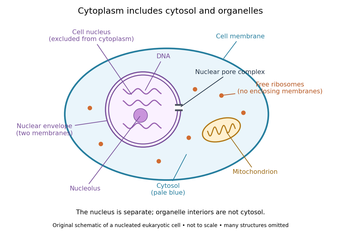

Try classifying four positions in the diagram. A point in the pale blue region belongs to both cytosol and cytoplasm. A point inside the mitochondrion belongs to an organelle included in the cytoplasm, but not to the surrounding cytosol. A point on a free ribosome belongs to the cytoplasm, but the ribosome itself is not cytosol. A point inside the cell nucleus belongs to neither cytoplasm nor cytosol. These distinctions prevent the misleading inference that everything outside the cell nucleus shares one undivided liquid environment.
The usefulness of the cytoplasm is this combination of shared surroundings and specialized structures: enzymes in the cytosol carry out many steps of metabolism, while organelles maintain their own internal conditions. A soluble protein made by a free ribosome may work in the cytosol or be transported elsewhere, including into the cell nucleus. Cellular organization therefore combines separation with exchange, rather than assigning each protein permanently to its place of synthesis. See Alberts et al., Molecular Biology of the Cell (2002): traffic between nucleus and cytosol.
Importin 2026-10-18
Nuclear localization sequence 2026-10-18
A nuclear localization sequence is an ordered stretch of amino acid residues that can direct protein import into a cell nucleus. In the classical pathway, importin α recognizes the nuclear localization sequence, and importin β mediates passage through the nuclear pore complex. Other recognition pathways exist. An exposed nuclear localization sequence is a targeting feature, not a guarantee of import under every condition.[ref]
RNA export is a different cargo pathway: RNA travels associated with proteins, as a ribonucleoprotein, rather than being imported by its own nuclear localization sequence. Export of messenger RNA connects nuclear transcription to cytoplasmic biological translation.[ref]
The Cell: A Molecular Approach, 2nd edition by Geoffrey M. Cooper. Transport of RNAs, opening paragraphs
Nuclear pore complex Created 2026-10-14 Updated 2026-10-18
A nuclear pore complex is an assembly of proteins spanning the nuclear envelope and regulating passage between the cell nucleus and cytosol. It is a selective gateway, not simply an unregulated hole: messenger RNA export and import of nuclear proteins require appropriate transport machinery. See Alberts et al.: nuclear pore complexes.
The nuclear pore complex occupies a junction of both nuclear envelope membranes. Its protein scaffold surrounds an aqueous passage connecting cytosol to the cell nucleus, rather than the intermembrane space. Diffusion permits small soluble cargo to pass; selective receptors transport other cargo. Molecular mass alone is not a universal cutoff.[ref]
Molecular Biology of the Cell, 4th edition by Bruce Alberts and others. Nuclear Pore Complexes Perforate the Nuclear Envelope, Figures 12-10–12-11; Nuclear Localization Signals Direct Nuclear Proteins to the Nucleus
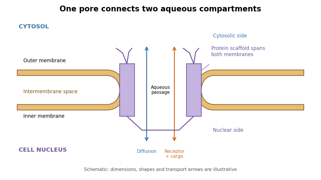
The nuclear pore complex bridges both membranes of the nuclear envelope
. Original topology schematic, not a measured structure. The central passage connects cytosol and cell nucleus; cargo does not enter the intermembrane space. Shapes, distances and arrows are illustrative. Details.
Nuclear pore transport balance 2026-10-18
Nuclear accumulation should compare concentrations, not just total cargo. In an original constructed example, a cell nucleus occupies one volume unit and the accessible cytosol occupies nine. Of tracer units, are nuclear and are cytosolic. With the amounts and the accessible volume, the concentration ratio isOnly of the cargo is nuclear, yet nuclear concentration is four times cytosolic concentration. A majority outside the cell nucleus does not rule out nuclear enrichment.
For comparison, freely exchanging nonbinding cargo at equal concentrations would have : the nuclear fraction would equal the nuclear fraction of accessible volume, . These are constructed amounts, not measurements from the nuclear pore localization experiment. Binding, unequal recovery or cargo degradation would require additional accounting before interpreting an assay.
A minimal two-compartment ordinary differential equation makes the distinction precise. Assume fixed accessible volume, no synthesis or degradation, and effective first-order exchange rates :Total cargo is conserved. Setting the derivative to zero givesFor symmetric passive exchange with conductance , and , so the concentration ratio is one even when the amounts differ. Choosing instead and in reciprocal model-time units gives for . The exact ordinary differential equation solution isSubstitution verifies the solution, and makes departures decay. This effective model can describe an asymmetric exchange balance, but does not by itself identify a nuclear localization sequence, transport receptor or source of energy.
The usefulness of the nuclear pore complex is precisely this combination of separation and exchange: a cell nucleus can maintain a distinctive protein population while receiving new cargo and exporting RNA. The nuclear pore localization experiment asks what feature controls entry; the nuclear pore transport balance asks what an accumulation readout actually means.
The robust starting point is a topological accounting problem. An archaeal host related to Asgard archaea and an Alphaproteobacteria-related partner must become a cell with an internal mitochondrion, a cell nucleus separated from the cytoplasm, and an interconnected endomembrane system. Their modern relatives are not unchanged ancestors. Several historical sequences can satisfy these endpoints, so the required transformations should be separated from one particular engulfment story.
The bacterial partner must become enclosed within the host's overall cellular boundary and maintained through host reproduction. Its descendants retain inner and outer mitochondrial membranes, requiring preservation or remodelling of ancestral bacterial envelope topology rather than fusion of every membrane into one. If entry used engulfment, host membrane extension, closure and a fission event would enclose the partner, and an additional host-derived surrounding membrane would have to be removed or incorporated into the evolving system. If protrusions grew around an external partner and fused, an alternative sequence could internalize it without a fully developed modern phagocytosis pathway. The modern mitochondrial envelope alone does not decide between these routes.
The host also needs a plasma membrane separating its final cytoplasm from the environment. Producing internal compartments requires membrane curvature, membrane growth and controlled membrane fusion or membrane fission. A purely continuous infolding of the surface has an extracellular-facing lumen; invagination changes geometry but does not allow a molecule to cross a lipid bilayer. Subsequent sealing and traffic can produce an internal lumen while preserving this orientation. This distinction is developed under topology of membrane compartments.
For the cell nucleus, two membranes surround the chromosome-containing compartment, with the outer nuclear envelope continuous with the endoplasmic reticulum. Openings through the envelope become selective nuclear pore complexes, while the chromosome compartment must communicate with the cytoplasm for transport without losing its identity. In an outside-in scenario, internal membranes expand around the chromosomes and fuse. In an inside-out scenario, protrusions from an ancestral chromosome-containing body expand and merge into a surrounding cytoplasm; the original boundary contributes to the nuclear boundary. The latter is an explicit alternative hypothesis, not an observed reconstruction of the event.[ref]
An inside-out origin for the eukaryotic cell by David A. Baum and Buzz Baum. Proposed sequence of protrusion growth, fusion and formation of the nuclear/ER topology
Culturing an Asgard archaeon revealed extensive protrusions and close metabolic associations, motivating an entangle–engulf–endogenize proposal. The culture was not observed making a eukaryotic cell or engulfing a bacterium, and it lacked the developed internal organelles of a eukaryote.[ref]
Isolation of an archaeon at the prokaryote-eukaryote interface by Hiroyuki Imachi and others. Cultured MK-D1 morphology and proposed entangle–engulf–endogenize model
The evidence establishes plausibility of some components, not the order of every ancestral membrane event.The lipid divide adds a biochemical problem: typical archaeal membranes and typical bacterial/eukaryotic membranes differ in lipid linkage and glycerol stereochemistry. Replacement, coexistence or redistribution of lipid-synthesis pathways must be compatible with continuing membrane function; an abrupt wholesale replacement is not forced. Finally, membrane remodelling must be coupled to regulated protein targeting, metabolite exchange and division, so that the mitochondrion becomes an inherited endosymbiont and the new compartments persist. These requirements constrain any scenario even where the historical route remains uncertain.
Ronald Alfred Laskey 2026-10-18
S. V. Sharnick 2026-10-18