Topics (384k) Articles (398k) Users (410) Discussions (242) Comments (383) Files (13k) New article

Willi Schaffner coauthored the 2002 investigation of interference by plant-derived substances and antioxidants in the MTT assay.[ref]
Interference of plant extracts, phytoestrogens and antioxidants with the MTT tetrazolium assay by Regina Bruggisser and others. 2002, byline and Abstract
The work illustrates why a biochemical test must distinguish its intended biological response from reactions of the test substance with its reagents.The authors observed direct redox conversion of the dye with compounds including resveratrol in the absence of cells.[ref]
Interference of plant extracts, phytoestrogens and antioxidants with the MTT tetrazolium assay by Regina Bruggisser and others. Abstract, cell-free experiments
Such a result establishes an alternative route to the assay signal; it does not establish whether the compound protects or harms a living cell.An original concentration model shows why a smooth response curve is insufficient evidence of cell proliferation. Suppose the cellular contribution to absorbance stays fixed at , while chemical interference adds , where is a dimensionless multiple of a chosen test concentration. At , measured absorbance is . This perfectly ordered trend contains no increase in viable cell number. A matching compound-only series tests the chemical explanation directly; a biological interpretation requires additional evidence.
Heide Tullberg-Reinert coauthored the 2002 study of chemical interference with the MTT assay. Washing treated cells before adding MTT reduced the direct redox interference observed with kaempferol.[ref]
Interference of plant extracts, phytoestrogens and antioxidants with the MTT tetrazolium assay by Regina Bruggisser and others. 2002, byline and final sentences of Abstract
That experimental comparison connects the readout to the presence of the test substance during measurement. Washing is an intervention on assay conditions, rather than proof that the earlier exposure had no biological effect. It may remove extracellular material while leaving changes in cell viability or cellular metabolism.
In an original model, let the cellular contribution to absorbance be and removable chemical interference be . Before washing the total is . Removing 90 percent of the interference leaves , still above the cellular contribution. A smaller signal after washing supports an interference explanation, but residual signal and loss of loosely attached cells must also be considered. Paired negative controls and independent counts make the interpretation stronger than washing alone.
Ezio Merler coauthored the 1971 study of angiogenic activity isolated from tumours, alongside Judah Folkman, Charles Abernathy and Gretchen Williams.[ref]
Isolation of a Tumor Factor Responsible for Angiogenesis by Judah Folkman and others. Byline and Abstract
The investigators coupled biochemical separation with a biological response, asking whether separated material could induce new capillaries.This is the logic of bioassay-guided fractionation: a fraction is interesting because a reproducible biological effect follows it through the separation procedure. An active fraction need not be a single pure molecule. The historical preparation should therefore not be identified retrospectively with vascular endothelial growth factor merely because both concern angiogenesis.
An original example distinguishes recovery from enrichment. Suppose a starting extract contains 100 arbitrary units of angiogenic activity in 20 milligrams of protein. Its activity per milligram is . A later fraction contains 40 units in 2 milligrams, giving units per milligram. The process has achieved fourfold enrichment with 40 percent activity recovery. Neither number establishes chemical purity. Here the activity units are assumed additive for illustration; an ordinal visual score cannot automatically be treated as an additive biochemical quantity.
Charles Abernathy coauthored the 1971 experimental study of a tumour-derived angiogenic fraction with Judah Folkman, Ezio Merler and Gretchen Williams.[ref]
Isolation of a Tumor Factor Responsible for Angiogenesis by Judah Folkman and others. Byline and Abstract
An important aspect of the experimental argument was spatial separation: blood vessels could respond outside filter chambers containing tumour cells.[ref]Isolation of a Tumor Factor Responsible for Angiogenesis by Judah Folkman and others. Printed p.275, membrane-chamber observations
A barrier experiment tests whether direct cellular contact is necessary. Its interpretation depends on membrane permeability: passage of a dissolved substance differs from passage of an intact cell. Exclusion of cells with persistence of a vascular response supports a diffusible signal, but does not uniquely identify the signal's molecular structure.
An original dimensional estimate shows why distance and time belong in the interpretation. A characteristic diffusion time over a distance is , where is a diffusion coefficient. Taking stipulated values and gives about , or 42 minutes. Doubling the distance multiplies that estimate by four. Binding, degradation and geometry can alter the actual response; the calculation is not a reconstruction of the experiment.
Gretchen Williams coauthored the 1971 investigation with Judah Folkman, Ezio Merler and Charles Abernathy that isolated angiogenic activity from tumours.[ref]
Isolation of a Tumor Factor Responsible for Angiogenesis by Judah Folkman and others. Byline, Abstract and printed p.275
The collaboration helped turn tumour angiogenesis into an experimentally testable interaction between tumours and surrounding blood vessels.The important distinction is between growth of a tumour's own cells and growth of host endothelial cells. A substance released by one population can change the behaviour of the other. Thus a vascular response outside an implanted tumour cannot be interpreted simply as movement of the implanted cells.
An original measurement example illustrates the distinction. Suppose a marked tissue region initially contains 20 vessel branch points, and later contains 32. The increase is , or 60 percent of the initial count. That observation alone does not give a cell proliferation rate: vessel branching, length and endothelial cell number are different measurements. It also does not establish effective perfusion. A convincing experiment therefore defines its vascular readout and compares it with a matched negative control. These numbers illustrate the reasoning, not the paper's data.
Maria Feoktistova is first author of a 2016 crystal violet staining protocol with Peter Geserick and Martin Leverkus. It quantifies material remaining attached to a culture surface, using a dye that binds cellular proteins and DNA.[ref]
Crystal Violet Assay for Determining Viability of Cultured Cells by Maria Feoktistova and others. 2016, byline and Abstract, maintained adherence and dye binding
The contribution concerns an experimentally accessible readout of changes in cell culture, rather than an intrinsic molecular count of viable cells.A crystal violet biomass response becomes a cell-number estimate only through a suitable calibration. In an original matched-condition model, letwhere is absorbance and the retained cell number. Standards containing 0, 2000 and 4000 cells give readings , and . A sample reading would imply under this model.
The slope incorporates stain retained per cell, extraction and optical geometry. If a treatment doubles that per-cell response while preserving the background, only 2500 retained cells produce the same sample reading. An accurate measurement of colour can therefore coexist with an inaccurate biological interpretation. Independent counting and a calibrated response range test assumptions that repeated optical readings alone cannot settle. The numerical standards here are illustrative, not measurements from Feoktistova's protocol.
Peter Geserick coauthored the 2016 crystal violet assay protocol with Maria Feoktistova and Martin Leverkus.[ref]
Crystal Violet Assay for Determining Viability of Cultured Cells by Maria Feoktistova and others. 2016, byline
The method uses retained attachment of cultured cells as an indirect readout relevant to survival and growth. The authors caution that identifying why staining declines requires additional methods.[ref]Crystal Violet Assay for Determining Viability of Cultured Cells by Maria Feoktistova and others. Abstract, interpretation of reduced staining
This distinction connects a biological endpoint with a measurement model. The observed crystal violet biomass response can change through cell number, attachment, stain binding or extraction. These contributions cannot generally be separated by one absorbance reading.
An original normalization example has background-corrected control and treatment signals and . Their ratio is . If each also includes an unremoved background , the ratio of raw readings is insteadBackground subtraction is therefore part of defining the compared quantity. Even the corrected ratio is not automatically the surviving fraction: the amount retained per living cell must also remain comparable, and handling must not remove different fractions of otherwise viable cells. A matching negative control and an independent observation address these different assumptions. These numbers illustrate the model rather than an experiment assigned to Geserick individually.
Sandra Prior researches bioassay standards for therapeutic monoclonal antibodies. Her institutional profile describes work linking antibody structure and biological function, including the development of cell-based assays.[ref]
Dr Sandra Prior by NIBSC. Research account, bioassay standards and monoclonal-antibody structure–function relationships
Her contribution is to making functional measurements comparable, rather than treating an antibody's chemical amount as a complete description of its biological activity.
Prior is first author of a 2017 WHO collaborative-study report evaluating a proposed international standard for the biological activities of rituximab.[ref]
Report on a Collaborative Study for Proposed Candidate 1st International Standard for the biological activities of Rituximab by Sandra Prior and others. WHO/BS/2017.2309, cover and study title
The report used comparisons of preparations and response-curve parameters to assess relative biological potency. Its coded duplicates supplied empirical information for study-specific bioassay parallelism criteria; the report distinguished these from routine laboratory validity limits.[ref]Report on a Collaborative Study for Proposed Candidate 1st International Standard for the biological activities of Rituximab by Sandra Prior and others. printed pp. 8 and 10–11
These are contributions of the collaborative team, not an attribution of every analytical step to one author.An original calibration example illustrates the scientific problem. Suppose two laboratories estimate the same test preparation's activity relative to a shared reference as and . Their geometric mean is , whereas their arithmetic mean is . The geometric comparison treats reciprocal departures symmetrically on a logarithmic scale. Agreement of that summary with one does not establish acceptable individual curves or prove that the laboratories share no bias. Assay similarity, experimental controls and uncertainty remain separate questions.
The report also explains why assigned biological units are not simply a conversion from measured antibody mass: first-standard unitage defines a conventional activity scale, while a replacement standard is calibrated against that scale.[ref]
Report on a Collaborative Study for Proposed Candidate 1st International Standard for the biological activities of Rituximab by Sandra Prior and others. printed p. 33, Appendix 3
This distinction connects Prior's standardization work with reference-standard uncertainty in relative potency and the interpretation of interlaboratory comparisons.A melanosome is a specialized, membrane-bounded organelle in which melanin is synthesized and stored. Melanocytes produce melanosomes; pigment can subsequently be transferred to neighbouring keratinocytes. The melanosome is the compartment, whereas melanin is its pigment cargo. Maturation involves changes in internal organization as well as accumulation of pigment. [ref]
Molecular Biology of the Cell, 4th edition by Bruce Alberts and others. Chapter 22, skin: melanocytes and pigment distribution
An experiment connected that organization to a particular protein. Expression of Pmel17 in nonpigmented cells produced internal striations resembling those of immature melanosomes, within multivesicular compartments. The result supported a structural role for Pmel17 in constructing a scaffold before extensive pigment deposition. It did not show that expression of one protein produces every function of a mature melanosome. [ref]
Pmel17 initiates premelanosome morphogenesis within multivesicular bodies by Joanne F. Berson and others. 2001, abstract and Pmel17 expression experiments
A vesicle is a small compartment enclosed by a lipid bilayer. Its vesicle lumen can carry soluble cargo, while its lipid bilayer carries lipids and membrane proteins. In a transport pathway, budding separates a carrier from one compartment and membrane fusion joins it to another. Orientation matters: fusion of a secretory vesicle with the cell membrane connects its vesicle lumen to the extracellular space while incorporating its membrane proteins into the surface. [ref]
Molecular Biology of the Cell, 4th edition by Bruce Alberts and others. Chapter 13, Intracellular Vesicular Traffic: topology of membrane transport
A vesicle is therefore more than a freely diffusing package. Recognition, movement and membrane fusion require distinct interactions. In a reconstitution experiment, complementary SNARE proteins placed in separate liposomes brought opposing lipid bilayers together and promoted fusion; the experiment isolated one part of transport from the rest of a living cell. [ref]
SNAREpins: minimal machinery for membrane fusion by T. Weber and others. 1998, abstract: separate v-SNARE and t-SNARE vesicles
A germ cell belongs to the cell lineage that produces gametes and transmits a genome to the next generation. The term includes immature precursors, so a germ cell need not already be a sperm or an ovum, or already be haploid. In a mammal, proliferating precursors precede meiosis and subsequent gamete differentiation. The distinction from a somatic cell concerns developmental fate and ancestry, not simply where a cell is located. [ref]
Molecular Biology of the Cell, 4th edition by Bruce Alberts and others. Chapter 20, Germ Cells and Fertilization
Germ cell specification can depend on signals from neighbouring tissues. In a mouse experiment, embryos lacking functional BMP4 lacked primordial germ cells; the study used mutants and chimaeras to identify an essential contribution from extraembryonic tissue. This makes the inherited lineage an outcome of developmental signalling, rather than a compartment that must always be isolated at the first cleavage. [ref]
A cell population is a defined collection of biological cells, for example the cells in a culture, tissue compartment or measured cell type. Its definition specifies which cells belong, rather than assuming that every included cell is identical. Cells can differ in cell cycle stage, differentiation, molecular abundance or future fate.
For a counted compartment, a binary cell division adds one cell to the total, while cell death removes one; migration and differentiation can also change membership. A net change in cell count does not reveal how many divisions and deaths occurred. A hundred divisions accompanied by ninety deaths gives the same net increase as ten divisions without death.
Cell lineage measurements resolve some of this ambiguity. In the primary reconstruction of Caenorhabditis elegans embryogenesis, investigators followed living cells with differential interference contrast microscopy and recorded division histories and cell fates.[ref]
The embryonic cell lineage of the nematode Caenorhabditis elegans by John Sulston and others. Introduction, lineage reconstruction and Figure 3: live Nomarski observations and the embryonic pedigree
Flow cytometry and single-cell RNA sequencing instead characterize sampled cells by measured properties. Their observed fractions need not represent the entire starting cell population if survival, dissociation or detection differs between cell types.Cell survival means that a biological cell remains alive over a specified interval and under specified conditions. It differs from cell proliferation: a neuron can survive for years without dividing, while an increasing cell population can conceal simultaneous cell division and cell death. A cell viability assay measures a chosen property, such as retention of an intact cell membrane or continuing metabolism, rather than every possible future function. [ref]
Molecular Biology of the Cell, 4th edition by Bruce Alberts and others. Chapter 17, Programmed Cell Death: survival signals and regulation of apoptosis
The balance is actively regulated. Survival signals can restrain apoptosis; loss of a required signal can permit a death programme. In a cell-free experiment, removing cytochrome c suppressed apoptotic activity and adding it back restored that activity in the presence of dATP. This linked a normally compartmentalized mitochondrial protein to activation of an apoptosis pathway, rather than identifying survival merely with a high metabolic readout. [ref]
Induction of apoptotic program in cell-free extracts: requirement for dATP and cytochrome c by X. Liu and others. 1996, abstract: immunodepletion, add-back and cytosolic release
An eyespot is a subcellular light-sensing apparatus that helps a motile organism determine the direction of illumination. In Chlamydomonas reinhardtii, it combines carotenoid-rich granules in the chloroplast with channelrhodopsin proteins in the adjacent cell membrane. Its optical arrangement gives the light-sensitive machinery a directional response; it does not form a visual image.[ref]
Eyespot-dependent determination of the phototactic sign in Chlamydomonas reinhardtii by Noriko Ueki and others. Introduction and Fig.1, eyespot granules and membrane photoreceptors
During swimming, flagella propel the rotating biological cell, so the eyespot samples changing orientations to the light. Differences in light response can then guide phototaxis. Experiments with mutants lacking the visible pigment layers found that they could still show phototaxis, but with the direction reversed under the tested conditions. Optical observations showed that the cell body could concentrate light onto the rear of the receptors; pigment-layer shielding helps determine the direction of the response.[ref]
Eyespot-dependent determination of the phototactic sign in Chlamydomonas reinhardtii by Noriko Ueki and others. Abstract, mutant comparisons and Fig.5, cellular lens effect
This distinguishes detecting light from correctly interpreting its direction. Pinned article: Introduction to the OurBigBook Project
Welcome to the OurBigBook Project! Our goal is to create the perfect publishing platform for STEM subjects, and get university-level students to write the best free STEM tutorials ever.
Everyone is welcome to create an account and play with the site: ourbigbook.com/go/register. We belive that students themselves can write amazing tutorials, but teachers are welcome too. You can write about anything you want, it doesn't have to be STEM or even educational. Silly test content is very welcome and you won't be penalized in any way. Just keep it legal!
Intro to OurBigBook
. Source. We have two killer features:
- topics: topics group articles by different users with the same title, e.g. here is the topic for the "Fundamental Theorem of Calculus" ourbigbook.com/go/topic/fundamental-theorem-of-calculusArticles of different users are sorted by upvote within each article page. This feature is a bit like:
- a Wikipedia where each user can have their own version of each article
- a Q&A website like Stack Overflow, where multiple people can give their views on a given topic, and the best ones are sorted by upvote. Except you don't need to wait for someone to ask first, and any topic goes, no matter how narrow or broad
This feature makes it possible for readers to find better explanations of any topic created by other writers. And it allows writers to create an explanation in a place that readers might actually find it.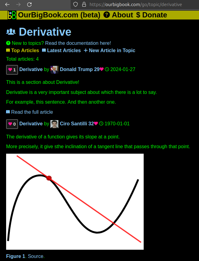
Figure 1. Screenshot of the "Derivative" topic page. View it live at: ourbigbook.com/go/topic/derivativeVideo 2. OurBigBook Web topics demo. Source. - local editing: you can store all your personal knowledge base content locally in a plaintext markup format that can be edited locally and published either:This way you can be sure that even if OurBigBook.com were to go down one day (which we have no plans to do as it is quite cheap to host!), your content will still be perfectly readable as a static site.
- to OurBigBook.com to get awesome multi-user features like topics and likes
- as HTML files to a static website, which you can host yourself for free on many external providers like GitHub Pages, and remain in full control

Figure 2. You can publish local OurBigBook lightweight markup files to either OurBigBook.com or as a static website.
Figure 3. Visual Studio Code extension installation.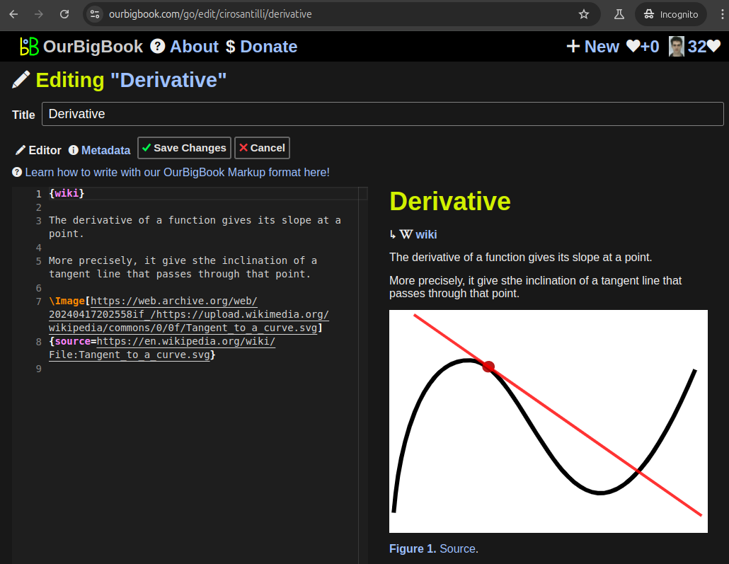
Figure 5. . You can also edit articles on the Web editor without installing anything locally. Video 3. Edit locally and publish demo. Source. This shows editing OurBigBook Markup and publishing it using the Visual Studio Code extension. 
- Infinitely deep tables of contents:
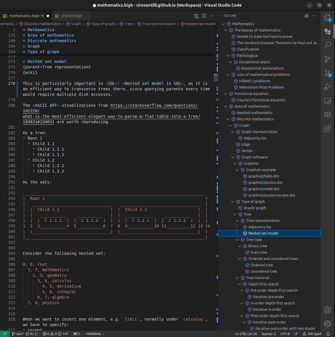

All our software is open source and hosted at: github.com/ourbigbook/ourbigbook
Further documentation can be found at: docs.ourbigbook.com
Feel free to reach our to us for any help or suggestions: docs.ourbigbook.com/#contact