Ronald R. Breaker is a biochemist at Yale University whose research investigates catalytic and regulatory functions of nucleic acids. His university biography connects his laboratory's work on deoxyribozymes with its investigations of riboswitches, in which RNA recognizes a molecular signal and affects gene expression.[ref]
Ronald Breaker profile at Yale School of Medicine. Biography: catalytic DNA and metabolite-binding messenger RNA
In the 1994 primary paper with Gerald F. Joyce, Breaker reported a DNA catalyst that cleaves an RNA linkage. The experiment used in vitro selection: molecules with useful activity were enriched through successive selection and amplification rounds. The selected chemistry depended on lead ions, so the result established catalytic capacity under the tested conditions, not the existence of a natural cellular DNA enzyme.[ref]
A DNA enzyme that cleaves RNA by Ronald R. Breaker and Gerald F. Joyce. Abstract, Background, Results and Conclusions
The paper's intermolecular catalyst had a reported turnover number of about . An original counting example explains the term: if of active catalyst each process one substrate per minute under sustained substrate-saturating conditions, the initial production rate is approximately . The catalyst can process more than its own molecular count over successive cycles. This differs from a reagent consumed once per cleavage. Binding, chemical reaction and product release can impose different kinetic limits.
Breaker later coauthored with Yingfu Li the primary study of chemically driven RNA degradation. That work supplies a baseline for interpreting catalysis: an apparent rate enhancement must compare a catalyzed reaction with the appropriate uncatalyzed reaction under specified conditions.[ref]
Kinetics of RNA Degradation by Specific Base Catalysis of Transesterification Involving the 2′-Hydroxyl Group by Yingfu Li and Ronald R. Breaker. Abstract: background cleavage rates and interpretation of catalytic enhancement

Yingfu Li is a biochemist at McMaster University who studies functional nucleic acids, including catalytic DNA and RNA and their use in molecular sensing. His institutional profile and laboratory biography verify the identity and research connection with Ronald R. Breaker.[ref]
Yingfu Li profile at McMaster University. Institutional specialization record; linked laboratory Biography and Research Interests
Li is first author, with Ronald R. Breaker, of the 1999 primary study measuring chemically driven cleavage of RNA phosphodiester bonds. Defined chimeric DNA/RNA substrates allowed the investigators to examine hydroxide-dependent transesterification involving the adjacent -hydroxyl group. This addresses a necessary reference question for studying ribozymes and deoxyribozymes: how rapidly does the corresponding linkage break without the proposed catalyst?[ref]
Kinetics of RNA Degradation by Specific Base Catalysis of Transesterification Involving the 2′-Hydroxyl Group by Yingfu Li and Ronald R. Breaker. Title, byline and Abstract: defined substrates and specific-base cleavage
An original kinetic example illustrates the comparison. Suppose an uncatalyzed linkage has observed rate constant and a tested catalytic condition gives . The rate ratio is . For a simple first-order reaction, the corresponding half-lives are and , approximately 69,315 minutes and 6.93 minutes. These are constructed values, not the study's measured constants. Changing pH, temperature or substrate structure between the two measurements can change the baseline and therefore the meaning of that ratio.
The connection to molecular sensing is that molecular recognition and reaction are distinct functions. A useful signal can depend on binding a target, changing a catalyst's activity and detecting the resulting product. Quantifying each contribution is more informative than observing an output change without identifying its origin.

Yingfu Li in the named McMaster University faculty portrait
. The official McMaster engineering faculty page identifies this external photograph as Yingfu Li.Andreas Schroeder is the first-listed author of the 2006 primary study introducing the RNA integrity number. The paper credits him with study design, methodology, experiments, validation, software implementation and manuscript preparation. These documented roles identify his contribution without inferring a later appointment or conflating him with namesakes.[ref]
The RIN: an RNA integrity number for assigning integrity values to RNA measurements by Andreas Schroeder and others. Author byline and Authors’ contributions, initials AS
The scientific problem was to make RNA integrity assessment more reproducible. An electropherogram contains a distribution of RNA fragments rather than a direct yes-or-no intactness reading. The team's approach combined trace features into a learned score; Schroeder contributed to both the experimental work and its software implementation.[ref]
The distinction between total RNA and intact RNA explains the motivation. Suppose one preparation contains ten strands of 1000 nucleotides each. Cleaving every strand once into two 500-nucleotide pieces doubles the number of strands from ten to twenty but leaves the total at 10,000 nucleotides. An analysis requiring an uninterrupted 1000-nucleotide template now fails, even though no nucleotide material has been removed. A distribution-sensitive measurement can expose this change in a way that total amount alone cannot.
A trained integrity score is consequently a measurement model: its features, training labels and validation data determine what its numerical scale supports. The RNA integrity number article describes the original apparatus and gives examples distinguishing score, signal amount and prediction error.
Chi Irg Yu is credited in the primary publisher byline as a coauthor, with Ruth E. Benesch and Reinhold Benesch, of the 1969 study of hemoglobin oxygenation in the presence of 2,3-Bisphosphoglycerate. The paper concerns how temperature, pH, ionic strength and hemoglobin concentration affect the oxygenation problem. The full spelling here follows that source; no additional career dates, appointments or identities are inferred from abbreviated citations.[ref]
Oxygenation of hemoglobin in the presence of 2,3-diphosphoglycerate. Effect of temperature, pH, ionic strength, and hemoglobin concentration by Ruth E. Benesch and others. Publisher byline and title, Biochemistry8(6), pp.2567–2571, June1969
The scientific question separates oxygen affinity from the amount of oxygen carried. An equilibrium oxygenation curve describes oxygen saturation as a function of oxygen partial pressure under fixed conditions. The number of available binding sites additionally depends on how much hemoglobin is present. Equal saturation does not imply equal total bound oxygen.
For an original stoichiometric example, take a preparation containing hemoglobin tetramers, each with four available oxygen-binding sites. Its site concentration is . At saturation , the concentration of bound oxygen molecules isA preparation with tetramers at the same saturation instead contains bound oxygen. These are constructed counts, not data from the 1969 paper. They show why protein concentration is a separate experimental variable from the fraction of occupied sites.
Regulator binding introduces another coupled equilibrium. 2,3-Bisphosphoglycerate preferentially binds deoxyhemoglobin, so a specified added amount of regulator need not equal its free concentration after protein binding. A comparison that changes protein concentration may change both available oxygen-binding capacity and regulator partitioning. The binding calculation in Ruth E. Benesch illustrates this free-versus-total distinction; the thermodynamic cycle in Reinhold Benesch explains reciprocal ligand coupling.
A controlled comparison therefore specifies protein amount, regulator amount, oxygen partial pressure, pH, temperature and solution composition, then identifies which readout was measured. Changing several of these together cannot isolate every individual influence from one saturation value. Yu's contribution is documented as part of the paper's coauthorship; that byline does not establish a separate allocation of particular experimental tasks.
Mathieu Clément-Ziza is a researcher named as first author of the 2008 study of RNA stabilization during laser capture microdissection. The primary byline associates that work with INSERM U781 and the Necker-Enfants Malades/Université René-Descartes setting in Paris. This is the affiliation printed for that study, rather than a claim about a current appointment.[ref]
Stabilization of RNA during laser capture microdissection by performing experiments under argon atmosphere or using ethanol as a solvent in staining solutions by Mathieu Clément-Ziza and others. Primary byline and affiliation, RNA14 (2008), pp.2698–2704
The study addressed a concrete measurement problem: selecting a recognizable cell population is not sufficient if the RNA changes during preparation and collection. The authors hypothesized that tissue rehydration permits endogenous ribonuclease activity. They tested both an argon atmosphere during collection and an ethanol-based fixation/staining approach, obtaining preservation over the tested interval up to 90 minutes. The two interventions address the preparation environment rather than merely adjusting a downstream expression value.[ref]
Stabilization of RNA during laser capture microdissection by performing experiments under argon atmosphere or using ethanol as a solvent in staining solutions by Mathieu Clément-Ziza and others. Abstract, proposed mechanism and two preparation approaches
Figure1 provides an illuminating experimental design. RNA from section scrapes was assessed at different times after staining; the caption reports at least three repeats and an Agilent Bioanalyzer readout using the RNA integrity number. Other panels show separation traces before and after preparation and following 30, 60 and 90 minutes of collection. These readouts test the state of the recovered analyte instead of assuming that a microscope image certifies molecular preservation.[ref]
The spatial part was separately documented: Figure2 shows human-colon ganglia and selected neurons before and after collection. That pairing illustrates the distinction between selecting the intended region and recovering suitable molecules from it. For example, two preparations with the same total RNA concentration could have different fragment-length distributions and different usefulness for a long-transcript assay. Concentration alone would not resolve that difference; RNA integrity supplies the missing measurement question.
The contribution described here is the collaborative method in the primary paper. Its byline does not allocate every apparatus task to one coauthor, and the reported preservation interval does not establish a universal lifetime for all tissues or preparation conditions.
Ruth E. Benesch was a biochemist whose work with Reinhold Benesch investigated hemoglobin and its interaction with small-molecule regulators. Their jointly authored 1969 account connected 2,3-Bisphosphoglycerate in red blood cells with lower oxygen affinity through preferential binding to deoxyhemoglobin. The published full byline uses Ruth E. Benesch; the middle initial is retained here.[ref]
Intracellular Organic Phosphates as Regulators of Oxygen Release by Haemoglobin by Reinhold Benesch and Ruth E. Benesch. Publisher byline and abstract, Nature221, pp.618–622, 1969
She was first author of a separate 1969 study of hemoglobin oxygenation in the presence of the same metabolite, with Reinhold Benesch and Chi Irg Yu. Its stated experimental variables were temperature, pH, ionic strength and hemoglobin concentration. Those variables matter because a ligand's effect is a property of the complete chemical equilibrium under specified conditions, not just the name of the ligand. The shared byline supports a collaborative contribution; it does not identify who performed every apparatus task.[ref]
Oxygenation of hemoglobin in the presence of 2,3-diphosphoglycerate. Effect of temperature, pH, ionic strength, and hemoglobin concentration by Ruth E. Benesch and others. Publisher title, byline and original June1969 issue information
An original binding calculation illustrates why protein concentration belongs in such a comparison. In a deliberately simplified one-site model, let be the total concentration of binding sites, the free concentration of 2,3-Bisphosphoglycerate, and its dissociation constant. The bound concentration isAt and , half the sites are occupied: . The total ligand concentration is , not . Substituting total concentration for free concentration would predict a different occupancy. These are stipulated numbers, not measured binding constants from her paper.
The biochemical insight extends beyond the small molecule: oxygenation and regulator binding are coupled properties of a protein. Their 1969 joint account describes one bound phosphate-metabolite molecule per hemoglobin tetramer, rather than one for every oxygen-binding subunit. This distinguishes regulatory stoichiometry from oxygen-carrying capacity.[ref]
Intracellular Organic Phosphates as Regulators of Oxygen Release by Haemoglobin by Reinhold Benesch and Ruth E. Benesch. Abstract: preferential deoxyhaemoglobin binding and one molecule per tetramer
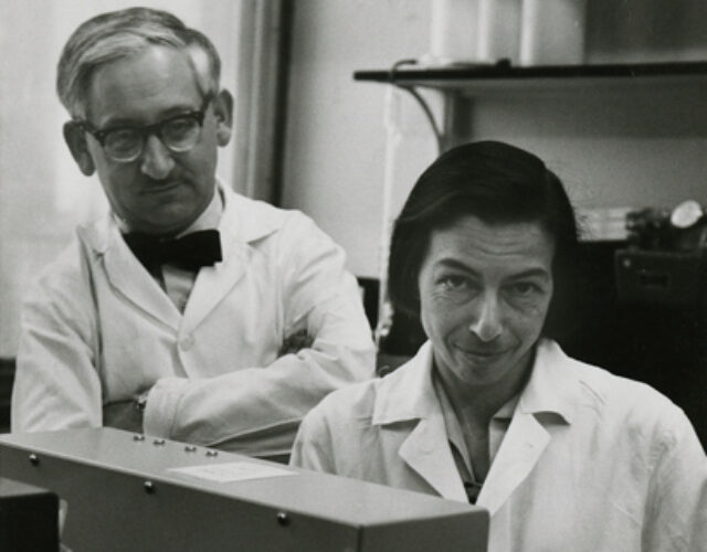
The historical photograph below is explicitly identified by the Science History Institute and credited to Columbia University's Department of Biochemistry.[ref]
Reinhold and Ruth Benesch photograph at the Science History Institute. Photograph caption and credit
Reinhold Benesch was a biochemist who investigated hemoglobin function in collaboration with Ruth E. Benesch. Their 1969 account of intracellular organic phosphates explained a mechanism by which 2,3-Bisphosphoglycerate changes oxygen affinity: the regulator binds preferentially to deoxyhemoglobin, making oxygen release more favourable at a specified oxygen partial pressure. This identifies a chemical regulator of oxygen transport, rather than treating hemoglobin affinity as an immutable property of the isolated protein.[ref]
Intracellular Organic Phosphates as Regulators of Oxygen Release by Haemoglobin by Reinhold Benesch and Ruth E. Benesch. Publisher byline and abstract, Nature221, pp.618–622, 1969
The discovery is an example of allostery: ligand binding at one part of a molecular system affects binding elsewhere. A short equilibrium argument shows why the effect is reciprocal. Consider an original four-state model with one oxygen ligand and one regulator , allowing states , , and . Define dissociation constantsMultiplying around the two possible binding paths givesThus, if regulator binding makes oxygen ten times harder to bind in this model, , oxygen binding must likewise increase the regulator's dissociation constant tenfold. An equilibrium coupling cannot act in only one direction. The four-state construction is an illustrative reduction, not a claim that the four oxygen-binding subunits of real hemoglobin behave as one physical site.
This reasoning motivates a controlled measurement: compare oxygen-equilibrium curves for matched hemoglobin preparations with different regulator concentrations, while controlling pH, temperature and solution composition. Measuring regulator binding as well tests the reciprocal side of the proposed mechanism. His coauthored 1969 paper with Ruth E. Benesch and Chi Irg Yu explicitly examined how several of these conditions enter the oxygenation problem.[ref]
The scientific contribution is collective in these sources. Their joint paper does not establish a separate personal allocation of the chemical preparation, measurements and interpretation. The photograph below is the institutionally identified joint image, credited to Columbia University's Department of Biochemistry.[ref]
Reinhold and Ruth Benesch photograph at the Science History Institute. Photograph caption and credit
Gabriele Varani studies the structural basis of RNA function and recognition. His university laboratory identifies NMR spectroscopy, crystallographic and biochemical methods among its approaches. His named publication list includes the 1991 study of an unusually stable RNA hairpin capped by a UUCG tetraloop.[ref]
Gabriele Varani faculty profile at the University of Washington. faculty Research summary; linked laboratory publication list, 1991 hairpin paper
The hairpin study determined a solution structure from interproton distances and NMR scalar coupling measurements, combining structural restraints with a calculation of molecular geometry. Its proposed stabilizing features include a noncanonical pair between guanine and uracil, a cytosine–phosphate contact and base stacking. These features connect the observed fold with the sequence-dependent stability of the RNA.[ref]
Structure of an unusually stable RNA hairpin by Gabriele Varani and others. Abstract, experimental restraints, structure calculation and loop contacts
An original geometry example illustrates what an NMR distance restraint contributes. Suppose three labelled sites A, B and C have distance intervals, in ångströangstroms,No molecular geometry can satisfy all three: the triangle inequality requires . A proposed conformation, an assignment or an assumed restraint would need reconsideration. The intervals are chosen to demonstrate consistency checking, not quoted experimental distances from the paper.
Compatible distance restraints still need not specify a unique three-dimensional arrangement. Mirror-related arrangements preserve all pairwise distances, while additional stereochemical information can distinguish them. Measurements of NMR scalar coupling provide further conformational information; the original study used them to constrain the backbone rather than relying on interproton distances alone.[ref]
Structure of an unusually stable RNA hairpin by Gabriele Varani and others. Abstract, backbone constraints from scalar-coupling measurements
The motivating experiment therefore does more than establish an RNA sequence. It measures interactions in solution that restrict how the nucleotides can be arranged. The resulting structural model explains specific contacts, while the measurement restraints define what the data actually support.

Gabriele Varani, identified by the University of Washington faculty profile.
H. A. Heus is an author of a 1991 solution-structure study of RNA hairpins containing GNRA tetraloops. The paper identifies a Chemistry and Biochemistry affiliation at the University of Colorado, Boulder. Its structures for the GCAA and GAAA sequences helped explain how loop contacts can stabilize a short RNA fold.[ref]
The experiment used NMR spectroscopy to determine the structures in solution. Both reported loops contain an unusual pair between guanine and adenine between the first and last loop residues, together with a base–phosphate hydrogen bond, a sugar–base hydrogen bond and extensive base stacking. The proposed stability therefore involves a network of interactions rather than only the count of canonical pairs in the stem.[ref]
Structural features that give rise to the unusual stability of RNA hairpins containing GNRA loops by H. A. Heus and others. Abstract, shared structural features of GCAA and GAAA
An original sequence-counting example clarifies the family name. In GNRA, G denotes guanine, N permits any of the four ordinary RNA bases, R permits either purine base, and A denotes adenine. Thus the motif admits nominal sequences. GCAA and GAAA differ at N while retaining the initial G and final A. Membership in that sequence family does not establish equal folding stability for all eight sequences; the paper reports structures of two particular members.
A separate thermodynamic illustration shows why a strong folding preference can still be sensitive to a modest energy change. In a stipulated two-state equilibrium, let the folded fractions be and under two different conditions. Their folding equilibrium constants areAt , the corresponding difference in folding Gibbs free energy iswhere is the molar gas constant. These are original illustrative populations, not measured values attributed to Heus's paper. They connect a molecular-interaction explanation with an experimentally measurable equilibrium preference.
The reported GNRA tetraloop structures give a molecular interpretation of sequence conservation: bases and sugar–phosphate groups have to support a compatible set of contacts. A sequence change can preserve the stem yet alter those loop interactions.
Justin P. Sheehy is first author of a2010 study measuring the thermodynamic stability of common RNA tetraloops. These four-nucleotide loops cap RNA hairpins, and their contribution to stability depends on sequence and structural context.[ref]
The study used RNA optical melting: measuring absorbance as temperature changed, then fitting the transitions to obtain thermodynamic parameters. Its Results distinguish33 stem–loop sequences from24 unique tetraloops, because some loops were tested in multiple stems. Repeated melting curves over an approximately50-fold concentration range showed single transitions and concentration independence, supporting an intramolecular hairpin interpretation.[ref]
Thermodynamic characterization of naturally occurring RNA tetraloops by Justin P. Sheehy and others. Thermodynamic parameters,Table2; Optical melting experiments and thermodynamics
An original two-state RNA melting model makes the measured quantities interpretable. If folding has temperature-independent enthalpy change and entropy change , then its folding Gibbs free energy isThe RNA melting temperature of this stipulated unimolecular model satisfies , givingabout60.2 degrees Celsius. At310 kelvin the model instead gives , favoring folding. These invented parameters illustrate the calculation; they are not reported values for one of Sheehy's sequences.
The experiment links a temperature-dependent optical signal with a model of RNA stability. An absorbance transition alone is not an atomic structure, and changing the stem can change the inferred loop contribution. The author identity here follows the primary scientific byline; it does not assume a career or portrait from a same-name directory entry.
Uttam L. RajBhandary studied how RNA structure determines recognition by proteins, particularly the different jobs of transfer RNA during biological translation. MIT's named profile connects Uttam RajBhandary with publications credited to RajBhandary, UL, including work on initiator transfer RNA.[ref]
Uttam RajBhandary faculty profile at MIT. Research Summary and Key Publications, especially Initiator transfer RNAs
With B. L. Seong, he tested whether conserved base pairs in the anticodon stem help distinguish an initiator transfer RNA from an elongator transfer RNA. The 1987 study replaced one, two or three G–C pairs with the corresponding elongator pattern and examined purified transfer RNA variants. Their initiation activity decreased even though they could still bind bacterial initiation factor IF2; AUG-dependent binding at the ribosomal P site was impaired.[ref]
Escherichia coli formylmethionine tRNA: mutations in GGGCCC sequence conserved in anticodon stem of initiator tRNAs affect initiation of protein synthesis and conformation of anticodon loop by B. L. Seong and U. L. RajBhandary. Abstract, stem substitutions and comparison of initiation-factor and P-site binding
The experiment separates carrying the right amino acid from being recognized at the right step. For an original counting example, suppose 100 charged carriers are presented to a simplified initiation assay, 80 reach a tested binding state, and 20 of those proceed to productive initiation. The productive fraction is . Equal charging or equal binding at an earlier state would not establish equal productive output. These stipulated counts illustrate sequential selection; they are not measured values from the paper.

B. L. Seong is the author credited with U. L. RajBhandary in a 1987 study of structural recognition of initiator transfer RNA. They tested how a conserved anticodon-stem pattern distinguishes an initiator transfer RNA from an elongator transfer RNA.[ref]
Separate assays compared charging, formylation, bacterial initiation factor IF2 binding, ribosomal P site binding and protein synthesis. Greater accessibility of the anticodon loop to S1 nuclease connected the functional change with altered RNA conformation.[ref]
For an original assay example, suppose two preparations each contain 100 units of charged transfer RNA, and an earlier binding assay reports 80 units for each. If the later ribosomal readout reports 60 and 15 units, its fourfold difference cannot be explained by the amount of charged carrier alone. The example motivates measuring distinct steps rather than labeling every loss of activity a failure of binding. The numbers are illustrative; the paper's contribution is the experimentally supported distinction between factor recognition, ribosomal positioning and productive biological translation.
Ben F. Luisi coauthored a 2000 X-ray crystallography study of polynucleotide phosphorylase. It revealed a protein trimer with a central channel, providing a structural model for how a multicomponent enzyme might retain and process RNA.[ref]
A duplicated fold is the structural basis for polynucleotide phosphorylase catalytic activity, processivity, and regulation by M. F. Symmons and others. Abstract, Results and Conclusions
The authors' 2002 account supplies the full byline Ben F. Luisi.[ref]The polynucleotide phosphorylase central channel was observed without bound RNA; a role in processivity was proposed rather than directly filmed.[ref]
An original probability model illustrates the proposed retention advantage. Suppose an enzyme completes one cleavage, then remains associated for another with probability , independently after each cleavage. The number of completed cleavages in one encounter hasThus changing from to changes the mean run from to cleavages without specifying how quickly an individual cleavage happens. This invented geometric distribution separates processivity from reaction rate; it is not a fitted model or measured result from Luisi's paper.
George H. Jones coauthored structural and biochemical work on polynucleotide phosphorylase. The initial-only 2000 byline G. H. Jones is supplemented by the full byline in the collaborators' 2002 account and in a 2003 paper on enzyme purification.[ref]
Running rings around RNA: a superfamily of phosphate-dependent RNases by Martyn F. Symmons and others. Full byline
[ref]The 2003 work separated purified enzyme activity from possible contaminating nuclease activity by testing the dependence of degradation on phosphate. That distinction matters because polynucleotide phosphorylase uses phosphorolysis, whereas an unrelated hydrolytic nuclease could also make intact RNA disappear.[ref]
For an original control calculation, suppose intact-RNA signal falls by units with phosphate and by without it. Subtraction gives an increment of units associated with the changed condition. Interpreting all units as phosphate-dependent activity would ignore the background. Even the subtraction requires comparable samples, linear response and unchanged background behavior; it is not automatically a molecular rate measurement.
The shared 2000 X-ray crystallography paper supplied another kind of evidence: the architecture of a three-subunit protein complex. A structure can suggest where substrate recognition occurs, while biochemical controls test what chemical reaction the preparation actually performs.[ref]
A duplicated fold is the structural basis for polynucleotide phosphorylase catalytic activity, processivity, and regulation by M. F. Symmons and others. Abstract: trimeric architecture and catalytic-site interpretation
Martyn F. Symmons is the first-named author of a 2000 structural study of polynucleotide phosphorylase. The collaborators' subsequent account gives his full byline, preserving the connection to the original M. F. Symmons credit.[ref]
The structure showed a duplicated core within each protein subunit and a three-subunit assembly. Bound tungstate, a phosphate analogue, helped identify a catalytic region; the structure did not contain bound RNA.[ref]
A simple counting example clarifies the architectural claim. A protein trimer with two related core protein domains per chain contains such domains but only three chains. The domain count alone does not establish six equivalent active sites. A repeated fold can retain a similar shape while acquiring a different catalytic or structural role.
This distinction suggests an experimental comparison: locate a bound chemical analogue, identify nearby amino acid residues, then compare functional measurements after controlled changes while independently checking protein folding. A loss of activity after global unfolding would not isolate the role of one active site. This is a general interpretation of the structural strategy, rather than an assignment of particular experiments to Symmons solely from author order.
L. J. Zatman coauthored a 1964 investigation of bacterial methanol dehydrogenase with Chris Anthony. The original byline gives the initials retained here. Their work investigated how an enzyme transfers reducing equivalents during methanol oxidation.[ref]
A particularly informative part of the study concerned product identity. The researchers separated material by steam distillation before testing for formaldehyde, because an assay reagent interfered with direct measurement. They explicitly distinguished detecting formaldehyde after analysis from proving that free formaldehyde was the immediate enzymic product.[ref]
The microbial oxidation of methanol. 2. The methanol-oxidizing enzyme of Pseudomonas sp. M 27 by C. Anthony and L. J. Zatman. p.620, Product of enzymic methanol oxidation and Table 2
The underlying oxidation can be represented by the balanced half-reactionBoth sides contain one carbon, one oxygen and four hydrogen atoms, and the total charge remains zero. This bookkeeping establishes the two-electron change for that net conversion. It does not identify the electron acceptor, the cofactor or every intermediate.
The same distinction applies to an analytical treatment that releases a detectable molecule from a bound precursor. Recovering the molecule establishes what the whole treatment yields; an independent observation is needed to establish its state before treatment. This makes the paper a useful example of how careful product analysis can support a reaction assignment while leaving a mechanistic question open.
Chris Anthony coauthored the 1964 study of a bacterial methanol dehydrogenase with L. J. Zatman. His own research account explicitly connects his name with the initial-only Anthony–Zatman publications.[ref]
Chris Anthony research account and personal homepage. Research career and linked publication history: 1964 methanol-oxidizing enzyme paper
The experiment measured an optical response associated with electron transfer, using a dye-linked enzyme assay. The paper describes a Unicam SP.500 spectrophotometer and cuvettes. Establishing the reaction product required separate chemical analysis rather than the optical signal alone.[ref]
The microbial oxidation of methanol. 2. The methanol-oxidizing enzyme of Pseudomonas sp. M 27 by C. Anthony and L. J. Zatman. p.614, Materials and Methods; p.620, product analysis
For an original illustration of the optical measurement, assume the Beer-Lambert lawwith absorbance , liquid path and a hypothetical molar coefficient . ThenA decrease of absorbance units would correspond to a decrease of the absorbing species under those assumptions. It does not by itself give the amount of methanol consumed: the chemical identity of the absorber and the coupling stoichiometry must also be known. The coefficient and measurements are invented for this example.

Chris Anthony, portrait linked from his named personal homepage
. [ref]Chris Anthony research account and personal homepage. Named homepage and linked personal portrait
Jeong Ho Chang is a researcher in structural biology and a coauthor of a 2011 study of Xrn1, an exoribonuclease that degrades RNA from its end toward its end. The paper's contribution statement explicitly credits Chang with mutagenesis, nuclease assays and thermal shift assays, as well as shared crystallographic work.[ref]
The team combined X-ray crystallography with biochemical measurements. A defined protein construct yielded structures at resolution. Additional protein domains lay away from the active site, yet alterations in these regions affected activity. Comparisons of RNA binding, degradation and thermal stability helped separate possible explanations for a defective enzyme.[ref]
Structural and biochemical studies of the 5′→3′ exoribonuclease Xrn1 by Jeong Ho Chang and others. Results, Structure determination and Domain D1 may stabilize the N-terminal segment; Figures 5–6
One important experimental distinction is that the crystallized E178Q construct was catalytically defective; its detailed protein structure was not itself a demonstration of normal Xrn1 activity. A structural model and a functional assay supply complementary evidence.[ref]
Structural and biochemical studies of the 5′→3′ exoribonuclease Xrn1 by Jeong Ho Chang and others. Structure determination and Figure 5b: E178Q structure and essentially absent nuclease activity
For an original measurement example, suppose arbitrary units of intact RNA remain in a control and after an assay. The disappearance fraction is . That observation alone does not identify the degradation products, the processivity, or whether binding became weaker. Each requires an additional readout; the invented numbers illustrate why several assays are useful.

Jeong Ho Chang research profile at Kyungpook National University. Named profile, linked portrait and explicit listing of the 2011 Xrn1 paper
M. Bycroft is the first-named author of a 1997 structural study of the S1 RNA-binding domain. The paper examined this protein domain from Escherichia coli polynucleotide phosphorylase using nuclear magnetic resonance. Its initial-only byline is retained here.[ref]
The resulting protein structure contained a five-stranded antiparallel beta barrel. Conserved amino acid residues and neighboring loops suggested a possible RNA-binding face. Comparing this fold with other proteins connected a small structural module with a broader family of nucleic-acid interactions.[ref]
The solution structure of the S1 RNA binding domain: a member of an ancient nucleic acid-binding fold by M. Bycroft and others. Abstract: solution structure, putative RNA-binding face and related proteins
The distinction between an observed fold and a proposed binding face is scientifically useful. Finding several conserved amino acid residues on the same exposed surface suggests an experiment: alter selected residues, then compare RNA binding while independently checking whether the protein still folds. Weaker binding after a change that also unfolds the protein would not establish a direct contact at that residue. This is an experimental interpretation of the structural hypothesis, not a report that every such test was performed in the 1997 paper.
For a simple architectural example, two proteins can each contain one S1 RNA-binding domain while having different additional protein domains. Sharing that module alone does not imply identical enzyme activity, the same RNA preference, or an equal number of active sites.
William N. Beavers is a researcher whose work combines analytical chemistry with the study of chemical changes to proteins. His institutional profile identifies post-translational modification as a research interest and directly lists his scientific publications.[ref]
William N. Beavers research profile at Louisiana State University. Named faculty profile, Research Interest and Select Publications
In a 2017 study, Beavers and collaborators investigated proteins modified by endogenously generated lipid electrophiles. Chemical labelling and selective capture enabled a broad analysis of modified targets. The results highlighted mitochondria as both a source associated with electrophile generation and a prominent location of modified proteins in that experimental system.[ref]
A useful quantitative distinction is the amount of a modified protein versus the fraction of that protein that is modified. For an original ideal measurement model, suppose response-corrected signals correspond to modified and unmodified molecules in sample A, and modified and unmodified molecules in sample B. The modified amount doubled, but the modified fraction is in both samples. These are invented counts, not results from the paper.
The response correction is essential. A chemically modified molecule may be captured or detected differently from an unmodified one, so raw signal intensities cannot automatically be substituted for molecule counts. Nor does more captured material alone establish a higher probability of modification if total protein abundance also changed. An informative interpretation therefore distinguishes chemical selectivity, total abundance and the denominator used for a reported fraction.
Beavers is also credited as a coauthor on a separate 2020 microbial-community study, illustrating the range of scientific contexts in which chemical measurements and biological comparisons meet.[ref]

- Clostridioides difficile proline fermentation in response to commensal clostridia by Christopher A. Lopez and others
- Protein Modification by Endogenously Generated Lipid Electrophiles: Mitochondria as the Source and Target by William N. Beavers and others
- William N. Beavers research profile at Louisiana State University
Sk Tofajjen Hossain is the published author name credited with biochemical studies of how RNase R processes structured RNA. The original 2016 paper gives this name and a University of Miami biochemistry affiliation; the initial element Sk is retained without expansion.[ref]
How RNase R Degrades Structured RNA: Role of the Helicase Activity and the S1 Domain by Sk Tofajjen Hossain and others. Published byline and affiliation
Hossain and collaborators compared the enzyme's RNA-unwinding and nuclease activities using designed substrates and changes in its S1 domain. They also used intrinsic tryptophan fluorescence to investigate changes associated with RNA binding. Measuring these different observables helped distinguish RNA recognition, structural change and degradation rather than treating all of them as one event.[ref]
A generic binding model makes one important distinction clear. If a single binding site has dissociation constant and free ligand concentration , its ideal equilibrium occupancy isAt , half the sites are occupied; at , the occupancy is , not twice as large. These are mathematical examples, not fitted values from the paper. They show why an increase in measured binding need not be proportional to ligand concentration, and why an equilibrium binding result alone does not measure chemical turnover.
In particular, detecting ATP bound to an enzyme is different from demonstrating ATP hydrolysis. A binding signal does not, by itself, identify how energy is used during RNA unwinding. Likewise, an altered fluorescence signal can report a changed local environment without directly counting phosphodiester bonds cleaved. The strength of the study lies in relating separate measurements to a model of how RNA secondary structure is handled, rather than inferring the entire mechanism from one signal.
D. R. Davies is the published author name credited on a structural study of Tn5 transposase. The original 2000 paper lists a University of Wisconsin biochemistry affiliation. Its initials are retained here because the scientific byline establishes this contribution without establishing a full personal identity.[ref]
Three-dimensional structure of the Tn5 synaptic complex transposition intermediate by D. R. Davies and others. Published byline, affiliation and Abstract; Science 289, 77–85
Davies and collaborators determined a protein–DNA assembly at ångströms crystallographic resolution. The protein dimer held two DNA ends, with each end contacted by both subunits. The arrangement explained how one subunit could bind an end while the other supplied the active site for cleavage. It supplied a structural framework for understanding coordinated DNA transposition.[ref]
Three-dimensional structure of the Tn5 synaptic complex transposition intermediate by D. R. Davies and others. Abstract: dimeric complex, shared DNA contacts and cleavage in trans
The geometry is more revealing than a count of components. Imagine two subunits A and B and two DNA ends L and R. Knowing merely that the assembly contains two subunits and two ends leaves open an arrangement in which A independently processes L and B independently processes R. Contacts that span the two subunits instead couple recognition of an end to the partner's catalytic machinery. Such an arrangement provides a concrete reason why an isolated, correctly bound DNA end need not behave like the complete transpososome.
The quoted distance is nanometres, since one ångström is nanometres. Crystallographic resolution describes the spatial detail supported by the diffraction data; it is not a frame interval in a movie and does not mean that every atomic coordinate has an uncertainty of precisely nanometres. A static structure constrains possible reaction mechanisms, while the sequence and rates of chemical steps require additional evidence. The distinction matters whenever an attractive structural model is used to explain a dynamic enzyme reaction.
Marina Serna is a researcher in structural biology whose work includes cryo-electron microscopy of large protein complexes. She was first author of the 2016 study determining the architecture of the complement system's membrane attack complex.[ref]
Structural basis of complement membrane attack complex formation by Marina Serna and others. Original author list; Results, Cryo-EM structure of the MAC
The experiment linked function to structure. The researchers assembled complexes on liposomes, checked release of a fluorescent marker to establish functional pores, then isolated the complexes for frozen-hydrated imaging. The reconstruction identified eighteen copies of complement C9 in the analyzed completed pore.[ref]
Structural basis of complement membrane attack complex formation by Marina Serna and others. Results, Cryo-EM structure of the MAC and Molecular architecture; Figure 1
A constructed fluorescence example explains why assay controls matter. Suppose a no-release control gives signal , a sample gives , and a complete-release reference gives , all in compatible instrument units. A normalized release readout would beThis is an invented linear-calibration example, not the paper's measured values. The raw sample signal is forty units, but the estimated fraction is thirty percent because the background and reference determine the denominator.
The functional check and structural reconstruction answer different questions. Marker release tests whether a preparation makes membranes permeable; the reconstructed density constrains the architecture of selected complexes. Eighteen C9 copies in the studied end product do not establish that eighteen are a universal minimum for membrane damage.[ref]
Structural basis of complement membrane attack complex formation by Marina Serna and others. Results, completed-pore stoichiometry distinguished from lysis by fewer C9 molecules

Marina Serna
. Portrait by Esther Sánchez / CNIO, explicitly identified in the institutional research profile: www.cnio.es/en/news/cnio-researcher-marina-serna-awarded-for-her-contribution-to-solving-a-crucial-biological-problem-2/ .Brian T. Wimberly is a researcher in RNA structure and ribosome function. The full byline of the 2000 paper on the bacterial small ribosomal subunit establishes the expansion of the credited initials B. T. Wimberly. The paper lists him among six equally contributing authors at the forefront of its structural work.[ref]
Structure of the 30S ribosomal subunit by B. T. Wimberly and others. Publisher author list, affiliations and equal-contribution note
The team determined a 30S ribosomal subunit structure refined to resolution using X-ray crystallography. It located the folded ribosomal RNA and associated ribosomal proteins in a molecular model, providing a framework for interpreting decoding, assembly and biochemical interactions.[ref]
Structure of the 30S ribosomal subunit by B. T. Wimberly and others. Abstract; Figures 1–2, electron-density maps and structure overview
The intellectual advance connected molecular arrangement to a function: how a ribosome reads a messenger RNA sequence. The small ribosomal subunit organizes the interactions of codons with transfer RNA anticodons, so the geometry of its RNA and proteins constrains explanations of decoding.
A constructed comparison clarifies what a structural measurement contributes. Two models could contain the same list of molecules yet place a functional contact apart in one and apart in the other. Composition alone cannot distinguish them; spatial evidence can. This is not a distance reported in the paper, and nominal spatial resolution should not be interpreted as identical coordinate precision for every atom.
Wimberly's institutional biography also describes using complementary structural methods to relate RNA organization to biological function.[ref]

Brian T. Wimberly
. Portrait identified by the New York Structural Biology Center profile: nysbc.org/people/brian-wimberly .Alexander E. Aleshin is a researcher in structural biology credited with studies of the complement system. He was first author of the 2012 determination of the crystal structure of complement C6, with a Sanford-Burnham Medical Research Institute affiliation in the original byline.[ref]
Structure of complement C6 suggests a mechanism for initiation and unidirectional, sequential assembly of membrane attack complex (MAC) by Alexander E. Aleshin and others. Author affiliations; Results, Overall Structure of C6
The biological problem was how soluble proteins assemble a membrane-damaging machine. Complement C6 is recruited during formation of the membrane attack complex. Its structure revealed a closed arrangement of protein domains. Comparing this arrangement with related components suggested how changes in domain orientation could help expose membrane-inserting regions and propagate assembly.[ref]
Structure of complement C6 suggests a mechanism for initiation and unidirectional, sequential assembly of membrane attack complex (MAC) by Alexander E. Aleshin and others. Abstract; Results, C6 Adopts a Default Closed Autoinhibited State; Discussion
A useful mechanical analogy is a folded tool whose working surface is inaccessible until a hinge opens. The analogy motivates two experimentally distinct questions: what configurations exist, and what causes transitions between them? Determining a crystal structure answers the first kind of question for the crystallized state; it does not directly film the assembly sequence.
The distinction is explicit in this work. The complement C6 coordinates and comparisons constrain a proposed model of directional membrane attack complex assembly. Testing the proposed succession of movements requires additional evidence about interacting components and assembly intermediates. Aleshin and colleagues thus connected a measured molecular shape to a testable explanation of a larger biological process.[ref]
Structure of complement C6 suggests a mechanism for initiation and unidirectional, sequential assembly of membrane attack complex (MAC) by Alexander E. Aleshin and others. Discussion, Model for MAC Initiation and Final Steps in MAC Assembly
Beom Seok Park is a researcher credited with structural studies of recognition in innate immunity. He was first author of the 2009 study of the Toll-like receptor 4–MD-2 complex with lipopolysaccharide; the original paper records his Department of Chemistry affiliation at KAIST.[ref]
The structural basis of lipopolysaccharide recognition by the TLR4–MD-2 complex by Beom Seok Park and others. Author byline and affiliation; Nature 458, 1191–1195
The crystal structure connected ligand recognition to assembly. Most of the lipopolysaccharide's lipid chains fit inside a pocket in MD-2, while an exposed chain helps make an interface with another Toll-like receptor 4. The assembly contains two receptor-complex units. Thus the ligand helps organize contacts between proteins, rather than merely occupying an isolated binding pocket.[ref]
The structural basis of lipopolysaccharide recognition by the TLR4–MD-2 complex by Beom Seok Park and others. Abstract; Figures 1–3, ligand pocket and dimerization interfaces
A simple comparison shows why this matters. Imagine two ligands that both bind a receptor-associated pocket, but only one presents an exposed surface in the orientation required to join two receptor units. Equal pocket occupancy would not guarantee equal assembly or signaling. This is a conceptual example, not a numerical affinity measurement from the paper.
Park and collaborators' contribution was to provide a molecular account linking the arrangement of lipopolysaccharide, MD-2 and Toll-like receptor 4 to receptor activation. A crystal structure reveals the contacts in the captured complex; claims about response strength or behavior in an intact organism require further experimental measurements.[ref]
N. Miyashita coauthored, with O. Habuchi, a 1982 biochemical study distinguishing chondroitin 6-sulfotransferase from chondroitin 4-sulfotransferase in chick embryo cartilage. The activities were separated by size-exclusion chromatography, allowing their properties to be compared in different preparations.[ref]
Separation and characterization of chondroitin 6-sulfotransferase and chondroitin 4-sulfotransferase from chick embryo cartilage by O. Habuchi and N. Miyashita. original byline and Abstract, separation of the two activities
The scientific question concerns position as well as amount. Both enzymes modify chondroitin, but they act at different positions of N-acetylgalactosamine. Equal total group incorporation could therefore conceal different products. Distinguishing the activities gave an experimental way to investigate that structural difference.[ref][ref]
The study also reported removing endogenous acceptors from the enzyme preparations.[ref]
Separation and characterization of chondroitin 6-sulfotransferase and chondroitin 4-sulfotransferase from chick embryo cartilage by O. Habuchi and N. Miyashita. Abstract, acceptor removal
That feature makes the assay logic illuminating: material carried through purification can participate in the measured reaction. Defining the added acceptor is easier when an uncontrolled acceptor pool has been removed. The work thus connects separation of proteins with interpretation of their reaction-specific activity, rather than treating purification as a measurement of protein amount alone.O. Habuchi coauthored a 1982 study, with N. Miyashita, separating two positional sulfotransferase activities from chick embryo cartilage. The work experimentally distinguished chondroitin 4-sulfotransferase and chondroitin 6-sulfotransferase, which modify different positions in the same kind of carbohydrate backbone.[ref]
Separation and characterization of chondroitin 6-sulfotransferase and chondroitin 4-sulfotransferase from chick embryo cartilage by O. Habuchi and N. Miyashita. original byline and Abstract, positional activities
The separated preparations differed in their biochemical behavior. Under the reported comparison without sulfhydryl compounds, the 4-sulfotransferase activity was very low, whereas the 6-sulfotransferase activity was essentially unaffected.[ref]
This is useful evidence because an assay condition can change the contribution of different activities within a preparation. In a hypothetical mixture with two activities, observing a decline in total signal would not establish that both activities declined equally. Separating them and measuring each under matched conditions supplies more specific information.
The distinction also links reaction chemistry to the meaning of an enzyme assay. Chondroitin modification can vary in the number and location of attached groups, while protein abundance supplies another measurement. The credited study provides a concrete example of using separable properties to investigate how a family of related reactions is implemented.
A. J. Padgett-McCue coauthored a 2000 study identifying a chondroitin synthase and establishing what polysaccharide it made. The work connected an enzyme with the alternating glucuronic acid and N-acetylgalactosamine units of chondroitin. The characterized protein carried both sugar-transfer activities required to extend this repeating chain.[ref]
Identification and molecular cloning of a chondroitin synthase from Pasteurella multocida type F by P. L. DeAngelis and A. J. Padgett-McCue. Abstract, Journal of Biological Chemistry 275 (2000), pp. 24124–24129; DOI 10.1074/jbc.M003385200.
The experimental reasoning combined nucleotide sugar specificity, size-exclusion chromatography and selective enzymatic digestion of the product. This gives a useful example of complementary measurements: a large molecular size establishes that a polymer was made, but does not identify its repeating chemical structure. Agreement between the sugar composition and the response to a chondroitin-cleaving enzyme supplied more specific evidence. The contribution is therefore a chemically tested biosynthetic assignment, rather than a prediction based only on similarity between protein sequences.[ref]
Identification and molecular cloning of a chondroitin synthase from Pasteurella multocida type F by P. L. DeAngelis and A. J. Padgett-McCue. Abstract: donor specificity, gel filtration and contrasting lyase susceptibility.
Carissa L. White coauthored a 2002 study identifying a heparosan synthase. The characterized glycosyltransferase combined two sugar-transfer activities in one protein, building a heparosan chain from glucuronic acid and N-acetylglucosamine units. The product was the unsulfated, unepimerized backbone related to heparin, rather than a fully modified heparin molecule.[ref]
Identification and molecular cloning of a heparosan synthase from Pasteurella multocida type D by P. L. DeAngelis and Carissa L. White. Abstract, Journal of Biological Chemistry 277 (2002), pp. 7209–7213; DOI 10.1074/jbc.M112130200.
The study tested which nucleotide sugars supported chain formation and compared the product's susceptibility to different polysaccharide-cleaving enzymes. An illuminating distinction is between building a repeating backbone and modifying that backbone: identifying a heparosan synthase establishes a particular polymerization activity, but does not establish all the later reactions needed to make a differently substituted glycosaminoglycan. The reported one-protein, two-activity mechanism also shows that two alternating chemical additions need not require two separate polypeptides.[ref]
Identification and molecular cloning of a heparosan synthase from Pasteurella multocida type D by P. L. DeAngelis and Carissa L. White. Abstract: dual-action transferase, donor specificity and product digestion.
Natallia Strushkevich is the first author of a 2011 structural study of the mitochondrial system that converts cholesterol toward pregnenolone. The team examined the cholesterol side-chain cleavage enzyme together with its electron-transfer partner adrenodoxin.[ref]
An engineered fusion protein helped hold the partners together for crystal structure determination. The structure related the iron-sulfur cluster of adrenodoxin to the haem group of the catalytic protein, while structures with different bound sterols addressed successive reaction stages.[ref]
The engineered connection was an experimental aid, not evidence that the native proteins form one continuous chain. Comparing fusion proteins with different linker lengths also tested how the imposed connection affected reduction of the catalytic partner.[ref]
Structural basis for pregnenolone biosynthesis by the mitochondrial monooxygenase system by Natallia Strushkevich and others. Methods and supporting Figure S8: linker-dependent electron transfer
The example connects a useful structure-stabilizing intervention to an explicit check on its functional consequences.Bert J. C. Janssen is the first author of a 2005 structural study of Complement C3 and its processed fragment C3c. The team determined crystal structures that connected the arrangement of protein domains to activation and regulation in the complement system.[ref]
The structures helped explain how a reactive thioester, needed for later covalent attachment, can remain protected against premature hydrolysis. Comparing the native and processed forms also revealed substantial changes in domain arrangement.[ref]
The comparison is specifically Complement C3 versus C3c. It is not a direct pair of structures showing native Complement C3 and active C3b. Keeping the fragments distinct matters because different stages of processing have different biological roles: an informative structural difference is not automatically the exact movement that creates an active intermediate.
L. Åberg coauthored a 1982 investigation of an enzyme involved in making dermatan sulfate. With A. Malmström, L. Åberg used an isotope labelling assay to follow hydrogen exchange during epimerization of residues in chondroitin.[ref]
The label was tritium attached at carbon five. Material released during incubation was distilled and quantified by liquid-scintillation counting. The study also compared conditions supporting the two epimerase activities present in microsome preparations.[ref]
Biosynthesis of dermatan sulphate. Assay and properties of the uronosyl C-5 epimerase by A. Malmström and L. Åberg. Abstract: isotope readout and different cofactor/pH requirements
The assay illustrates a distinction between a chemical event and the signal used to detect it. Radioactive counts report movement of a label under specified conditions; interpreting them as an enzyme activity requires an appropriate labelled substrate and comparisons for background release. Counting the signal alone would not identify which enzyme produced it.
A. Malmström coauthored, with L. Åberg, a 1982 assay for the chondroitin-glucuronate 5-epimerase activity involved in dermatan sulfate biosynthesis. The reaction changes the stereochemical configuration of sugar residues already attached to a polysaccharide, rather than synthesizing a free sugar first.[ref]
Biosynthesis of dermatan sulphate. Assay and properties of the uronosyl C-5 epimerase by A. Malmström and L. Åberg. Original byline and Abstract: polymer-level reaction
The experiment used chondroitin carrying tritium at the reacting carbon. Release of the label during incubation was measured with a scintillation counter, converting a hard-to-see molecular exchange into a countable assay signal.[ref]
Biosynthesis of dermatan sulphate. Assay and properties of the uronosyl C-5 epimerase by A. Malmström and L. Åberg. Abstract: labelled substrate, distillation and liquid-scintillation counting
The preparations also distinguished activities associated with dermatan sulfate and heparan sulfate biosynthesis through different requirements for pH and cofactors. Similar overall epimerization chemistry therefore did not establish that the same enzyme performed both biological tasks.[ref]
Biosynthesis of dermatan sulphate. Assay and properties of the uronosyl C-5 epimerase by A. Malmström and L. Åberg. Abstract: two microsomal epimerase activities
Carola Hunte is a structural biologist and first author of a 2005 study of NhaA, the sodium/proton antiporter of Escherichia coli. The team determined a crystal structure in a state downregulated by pH, revealing a path from the cytoplasm toward a proposed ion-binding region within the cell membrane.[ref]
The arrangement of short helices and charged groups motivated an alternating access model: changes in the transporter would expose the relevant region toward different sides of the membrane. The paper distinguished the observed structure from the proposed movements and pH-regulatory mechanism.[ref]
That distinction connects two kinds of experiment. Transport measurements count movement of ions; a protein structure supplies candidate routes and interactions. Seeing a possible route in one conformation does not by itself measure transport rate or establish the complete cycle.

Claire Debarnot is the first author of a 2019 structural study of human heparosan-N-sulfate-glucuronate 5-epimerase, an enzyme that modifies heparan sulfate. The reaction changes glucuronic acid residues into iduronic acid residues by epimerization at carbon five.[ref]
The team combined crystal structures with real-time nuclear magnetic resonance measurements. An inactive mutant could bind defined carbohydrate substrates and products, allowing their positions to be examined without rapid conversion obscuring the comparison. The structures showed a bent chain and constrained sugar-ring conformations at the active site.[ref]
Substrate binding mode and catalytic mechanism of human heparan sulfate d-glucuronyl C5 epimerase by Claire Debarnot and others. Abstract; Results, substrate/product complexes; Figures 1A and 2
This approach connects shape and chemical activity. Binding a carbohydrate does not establish that an enzyme catalyses its conversion; the independent activity measurement tested that distinction. Conversely, the bound structures helped explain which local arrangements accompany the reaction.
Natalia Mast is the first author of a 2011 study explaining how the cholesterol side-chain cleavage enzyme carries out successive reactions converting cholesterol toward pregnenolone. The investigators studied the enzyme bound to the first oxygenated intermediate, combining a crystal structure with solution measurements.[ref]
The active site forms an elongated cavity leading toward the haem group. The intermediate's placement and interactions suggested that movement within this cavity permits successive reactions while the sterol remains associated with the enzyme. Solution spectroscopy tested changes in its position.[ref]
Structural basis for three-step sequential catalysis by the cholesterol side chain cleavage enzyme CYP11A1 by Natalia Mast and others. Abstract and Figure 3: structure and spectral comparisons
The example illustrates why one catalyst can support a multistep transformation without requiring the intermediate to diffuse freely after every step. A bound structure proposes spatial constraints; measurements in solution test whether the arrangement can change, rather than assuming that a crystal captures the only functional position.
M. Konrad authored a 1992 study isolating and characterizing the Saccharomyces cerevisiae gene for guanylate kinase. The enzyme transfers a terminal phosphoryl group from ATP to guanosine monophosphate, producing ADP and guanosine diphosphate in a reversible reaction.[ref]
Cloning and expression of the essential gene for guanylate kinase from yeast by M. Konrad. Abstract: reaction and gene isolation
Disrupting the gene caused recessive lethality, supporting its necessity for vegetative growth under the tested conditions. Inducible gene expression also produced large quantities of guanylate kinase in both Saccharomyces cerevisiae and Escherichia coli.[ref]
Cloning and expression of the essential gene for guanylate kinase from yeast by M. Konrad. Abstract: insertional mutation and expression systems
These interventions addressed complementary questions. Loss of function tested whether the native system could grow without the gene's contribution; increased expression made the protein available for biochemical measurements. The ability to express a protein abundantly does not by itself establish that the corresponding gene is essential.
G. R. Stark coauthored, with G. E. Davies, a 1970 experiment using protein cross-linking to investigate the organization of oligomeric proteins. The method connected a chemical intervention to a measurable series of protein subunit masses.[ref]
After reaction with dimethyl suberimidate, detergent gel electrophoresis separated individual chains and covalently linked combinations. The experiments included assemblies with identical subunits and assemblies containing different kinds of subunits; the latter showed unequal chemical reactivities.[ref]
The unequal-reactivity observation gives an important interpretive control. Failure to observe a linked pair need not mean that its members were far apart in the native protein: reactive groups may have been inaccessible or poorly reactive. Chemical proximity measurements must therefore be interpreted together with the chemistry used to record them.
G. E. Davies coauthored a 1970 method for investigating protein quaternary structure through protein cross-linking. With G. R. Stark, G. E. Davies used dimethyl suberimidate, a reagent with two reactive ends, to join nearby protein subunits before analysing the resulting products.[ref]
SDS-PAGE resolved products with masses equal to multiples of the individual protein subunit mass. A series of linked products could therefore reveal an assembly's subunit organization after treatment that would otherwise separate its components.[ref]
For an invented three-subunit assembly with per subunit, partial linking could produce species near , and . Seeing only the middle species would not establish that the original assembly contained two subunits: the completeness and selectivity of the linking reaction matter.
J. A. Huntington is the first author of a 2000 structural study explaining how an inhibitory serpin can trap and deform a protease. The team determined a crystal structure of the joined serpin–protease complex, connecting a large conformational change to loss of catalytic function.[ref]
Attack on the reactive-center loop was associated with movement of the tethered region about across the serpin. The attached protease was distorted, including disruption of its active site.[ref]
This gives a physical explanation for inhibition beyond simple obstruction of an active site. The protein scaffold changes shape after attack and carries the reacting partner into an unfavourable arrangement. The structural result identifies the trapped arrangement; the detailed time course of the moving complex remains a separate experimental question.
Jin Wu is the first author of a 2009 structural study of C3b bound to the first four domains of complement factor H. The paper's contribution statement credits Jin Wu with purifying Complement C3 and C3b, crystallizing the complex, collecting data and determining and analysing the protein structure.[ref]
The study combined that structure with team experiments testing C3 convertase decay and complement factor I-dependent cleavage. These connected a binding arrangement to two distinct regulatory effects in the complement system.[ref]
Structure of complement fragment C3b-factor H and implications for host protection by complement regulators by Jin Wu and others. Figure 1d–e: functional characterization of FH(1–4)
The division of measurements matters. A structure shows how partners fit together; an activity assay asks what changes when they interact. Agreement between these forms of evidence supports a mechanism more strongly than a binding image alone.
Kevin P. Williams is a researcher in enzyme assay methodology and coauthor, with John E. Scott, of guidance on assessing enzyme preparations. Their chapter distinguishes enzymatic purity from both protein mass purity and specific activity.[ref]
Validating Identity, Mass Purity and Enzymatic Purity of Enzyme Preparations by John E. Scott and Kevin P. Williams. Original byline; Definitions and Interrelationships
The practical contribution is a strategy for testing where an assay signal comes from. Inhibitor-based comparisons, changes of substrate and comparison of assay formats can expose unwanted activities that a clean-looking SDS-PAGE band would miss.[ref]
Validating Identity, Mass Purity and Enzymatic Purity of Enzyme Preparations by John E. Scott and Kevin P. Williams. Validating Enzymatic Purity: inhibitor, substrate and comparison studies
For example, suppose a control selectively suppresses the intended enzyme but a substantial signal remains. That observation motivates checking for another signal source; it does not, by itself, identify a second enzyme. Pairing that control with an independent enzyme assay or a different substrate helps distinguish biochemical contamination from a measurement artefact.
John E. Scott is an enzyme assay researcher and coauthor, with Kevin P. Williams, of guidance on validating enzyme preparations. The original byline associates both authors with the BRITE institute at North Carolina Central University when the chapter was prepared.[ref]
Validating Identity, Mass Purity and Enzymatic Purity of Enzyme Preparations by John E. Scott and Kevin P. Williams. Author affiliations and byline, 2012
Their methodological contribution separates three questions: is the intended enzyme present, what fraction of the protein material does it represent, and what fraction of the measured activity actually comes from it? Protein mass purity and enzymatic purity are different because a small contaminant can contribute disproportionately to an enzyme assay.[ref]
Validating Identity, Mass Purity and Enzymatic Purity of Enzyme Preparations by John E. Scott and Kevin P. Williams. Definitions; Interrelationships between Identity, Mass and Enzymatic Purity
Consider an invented preparation containing mass units of the intended enzyme and one of a contaminant. If that contaminant produces times as much assay signal per unit mass, the two components contribute and signal units. The intended enzyme supplies only of the measured signal despite protein mass purity.
J. G. Stavrianopoulos is the first author of a 1973 study purifying and characterizing calf-thymus Ribonuclease H. The investigators enriched the activity more than 3000-fold, then tested the preparation on defined RNA-DNA hybrids and on other RNA material. Activity required suitable metal ions, including magnesium or manganese under the reported conditions.[ref]
Purification and properties of ribonuclease H of calf thymus by J. G. Stavrianopoulos and others. Abstract: purification, activating ions and substrate tests; PNAS 70, 1959–1963, 1973
The preparation cleaved the tested RNA-DNA hybrids but did not attack ribosomal RNA. A negative substrate result is informative here: the name ribonuclease alone does not specify which molecular arrangement an enzyme recognizes. Comparing paired nucleic-acid substrates with an RNA preparation lacking the DNA partner helped characterize specificity rather than merely establishing that some cleavage occurred.[ref]
Purification and properties of ribonuclease H of calf thymus by J. G. Stavrianopoulos and others. Abstract: synthetic hybrid, phage-DNA/RNA hybrid and ribosomal-RNA negative result
T. D. Meek is the first author of a 1989 study characterizing HIV-1 protease produced in Escherichia coli. The purified enzyme processed a viral precursor protein into fragments consistent with the expected mature products. The investigators also compared molecular masses under denaturing and nondenaturing conditions and used protein cross-linking to probe subunit association.[ref]
The apparent mass changed from about 11,000 to 22,000 between dissociated and associated conditions, consistent with a dimer. Cross-linking supplied a separate line of evidence for association. Pairing such structural measurements with an activity assay was important: a single chain observed under denaturing conditions need not be the functional unit of an active enzyme.[ref]
I. Eronen is the first author of a 1985 study isolating and characterizing keratan sulfate from human brain. The researchers identified a sulfated carbohydrate fraction with repeating galactose and N-acetylglucosamine units. Its susceptibility to a keratan-sulfate-cleaving enzyme supplied a second test of the proposed chemical identity.[ref]
Isolation and partial characterization of keratan sulphate from human brain by I. Eronen and others. Abstract: isolated fraction, disaccharide backbone and enzyme susceptibility; Biochimica et Biophysica Acta 843, 155–158, 1985
The fraction also spanned a range of molecular sizes in size-exclusion chromatography. A material can therefore share a characteristic backbone while containing chains of different lengths. Combining chemical composition, size separation and a cleavage test makes the identification more informative than simply detecting a negatively charged substance in a tissue sample.[ref]
Isolation and partial characterization of keratan sulphate from human brain by I. Eronen and others. Abstract: gel-filtration heterogeneity and repeating sulfated structure
J. Choay is the first author of a 1983 study testing which defined fragments of heparin support binding to antithrombin. The team synthesized related four- and five-sugar structures, checked their chemical structures by nuclear magnetic resonance, and measured binding and enhancement of factor Xa inhibition.[ref]
Structure-activity relationship in heparin: a synthetic pentasaccharide with high affinity for antithrombin III and eliciting high anti-factor Xa activity by J. Choay and others. Abstract; Biochemical and Biophysical Research Communications 116, 492–499, 1983
The tested five-sugar fragment formed an equimolar complex with antithrombin and enhanced its inhibitory activity; the four-sugar fragment did not show the same behavior. Comparing chemically defined fragments helped separate a particular residue arrangement from the heterogeneous mixture of chains in a heparin preparation. Structure confirmation and activity measurement answered different questions, and both were needed for the argument.[ref]
Structure-activity relationship in heparin: a synthetic pentasaccharide with high affinity for antithrombin III and eliciting high anti-factor Xa activity by J. Choay and others. Abstract: synthetic tetrasaccharide/pentasaccharide comparison, binding and anti-factor Xa activity
G. Kartha worked with G. N. Ramachandran on the structure of collagen. Their 1955 paper developed a model in which three polypeptide chains wind around one another, contributing to the explanation of the collagen triple helix. The proposed arrangement had to accommodate the amino acid composition, hydrogen bonding and the pattern of experimental X-ray diffraction.[ref]
Structure of collagen by G. N. Ramachandran and G. Kartha. Nature 176, 593–595, 1955; original paper reprinted in National Medical Journal of India, Classics in Indian Medicine
The history illustrates how a structural model is tested. A geometrically plausible arrangement can still require revision if its predicted repeat disagrees with measured X-ray diffraction. The value of the model lies in connecting molecular packing with several independent observations, while leaving its detailed geometry open to correction. A proposed protein structure and a later experimentally refined atomic structure represent different stages of that process.[ref]
Structure of collagen by G. N. Ramachandran and G. Kartha. Opening discussion of the revised 1955 model and disagreement of the earlier proposal with diffraction data
J. Bella is the first author of a 1994 X-ray crystallography study of a collagen-like peptide. The structure, determined at 1.9 angstrom resolution, resolved a collagen triple helix and the surrounding water network. It connected the overall three-chain organization with individual hydrogen bonds and interactions involving hydroxyproline residues.[ref]
The peptide contained a single glycine-to-alanine substitution. Near that position, the three-chain structure untwisted locally and water-mediated connections replaced some direct interchain hydrogen bonds. This gives a concrete example of how one change in protein primary structure can alter a local structural arrangement without erasing the whole fold. The observation concerns the studied peptide crystal; proposed parallels with mutant collagen in tissues are a further biological interpretation.[ref]
Crystal and molecular structure of a collagen-like peptide at 1.9 angstrom resolution by J. Bella and others. Abstract: substitution site, local untwisting and water bridges
Articles were limited to the first 100 out of 120 total.
Articles by others on the same topic
There are currently no matching articles.