Past exam of the genetics course of the University of Cambridge 2024 Paper 2 4 Solution by  Codex 0 2026-10-20
Bicoid and Nanos are opposed maternal inputs with different principal molecular outputs: Bicoid directly helps activate anterior zygotic transcription, whereas Nanos prevents inappropriate posterior translation of maternal hunchback messenger RNA. They cooperate with, rather than replace, other maternal and zygotic regulators. It is misleading to describe them as two otherwise equivalent transcription-factor gradients.
The first shared feature is spatial control of a maternally produced transcript. During oogenesis, bicoid messenger RNA is supplied from the maternal germline and concentrated at the anterior of the oocyte through cis-acting localization information and transport/anchoring machinery. Its translation after egg activation establishes an anterior source of Bicoid protein. Specific antibody detection showed nuclear accumulation and an approximately declining anterior-to-posterior distribution, with mutant embryos providing specificity controls.[ref]
A gradient of bicoid protein in Drosophila embryos by Wolfgang Driever and Christiane Nüsslein-Volhard. 1988, pp. 83–93, protein detection, mutant controls and spatial distribution
Nuclear localization is functionally relevant because Bicoid is a homeodomain DNA-binding protein, acting on enhancers in the syncytial blastoderm before cellularization.Nanos is also maternally supplied, but its effective source is posterior. Oskar-organized germ plasm provides a posterior localization and translational environment for nanos messenger RNA. RNA retained elsewhere is normally translationally repressed, preventing a broadly distributed transcript population from producing an inappropriate embryo-wide Nanos signal. Posterior localization should not be reduced to directed transport of every molecule: live imaging of endogenous nanos messenger RNA demonstrated diffusion and entrapment as an important mechanism.[ref]
Live imaging of endogenous RNA reveals a diffusion and entrapment mechanism for nanos mRNA localization in Drosophila by Kevin M. Forrest and Elizabeth R. Gavis. 2003, live-RNA imaging and posterior trapping
Thus transcript localization, local translation and repression outside the source together determine where Nanos protein is produced. Bicoid and Nanos gradients are protein/activity distributions, not claims that the entire maternal RNA pool is itself a matching exponential gradient.Their main readouts occur at different regulatory levels. Bicoid binds enhancer DNA and contributes concentration-dependent activation of anterior targets, including zygotic hunchback gene expression. Different target responses depend on binding-site organization, other transcription factors, chromatin accessibility and time available for transcription. A nucleus does not merely read one Bicoid concentration in isolation: maternal Hunchback, gap-gene interactions and temporal integration sharpen or modify its response. Bicoid also represses translation of caudal messenger RNA in the anterior by recognizing RNA regulatory information, helping generate an opposing Caudal protein distribution from broadly available maternal RNA. Reporter and RNA-binding experiments establish this second, post-transcriptional role of a homeodomain protein.[ref]
RNA recognition and translational regulation by a homeodomain protein by Josh Dubnau and Gary Struhl. 1996, pp. 694–699, caudal RNA recognition and translation-initiation assays; publisher-linked corrigendum
Nanos is a zinc finger RNA-binding protein rather than a sequence-specific transcriptional activator of posterior gap genes. In cooperation with Pumilio and other repression machinery, it acts on Nanos response elements in the Hunchback 3′ untranslated region, reducing maternal Hunchback production posteriorly. Repression can involve inhibition of translation and regulated RNA stability; measuring protein alone would not distinguish their contributions. Response-element mutation and transfer into reporters test whether regulation depends on a particular RNA sequence rather than on posterior position alone. Such experiments showed that the quality and number of response elements influence Nanos-dependent regulation.[ref]
RNA regulatory elements mediate control of Drosophila body pattern by the posterior morphogen nanos by Robin P. Wharton and Gary Struhl. 1991, pp. 955–967, necessary/sufficient response elements and concentration-dependent reporter regulation
The distinction between maternal and zygotic Hunchback explains how the two inputs fit together. Maternal hunchback messenger RNA is broadly present, but posterior Nanos prevents it from giving broadly high Hunchback protein. Anteriorly, where Nanos is low, maternal Hunchback remains available and Bicoid activates additional zygotic hunchback gene transcription. Posteriorly, suppression of maternal Hunchback removes an inhibitory influence on abdominal gap-gene programmes, while Caudal and other inputs support posterior patterning. Bicoid's repression of anterior Caudal is complementary: an anterior transcriptional activator also helps keep a posterior-promoting protein out of the wrong region. These interactions build distinct regulatory environments at the two ends, rather than assigning each end with one independent positive instruction.
An idealized concentration model makes the difference between a gradient and its readout explicit. With a localized Bicoid source, uniform diffusion coefficient and first-order loss , a one-dimensional steady-state approximation away from the source givesA hypothetical target responding at threshold has boundary . If the source amplitude doubles without changing the other parameters, the boundary shifts by . This is a model prediction, not a fitted measurement from the exam. Source-distribution changes, nuclear trapping, protein lifetime, non-steady dynamics and enhancer regulation can invalidate its simplest assumptions. The expression makes clear why source dosage can move a boundary without changing the identity of the target enhancer. In the classic tests, genetic changes in Bicoid source amount or distribution altered the protein gradient and displaced anterior fate-map positions, supporting an instructive concentration-dependent role.[ref]
The bicoid protein determines position in the Drosophila embryo in a concentration-dependent manner by Wolfgang Driever and Christiane Nüsslein-Volhard. 1988, pp. 95–104, source manipulation and fate-map shifts
Nanos positional activity can instead be read through the fraction of Hunchback transcripts repressed at a given position. Its effect depends on protein concentration, RNA-element affinity and abundance of its partners. Calling it a morphogen is compatible with an indirect transcriptional outcome: its graded activity is translated first into an opposing Hunchback distribution, which then influences zygotic transcription. That does not imply that Nanos supplies the same number of independently interpreted concentration thresholds as Bicoid, or that it alone specifies every abdominal identity.
Both inputs are necessary in the normal regulatory context because they correct different problems. Without maternal Bicoid, normal anterior zygotic activation and anterior repression of Caudal fail, causing major anterior-pattern defects. Without Nanos, maternal Hunchback persists posteriorly and interferes with abdominal development despite the presence of Bicoid anteriorly. The decisive genetic test is that reducing maternal Hunchback can suppress the posterior-pattern defect of Nanos mutants: this places much of Nanos's abdominal function in removal of an inhibitor rather than direct activation of every posterior target.[ref]
The Drosophila posterior-group gene nanos functions by repressing hunchback activity by Vivian Irish and others. 1989, pp. 646–648, maternal-hunchback genetic interaction
Such suppression does not restore every Nanos-dependent function, particularly its separate roles in germ-cell development.A further discriminating design would compare localized wild-type RNAs with localization-defective but equally expressed versions; use separate enhancer and 3′-UTR reporters; quantify target RNA and protein simultaneously; and test target-site mutants. Ectopic posterior Bicoid should impose inappropriate anterior-like regulatory activity where competent targets remain, while ectopic anterior Nanos should repress its susceptible maternal targets rather than operate as a conventional DNA-binding anterior repressor. Appropriate controls include RNA abundance, protein stability, maternal genotype and developmental stage. Their complementarity is therefore an anterior transcriptional programme reinforced by selective translational repression at both ends, with Nanos additionally protecting a posterior environment in which abdominal patterning can proceed.
Past exam of the genetics course of the University of Cambridge 2024 Paper 2 3 Solution by Codex 0 2026-10-20
Codex 0 2026-10-20Mouse epiblast formation involves two lineage separations, followed by a coordinated change in pluripotent state and epithelial architecture. Subsequent gastrulation then converts a patterned epiblast into embryonic germ-layer derivatives. The primitive endoderm is an earlier extraembryonic lineage and must not be conflated with definitive endoderm generated during gastrulation.
During cleavage and compaction, differences in position and polarity help separate outer trophectoderm from inner cells. Polarized outer cells have relatively active nuclear YAP–TEAD4 and promote a trophoblast programme including CDX2. In inner cells, contact-associated Hippo signalling and LATS kinase activity favour YAP phosphorylation and exclusion from the nucleus, supporting inner-cell-mass identity rather than the outer programme. Experimental manipulation of these factors changes lineage-associated expression, linking position-dependent signalling to fate.[ref]
The Hippo signaling pathway components Lats and Yap pattern Tead4 activity to distinguish mouse trophectoderm from inner cell mass by Noriyuki Nishioka and others. 2009, pp. 398–410, YAP localization and YAP/TEAD4 perturbation
Position is therefore interpreted through regulatory machinery, rather than being an immutable label attached at the first cleavage.Within the inner cell mass, initially overlapping expression of NANOG and GATA6 resolves into an interspersed population of epiblast-biased and primitive-endoderm-biased cells. FGF4 from epiblast-biased cells acts through FGF receptors and ERK signalling to promote primitive-endoderm maturation, including SOX17 and GATA4. Lower FGF/ERK activity favours the NANOG-positive epiblast programme. This is a combination of cell-intrinsic differences and communication that adjusts the relative lineage allocation. FGF4-null analysis shows that initial primitive-endoderm-associated expression can occur without normal later restriction and maintenance; FGF4 should not simply be described as creating all early GATA6 expression.[ref]
FGF4 is required for lineage restriction and salt-and-pepper distribution of primitive endoderm factors but not their initial expression in the mouse by Minjung Kang and others. 2013, pp. 267–279, early coexpression versus later lineage restriction
Grb2/MAPK-pathway mutants and pathway manipulation independently support its role in the mouse segregation process.[ref]Early lineage segregation between epiblast and primitive endoderm in mouse blastocysts through the Grb2-MAPK pathway by Claire Chazaud and others. 2006, pp. 615–624, lineage markers and Grb2-dependent signalling
Subsequent cell sorting and positional stabilization then place primitive endoderm on the blastocyst-cavity-facing surface, with epiblast deeper inside and trophectoderm outside. Active cell movement, changes in adhesion and removal of some misplaced cells contribute to the final arrangement; the initial salt-and-pepper molecular pattern is not already a complete layered tissue. The preimplantation epiblast is a pluripotent population with OCT4, Sox2 and NANOG-associated circuitry. Its normal descendants supply the embryo proper and some extraembryonic mesoderm, whereas trophectoderm and primitive endoderm establish distinct supporting tissues.
Around implantation, roughly E4.5–E5.5, epiblast cells acquire an organized apical–basal axis and a central lumen. Matrix supplied around the epiblast provides a basal cue through integrins. The cells arrange into a polarized rosette, with apical domains directed toward the centre and basal surfaces toward the basement membrane. Apical membrane separation and lumen opening produce the proamniotic cavity, followed by expansion into the cup-shaped epiblast of the egg cylinder. This architecture places epiblast adjacent to extraembryonic ectoderm and enveloped by visceral endoderm, creating a geometry for reciprocal signalling. Live embryo observations and matrix-dependent pluripotent-cell cultures demonstrated that obligatory central apoptosis is an inadequate explanation of normal cavity formation.[ref]
Self-organizing properties of mouse pluripotent cells initiate morphogenesis upon implantation by Ivan Bedzhov and Magdalena Zernicka-Goetz. 2014, pp. 1032–1044, embryo imaging, integrin/matrix dependence and rosette formation
apoptosis can occur and affect abnormal cultures, but its presence does not show that the physiological lumen is carved by cell death.The morphological change accompanies exit from naive pluripotency, through acquisition of formative competence and toward a primed epiblast state. Naive regulators are downregulated, signalling dependence and chromatin accessibility change, and the cells become responsive to germ-layer-inducing cues. These states are developmental transitions within a lineage, not three separate cell ancestries.[ref]
Pluripotent state transitions coordinate morphogenesis in mouse and human embryos by Marta N. Shahbazi and others. 2017, pp. 239–243, state manipulation and epithelial organization
Embryonic stem-cell and epiblast-stem-cell cultures model parts of the transition, but culture media and matrix can uncouple molecular state from embryonic timing. Competence should therefore be tested by response to defined differentiation signals, with marker expression used as a readout rather than the sole definition of pluripotency.By approximately E5.5–E6.5, communication among epiblast, visceral endoderm and extraembryonic ectoderm establishes an anterior–posterior asymmetry. Nodal signalling participates both in posterior epiblast specification and in patterning the overlying visceral endoderm. Distal visceral endoderm cells become positioned anteriorly, establishing the anterior visceral endoderm. Antagonists including CER1, LEFTY1 and DKK1 limit posteriorizing Nodal/Wnt activity anteriorly; extraembryonic BMP signalling, Wnt and Nodal interactions favour the posterior primitive-streak region. Genetic analysis demonstrates that Nodal has different effects in the embryonic and neighbouring extraembryonic compartments, rather than acting as one uniform instruction everywhere.[ref]
Nodal signalling in the epiblast patterns the early mouse embryo by Jane Brennan and others. 2001, pp. 965–969, epiblast signalling and extraembryonic patterning
A simple isolated epiblast culture therefore lacks important spatial inputs of the embryo.At gastrulation, a cell's position, competence, signal history and time of passage through the primitive streak jointly influence its fate. Posterior epiblast cells activate mesendoderm-associated programmes, locally dismantle basement membrane and undergo an epithelial-to-mesenchymal transition. They lose or reorganize apical junctions, cross the streak and migrate between existing layers. FGF signalling through FGFR1 helps coordinate this behaviour by regulating Snail, E-cadherin and mesoderm-associated genes including Brachyury and Tbx6. Fate and movement are experimentally separable, even though a regulator can influence both.[ref]
FGF signaling regulates mesoderm cell fate specification and morphogenetic movement at the primitive streak by Brian Ciruna and Janet Rossant. 2001, pp. 37–49, FGFR1, Snail/E-cadherin and mesodermal marker analysis
The cells contributing to definitive endoderm activate factors including SOX17 and FOXA2 and insert into the outer endodermal epithelium; mesoderm spreads between endoderm and the remaining epiblast. Earlier or more anterior streak contributions include axial and anterior mesendoderm, while other positions and times supply paraxial, lateral, cardiac and extraembryonic mesodermal populations. These associations are a dynamic fate map, not proof that every pre-streak cell is irreversibly determined. Stronger or more prolonged Nodal/SMAD activity supports particular anterior mesendoderm and definitive-endoderm contributions in a context-dependent manner. Conditional SMAD2 loss can preserve streak formation and gastrulation movements while disrupting anterior definitive endoderm and prechordal progenitor specification, directly distinguishing fate choice from a general inability to gastrulate.[ref]
Cell fate decisions within the mouse organizer are governed by graded Nodal signals by Stéphane D. Vincent and others. 2003, pp. 1646–1662, epiblast-conditional Smad2 perturbation and organizer-derived fates
The epiblast remaining outside the ingressing populations forms ectoderm. Its neural versus surface-ectoderm choices depend on the local balance of BMP, FGF and antagonistic organizer-derived signals, alongside the timing of competence. Posterior progenitors can continue to supply neural and mesodermal tissues during axis elongation, so gastrulation is not one instantaneous partition of a homogeneous sheet. Nor does primitive endoderm simply become the entire embryonic gut: definitive endoderm formation and intercalation must be followed by lineage tracing. Conversely, visceral endoderm descendants are not all excluded from the gut epithelium: direct tracing demonstrates intercalation of embryonic and extraembryonic histories.[ref]
The endoderm of the mouse embryo arises by dynamic widespread intercalation of embryonic and extraembryonic lineages by Gloria S. Kwon and others. 2008, pp. 509–520, live imaging and lineage labels
To test the proposed sequence, timed genetic perturbations should be paired with live imaging, endogenous lineage-marker combinations and descendant tracing. Mosaic or tissue-restricted perturbation distinguishes an epiblast-autonomous requirement from loss of a supporting extraembryonic signal, and measurements of proliferation and apoptosis prevent altered cell numbers from being mistaken for fate conversion. The central mechanism is reciprocal signalling acting on a changing, polarized and competent epiblast, with cell movements both implementing and modifying the resulting pattern. Past exam of the genetics course of the University of Cambridge 2024 Paper 2 2 Solution by Codex 0 2026-10-20
Codex 0 2026-10-20I would use zebrafish and combine a physical record of divisions, an inherited DNA record, and independent tests of predicted progenitor outputs. The intended result is a set of lineage trees for individual animals, registered to brain regions and mature cell identities, with confidence limits. Expression-based developmental pseudotime orders molecular states; neither it nor a patch of cells sharing a colour is sufficient on its own to establish ancestry.
I would begin by defining the sampled interval and outputs: neural-plate progenitors, their expansion into forebrain, midbrain and hindbrain, and later neuronal and glial descendants. Sampling only neurons would miss persistent progenitors and glia; sampling one brain region would make an apparent restriction difficult to interpret. Nuclear and membrane fluorescent reporters, anatomical landmarks and a staging series would anchor the map. I would establish a molecular reference with single-cell RNA sequencing, then verify selected cell identities by spatial transcript detection and morphology. Expression identifies a state at the observation time; a neural progenitor can switch off its original marker while all its descendants remain in the lineage.
Direct imaging would provide the earliest ground truth. Multiview light-sheet fluorescence microscopy would image labelled nuclei and membranes from neural specification through the tractable early period. The time interval must resolve divisions and rapid cell rearrangements; multiple views reduce missed cells behind opaque regions. Automated segmentation and tracking would be manually checked at division events, crowded boundaries, apoptosis and apparent long jumps. I would maintain alternative branch assignments where the images do not resolve a mother–daughter relation. Unimaged siblings and lower-light controls would test whether mounting or illumination changes proliferation, survival or brain morphology. Whole-embryo light-sheet tracking has been demonstrated in early zebrafish development, but that does not guarantee an error-free record through all later brain growth.[ref]
Reconstruction of zebrafish early embryonic development by scanned light sheet microscopy by Philipp J. Keller and others. 2008, pp. 1065–1069, multiview nuclear tracking and digital embryo reconstruction
For a small set of progenitors, I would photoconvert a stable nuclear fluorescent protein in a single verified cell and follow its daughters continuously. Imaging immediately before and after conversion establishes whether neighbouring cells were also labelled. Repeated independent single-cell conversions provide a spatially interpretable test of the large-scale molecular map. Subsequent protein turnover and dilution eventually limit this record, so a permanent inherited label is preferable for long chase times. Sparse induction of a validated recombination reporter is another option, but the actual induction window and the chance of multiple independently labelled founders must be measured. Labelling based on a promoter reports the descendants of cells that expressed that promoter during the pulse, not necessarily one unique anatomical progenitor.
An inducible CRISPR recording system would extend the map to later stages. A neutral genomic barcode array would receive early edits, followed by separately timed editing pulses during neural development. Descendants inherit an ancestor's edits and can acquire additional scars. Recovering barcode sequences together with single-cell transcriptomes then links the inherited record to cell identity. A constitutive founder label or an independent array would help distinguish unrelated founders with recurrent common edits. The feasibility of coupling temporally induced scars to cell-type classification in the zebrafish brain was demonstrated by scGESTALT.[ref]
Simultaneous single-cell profiling of lineages and cell types in the vertebrate brain by Bushra Raj and others. 2018, Figures 1 and 3–6, inducible editing, lineage reconstruction and transcriptome readout
I would validate the operating parameters in the chosen line rather than assume the published recovery rate or editing spectrum transfers unchanged.Each pulse requires no-inducer, no-guide or no-Cas9 controls as appropriate, a measurement of editing duration, and tests that the recorder does not alter cell-cycle progression, survival or cell-type composition. Too little editing yields unresolved branches; too much editing saturates sites and can delete earlier information. I would sequence genomic DNA from a subset of cells to estimate how often a barcode is present but absent from the transcriptome readout: this estimates lineage-tracing dropout. Independent target sites and longer sequence tags reduce, but do not eliminate, recurrent-edit homoplasy. The same small deletion arising twice is not strong evidence for a common progenitor. Editing probabilities should be measured in each recording interval, because a large nominal space can contain only a few common outcomes.
An explicit barcode collision calculation would guide labelling density. If independently labelled founders receive barcode with probability , the pairwise match probability is . Among founders the expected number of matching pairs isFor a uniform library this becomes for possible barcodes. These are design calculations, not measurements from the paper. They explain why sparse labelling and empirically measured diversity matter more than calling a recorder “high complexity”. Shared common scars, missing earlier scars and sequencing errors would be represented as uncertain branches rather than forced into a completely resolved tree. The root and temporal order of new edits must agree with the actual induction schedule.
I would reconstruct ancestry from the inherited sequences independently of cell-type assignment, then ask which molecularly defined outputs occur on each branch. Otherwise using expression similarity to construct the tree and then declaring related cell types to share ancestry would be circular. Closely related cells may differentiate into different types, while the same type may arise from multiple progenitor pools. Spatial recovery by in situ barcode readout or matched tissue sections would test whether a sequence-defined clone occupies the predicted regions. A pooled brain atlas can compare homologous populations across fish, but independently generated barcode trees from different animals cannot simply be merged as though identical scars named the same historical cell.
A concrete prediction might be that one early progenitor population yields both a particular neuronal class and a glial class, before later progenitors become restricted. I would sparsely label one cell in that population, confirm the single founder by imaging, and score all its descendants for the two identities using several markers and anatomy. A true mixed-output clone supports clonal multipotency. Several cells labelled by the same promoter do not. Earlier and later pulses would test when the mixed output disappears. Apparent restriction after a late pulse would be compared with the probability of missing rare outputs: if each of descendants of a class is recovered independently with probability , the probability of recovering none is . Nonindependent dropout would need a more realistic estimate. Complete imaging of selected clones would therefore be especially valuable.
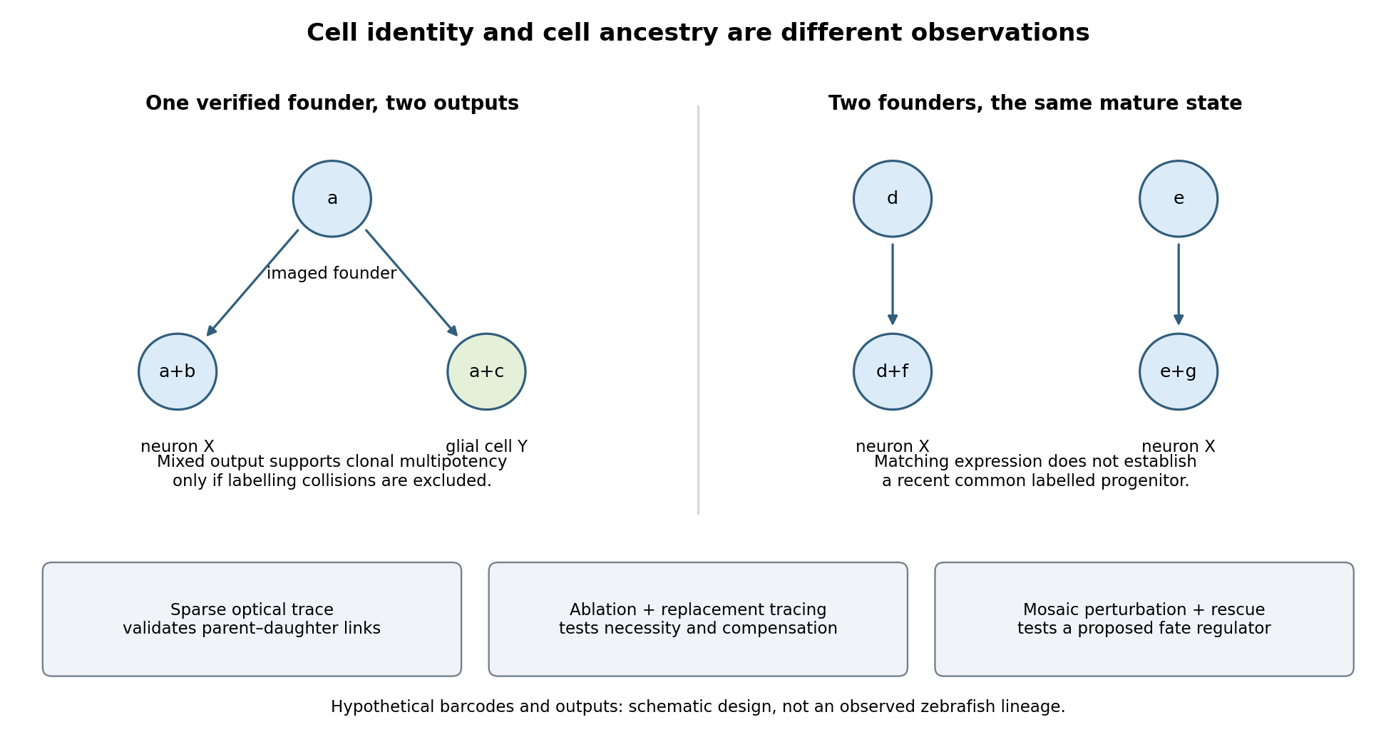

Functional tests would be designed to distinguish ancestry, necessity and developmental potential. Selective laser ablation of the labelled founder before its predicted bifurcation should remove or reduce both outputs if there is no compensation. A sham exposure, ablation of an adjacent equally sized population, and immediate assessment of collateral damage are essential controls. If outputs recover, I would trace the replacing cells: recovery can reveal regulative development rather than refute the original ancestry. Conversely, loss of a neuronal population after ablation could reflect loss of a required signal from the targeted cells, so it cannot alone prove that they were its ancestors.
I would then transplant labelled progenitors between matched hosts, including orthotopic controls and a heterotopic or temporally altered environment. Retention of both outputs under a viable alternative environment would support commitment; adoption of the host's outputs would show environmental instruction. Failure to differentiate after poor engraftment is not evidence of restriction. To test a proposed branch regulator, I would perturb it in sparse, permanently labelled clones and measure descendant identities, clone size, proliferation, apoptosis and position in parallel. A change in fate proportions at comparable survival and clone size is more persuasive than disappearance of the entire clone. Rescue by a perturbation-resistant allele, independent perturbations and comparison with neighbouring unperturbed clones would test specificity and cell autonomy. The lineage label should persist even if the cell-state marker used to select the progenitor is later lost.
Replication would be at the level of independently treated embryos and parents as well as clones; thousands of cells from one fish are not thousands of independent developmental replicates. I would report label specificity, collision and dropout estimates, alternative tree resolutions, and the sampled developmental window. A robust map is one in which sequence-based ancestry predicts independently observed divisions and clone outputs, while perturbations reveal which developmental choices are causal and which remain plastic.
Past exam of the genetics course of the University of Cambridge 2024 Paper 2 1 Solution by Codex 0 2026-10-20
Codex 0 2026-10-20The intestine comes from the E blastomere, but its identity is established by a network combining inherited regulators with induction from a neighbour. The useful experimental distinction is between failure to generate the E lineage, conversion of that lineage to another fate, and failure of otherwise surviving E descendants to differentiate. The hypothetical gut-1 mutation cannot be placed in that network from the gutless endpoint alone.
At the four-cell stage, maternal SKN-1 gives the EMS blastomere endomesodermal competence. EMS divides into anterior MS and posterior E. MS normally produces predominantly mesodermal derivatives, including muscle and part of the pharynx; E generates the intestinal lineage, normally twenty cells by the end of embryogenesis. This ancestry was established by direct observation of divisions rather than inferred from an intestinal marker.[ref]
The embryonic cell lineage of the nematode Caenorhabditis elegans by John E. Sulston and others. 1983, pp. 64–119, embryonic lineage and E descendants
The distinction matters because a missing differentiation marker need not mean that the corresponding ancestors never existed.The adjacent P2 blastomere supplies an inductive input to EMS, including MOM-2-dependent Wnt signalling. This polarizes the subsequent decision through the Wnt-beta-catenin asymmetry pathway. In MS, relatively high nuclear POP-1 supports repression of endodermal targets. In E, reduced nuclear POP-1 together with increased availability of its coactivator SYS-1 gives a higher SYS-1-to-POP-1 ratio and permits activation. Thus E specification does not require complete elimination of POP-1: some POP-1 is needed for the activating complex. Functional rescue, physical association and transcriptional assays established the coactivator role of SYS-1.[ref]
A beta-catenin identified by functional rather than sequence criteria and its role in Wnt/MAPK signaling by Ambrose R. Kidd III and others. 2005, pp. 761–772, SYS-1 rescue, POP-1 binding and coactivation
SKN-1 activates the divergent GATA transcription factors MED-1 and MED-2, and supplies additional input toward the transient endoderm regulators END-3 and END-1. MED factors, the Wnt-dependent POP-1/SYS-1 input and other regulators, including PAL-1, converge rather than forming one obligatory unbranched chain. END-3 helps activate END-1, and the two have overlapping specification functions. Their output includes ELT-7 and ELT-2, which stabilize and execute an intestinal differentiation programme through feedback and activation of differentiated targets. Perturbing MED-binding sites in the end genes produces graded, variable gut defects, demonstrating why a binary gut/no-gut score can conceal network robustness.[ref]
MED GATA factors promote robust development of the C. elegans endoderm by Morris F. Maduro and others. 2015, pp. 66–79, end-gene binding-site perturbations and quantitative intestinal phenotypes
Combined ELT-factor defects further show that specification and differentiated morphology must be assessed separately.[ref]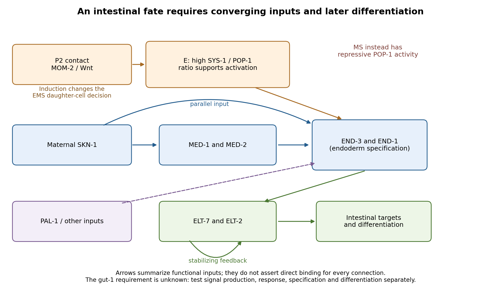
Parallel inputs and successive regulators in intestinal specification, with experimental entry points
. Details. 
First establish what the mutant actually loses. I would backcross the temperature-sensitive allele, measure penetrance at permissive and restrictive temperatures, and compare mutant embryos with wild-type siblings exposed to the same temperatures. Differential-interference-contrast imaging combined with a nuclear lineage label would follow EMS, its division orientation, E-cell birth, subsequent divisions, internalization and survival. I would score cell death and cell-cycle delay, not simply missing gut granules. Independent readouts would include endogenous transcripts encoding END-1 and END-3, ELT-2/ELT-7 expression, an intestinal structural marker and later intestinal morphology or lumen formation. Endogenous transcript detection controls for a transgene being selectively silenced or delayed. Stage matching by division history prevents temperature-dependent developmental rate from masquerading as altered expression timing. Loss of E-specific expression with ectopic MS-like descendants would support a fate transformation; normal early E identity with later degeneration or failed lumen formation would support a maintenance or differentiation defect.
Reciprocal temperature-shift experiments locate a functional interval. embryos maintained permissively would be shifted up at successive lineage-defined stages; a second series would start restrictively and be shifted down. Short restrictive pulses would refine any interval around the EMS division, early E divisions or later differentiation. An early upshift that causes gutlessness, a later upshift that does not, and rescue by a sufficiently early downshift would identify a temperature-sensitive period. If late shifts still damage an already marked intestine, an additional maintenance role is likely. I would measure recovery of a functional tagged GUT-1 protein, if available, because inactivation, refolding and protein perdurance broaden the observed interval. These experiments estimate when function is required, not necessarily when the gene is first transcribed. Wild-type pulse controls, matched handling and multiple clutches distinguish conditional loss of function from heat damage. Reciprocal crosses and separate shifts of mothers during oogenesis versus embryos would test maternal contribution and distinguish maternal-effect from zygotic requirements.
The site of expression and the site of requirement need separate tests. A rescuing endogenous knock-in could reveal whether GUT-1 is present in P2, EMS/E or surrounding tissue, but expression is not proof of cell autonomy. I would test rescue restricted to the signalling lineage versus EMS/E using promoters active before the inferred requirement; each construct must demonstrably produce functional protein at the relevant time. Late intestinal promoters cannot exclude an early E-lineage requirement if they fail to rescue. Marked genetic mosaics provide an independent test, provided loss of the rescuing element occurs before the sensitive interval and residual maternal protein is considered. With a suitable conditional system, local inactivation in P2 versus EMS would complement these experiments. Systemic RNA interference alone would not establish which cell requires the gene.
The most direct induction experiment would isolate EMS and recombine it with P2 in four genotype combinations. With otherwise viable blastomeres, mutant P2 failing to induce wild-type EMS suggests a signalling defect; wild-type P2 failing to induce mutant EMS suggests a responding-cell or downstream defect. Wild-type P2 plus wild-type EMS is the positive control; isolated EMS and EMS recombined with an irrelevant blastomere are negative controls. Contacts must be established at equivalent stages and orientations, with cleavage and survival recorded. The classic observation that P2, but not the other early neighbours, restores gut differentiation to isolated EMS establishes the logic of this assay.[ref]
Induction of gut in Caenorhabditis elegans embryos by Bob Goldstein. 1992, pp. 255–257, blastomere isolation and recombination
A mutant defect in division orientation or physical contact could abolish induction without changing synthesis of the signal itself.Finally, I would combine temporal readouts with epistasis and controlled genetic bypass experiments. Compare POP-1/SYS-1 asymmetry, MED activity, END-1/END-3 activation and ELT output in gut-1 embryos. Then express an END factor transiently in the correct early lineage, or an ELT factor at an appropriate later stage, while keeping endogenous controls and matched expression levels. Rescue by END-1 of missing end-gene activation would place the defective function upstream of, or parallel to an input bypassed by, END-1. Normal end-gene activation with failure of ELT induction would point later; persistence of early regulators with loss only of structural targets would point later still. Lack of rescue is interpretable only if the transgene is functional and the cells retain developmental competence. Because POP-1 can both repress and activate, simple POP-1 removal can produce misleading suppression or enhancement; its molecular output must be measured. Double-mutant enhancement could reveal parallel inputs rather than a linear order. Mapping, sequence identification, independent alleles and rescue by a wild-type genomic copy would establish the responsible locus. Direct promoter occupancy, a rapid transcriptional response and endogenous binding-site mutation would be needed before claiming direct regulation of a known target. The outcome should be a tested temporal and cellular requirement with constrained network relationships, not a position assigned from a single rescue.
Past exam of the genetics course of the University of Cambridge 2024 Paper 1 6 Solution by Codex 0 2026-10-20
Codex 0 2026-10-20Susceptibility of an isolated bacterium and failure to clear an infection measure different things. The scenario does not establish a particular resistance gene or a real species-specific mechanism. First distinguish antibiotic resistance, an increased ability to grow at inhibitory drug concentrations, from antibiotic tolerance, slower killing without the corresponding increase in the growth-inhibiting concentration, and bacterial persistence, survival of a subpopulation in an otherwise susceptible population.
One explanation is a reversible physiological state. Slowly growing or metabolically altered bacteria can survive an antibiotic whose lethal action depends on the active process it targets. Regrown survivors can then recover the original susceptibility. Nathalie Q. Balaban and colleagues directly followed individual bacteria and showed switching between growth states associated with persistence. The important inference is phenotypic heterogeneity, not that all survivors must share one dormant state or one toxin-antitoxin pathway.[ref]
Bacterial persistence as a phenotypic switch by Nathalie Q. Balaban and others. Single-cell observations and susceptibility after regrowth
A second explanation is the local infection environment. Biofilms can generate heterogeneous growth conditions, altered penetration and protected reservoirs; intracellular localization can separate bacteria from otherwise adequate extracellular drug exposure. Nutrient limitation, oxygen conditions, pH and host stress can change physiology or drug activity. Conversely, a laboratory sample may miss a resistant minority, mixed infection or an inducible phenotype, so apparent susceptibility should not be treated as proof that every bacterium at every site has the same response.
Triggers can include changes imposed by the host environment, entry into a slow-growth state, or stress associated with prior exposure. These are alternative hypotheses: the question supplies no data selecting one. Bacteria surviving because exposure never reached an effective level are also different from bacteria surviving an adequately delivered exposure. Persistence may pre-exist exposure rather than being induced by it.
At the level of investigation, compare inhibition of growth with survival over time, examine whether the phenotype persists after regrowth, and assess population heterogeneity and protected infection sites. A high-level response to treatment failure is to identify the reservoir and exposure problem, establish which organism and phenotype are actually present, and consider approaches addressing that mechanism rather than assuming that simply increasing the same drug will solve it. Removal of a colonized foreign-body reservoir, where relevant, addresses a different cause from a change in antimicrobial choice. The information supplied is insufficient to name an effective drug regimen or infer a single molecular trigger.
Past exam of the genetics course of the University of Cambridge 2024 Paper 1 5 Solution by Codex 0 2026-10-20
Codex 0 2026-10-20Genomic instability means an elevated propensity to acquire genetic alterations. It includes changes in nucleotide sequence, repeat length, chromosome structure and chromosome number. A genome carrying one fixed alteration is not, by that fact alone, genomically unstable: instability concerns the process that generates further alterations.
At the sequence level, DNA replication introduces mismatches and small insertion-deletion errors. Polymerase proofreading and DNA mismatch repair normally suppress their fixation. Failure of mismatch repair is particularly evident as microsatellite instability. Damaged bases, stalled replication forks and collisions with transcription can increase replication stress; unrepaired or improperly repaired lesions can produce substitutions and rearrangements. Homologous recombination and non-homologous end joining solve different repair problems, and inappropriate use or incomplete repair can alter genome structure.
At the chromosome level, failed chromosome-end protection can allow end fusion. Subsequent separation of fused chromosomes can initiate breakage-fusion-bridge cycles, producing repeated rearrangement and copy-number change. Faulty kinetochore attachment, weakened sister chromatid cohesion or abnormal spindle organization can cause chromosome missegregation and aneuploidy. The resulting imbalances may themselves disturb cell physiology and DNA replication. DNA confined in an aberrant micronucleus can experience defective replication and fragmentation; repair of clustered fragments is one route to chromothripsis, rather than every rearrangement arising through that mechanism.[ref]
Chromothripsis from DNA damage in micronuclei by Cheng-Zhong Zhang and others. Live-cell tracking linked to sequencing of descendants
The experimental signatures distinguish mechanisms. Mutation spectra and mismatch-repair status address sequence errors; repeat-length distributions address microsatellite instability; karyotyping, copy-number profiles and live chromosome imaging address numerical or structural chromosome instability. Single-cell or lineage-resolved measurements can show ongoing production of new states. A mixed endpoint population alone cannot establish whether variation arose rapidly during the experiment or was selected from pre-existing diversity.
In cancer, instability can generate selectable combinations that promote growth, escape from controls or resistance to treatment. It is neither automatically beneficial to the tumour nor sufficient for malignant transformation: excessive instability can impair proliferation or trigger apoptosis and cellular senescence. Examples of mechanistic connections include mismatch-repair deficiency in Lynch syndrome, homologous-recombination defects involving BRCA1 or BRCA2, and inherited replication or repair disorders. Chromosome missegregation can also contribute to developmental disease, pregnancy loss and infertility; instability in long-lived somatic tissues can impair function without producing a tumour.
A useful causal analysis asks what fails first, what alterations it produces, and which cellular responses restrain or select the affected cells. Restoring a repair factor or correcting a segregation defect should reduce the appropriate new-error rate, not merely change which clone grows best. The same reasoning explains why a genomic-instability phenotype can be informative in disease while remaining insufficient to diagnose a particular condition from morphology alone.
Past exam of the genetics course of the University of Cambridge 2024 Paper 1 4 Solution by Codex 0 2026-10-20
Codex 0 2026-10-20Both checkpoints couple cell-cycle timing to spatial organization, but they act at different transitions. The budding-yeast spindle position checkpoint prevents premature mitotic exit when the anaphase spindle has not been appropriately placed across the mother-bud axis. The fly centrosome orientation checkpoint delays entry into mitosis until interphase centrosomes have the orientation needed for a properly oriented division.
In Saccharomyces cerevisiae, one product of division must enter the bud. If both spindle poles and chromosome masses remain in the mother, cytokinesis would generate the wrong nuclear distribution. The mitotic exit network promotes sustained activation of Cdc14 and the decline of mitotic CDK activity. Its upstream GTPase Tem1 is restrained by the Bub2-Bfa1 complex. When the spindle is mispositioned, Kin4 helps preserve that inhibition and opposes the checkpoint-inactivating action of Cdc5 on Bfa1. Thus spindle mispositioning is connected to continued mitotic arrest rather than simply to an attempt to pull harder on chromosomes.[ref]
The Protein Kinase Kin4 Inhibits Exit from Mitosis in Response to Spindle Position Defects by Katharine E. D'Aquino and others. Kin4-dependent inhibition of mitotic exit
Spatial regulation makes this pathway sensitive to which compartment a spindle pole occupies. Kin4 activity is associated with the mother-cell side, whereas the bud environment favours release of the inhibitory state when a pole enters appropriately. Elm1 contributes to activation of Kin4. This is a regulated network involving localization and phosphorylation, rather than an infallible single switch at the bud neck. It is also distinct from the spindle checkpoint, which monitors kinetochore attachment and restrains anaphase; a spindle can have attached chromosomes yet be in the wrong position.[ref]
Elm1 kinase activates the spindle position checkpoint kinase Kin4 by Ayse Koca Caydasi and others. Kin4 activation and regulation of Bub2-Bfa1
In male Drosophila melanogaster germline stem cells, contact with the hub defines the stem-cell niche. Normally one centrosome is retained toward the hub and the other moves away, preparing a division in which one daughter retains niche contact. Live imaging showed that misoriented centrosomes can delay division until orientation is restored, including in aged or dedifferentiated cells. Counting misoriented interphase centrosomes alone therefore overestimates the frequency of misoriented mitoses.[ref]
Centrosome misorientation reduces stem cell division during aging by Jun Cheng and others. Interphase misorientation, delayed cycling and reorientation before mitosis
The fly checkpoint involves Centrosomin, Par-1, and regulation of cyclin A localization. Par-1 and the germline spectrosome help restrain the mitotic programme; perturbing this organization can uncouple orientation from mitotic entry. Bazooka provides a cortical platform associated with centrosome anchoring. These observations support spatial control of mitotic competence, not identity of the molecular pathway with yeast Tem1 signalling.[ref]
Cyclin A localization and the centrosome orientation checkpoint by Hebao Yuan and others. Par-1, spectrosome-associated cyclin A and checkpoint defects
[ref]A decisive experimental comparison measures both position and transition timing in live cells. A pure orientation defect raises misorientation and prolongs the relevant pause; loss of the checkpoint permits the transition despite misorientation. Rescue with an appropriate wild-type component helps establish specificity. In yeast, score mitotic exit and nuclear inheritance; in fly stem cells, score entry into mitosis and daughter contact with the niche. This separates the mechanism that constructs the correct geometry from the mechanism that waits for it.
Past exam of the genetics course of the University of Cambridge 2024 Paper 1 3 Solution by Codex 0 2026-10-20
Codex 0 2026-10-20Essential cell cycle genes cannot generally be discovered by selecting viable complete deletions alone. A temperature-sensitive mutation solves this problem: its product supports growth under permissive conditions but fails under restrictive conditions. Screen conditional mutants for a reproducible division defect, then distinguish a specific cycle block from nonspecific loss of viability. A cdc mutant is defined by its cycle phenotype, not simply by poor growth at high temperature.
In Saccharomyces cerevisiae, bud size and nuclear morphology provide convenient stage markers. After shifting an asynchronous culture, mutants defective at a particular step tend to accumulate with a characteristic morphology. Leland H. Hartwell and colleagues used time-lapse observations to distinguish the time a function must act from the stage at which its absence becomes visible. Temperature-shift timing, DNA-content measurements and recovery after return to permissive conditions strengthen the interpretation. A delayed terminal phenotype need not identify the moment of the original molecular defect.[ref]
Genetic control of the cell-division cycle in yeast. I. Detection of mutants by Leland H. Hartwell and others. Conditional mutants, execution points and termination phenotypes
In Schizosaccharomyces pombe, elongated cells that continue growing without dividing are especially useful candidates; nuclear number, septum formation and DNA content separate blocks in replication, mitosis and division. Paul Nurse, Pierre Thuriaux and Kim Nasmyth isolated conditional mutants defining multiple essential functions. Complementation testing groups recessive mutations affecting the same function, and segregation analysis distinguishes linked from independent loci. Molecular identification, rescue and biochemical tests then establish what the affected products do.[ref]
Genetic control of the cell division cycle in the fission yeast Schizosaccharomyces pombe by Paul Nurse and others. Conditional mutants affecting DNA synthesis, nuclear division and septation
Three contrasting examples explain why similar-looking division blocks need not have the same cause. In fission yeast, Cdc2, the cyclin-dependent kinase 1 catalytic subunit, is required for key cycle transitions. A restrictive loss-of-function allele can leave cells growing without entering mitosis; some alleles also reveal the earlier requirement at Start. Cdc25 is an activating protein phosphatase that removes inhibitory phosphorylation from Cdc2. Loss of Cdc25 therefore leaves the mitotic kinase inadequately activated and commonly produces an elongated G2-arrested phenotype. In budding yeast, Cdc6 participates in DNA replication licensing: failure to establish competent origins prevents successful genome duplication and hence normal division. Species and allele must be stated: identically numbered CDC names in different yeasts are not automatically homologous proteins.
The importance for cancer is that an unbiased genetic screen exposed conserved mechanisms connecting growth, DNA duplication and division. The yeast cross-species complementation experiment showed that central regulatory functions could be conserved across large evolutionary distances. Human cancers can alter cyclin-CDK regulation, DNA-replication control and checkpoint responses, providing a mechanistic connection to uncontrolled proliferation and genomic instability. This does not mean that every yeast cdc gene is a human oncogene, nor that any observed arrest is necessarily a checkpoint response: direct loss of an essential activity can itself stop progression.
Past exam of the genetics course of the University of Cambridge 2024 Paper 1 2 Solution by Codex 0 2026-10-20
Codex 0 2026-10-20The first task is to map the DNA associated with the centromeric histone variant CENP-A; the second is to determine how that occupancy is maintained. These require different evidence. A shared DNA sequence is not sufficient evidence for an inherited centromeric state.
Use validated anti-CENP-A chromatin immunoprecipitation sequencing with input DNA and an appropriate negative immunoprecipitation control. Check antibody recognition separately in human, horse and chicken; an endogenous epitope-tagged allele can provide an orthogonal control if its chromosome-segregation function is retained. Native digestion can recover CENP-A-containing nucleosomes, whereas crosslinking can retain indirect associations. CUT&RUN provides a complementary occupancy assay with different recovery biases. Establish centromeric function by colocalization with kinetochore proteins and by normal chromosome segregation, not merely by calling a sequencing peak.
Mapping repetitive DNA is the main analytical obstacle. Use centromere-resolved assemblies where possible, retain information from multimapping reads, and analyse satellite-family enrichment as well as uniquely mappable sequence. Absence of a peak in an incomplete reference is not evidence of absent CENP-A. Long-read sequence assemblies, informative single-nucleotide polymorphisms, and DNA fluorescence in situ hybridization can connect a recovered sequence to a particular chromosome and distinguish homologues.
For precise positioning, compare enriched domains with input-normalized background at several resolutions. Map nucleosome-protected fragments and use fibre immunofluorescence or sequence probes to check domain extent and heterogeneity. A broad population peak may represent alternative positions in different cells or homologues, rather than one broad CENP-A domain on every chromosome.
The expected outcome is a common centromeric function without a universal centromeric DNA sequence. Human CENP-A commonly occupies selected portions of alpha-satellite arrays, but human neocentromeres show that such arrays are not obligatory. Horse chromosome 11 provides a normal satellite-free centromere. Chicken chromosomes include both repeat-rich centromeres and comparatively short non-tandem-repetitive centromeric regions.[ref]
Chickens possess centromeres with both extended tandem repeats and short non-tandem-repetitive sequences by Wei-Hao Shang and others. Comparative chicken centromere mapping
[ref]Birth, evolution, and transmission of satellite-free mammalian centromeric domains by Solomon G. Nergadze and others. Satellite-free mammalian centromeres and inherited positional variation
To test mitotic stability, start from single-cell clones and remap CENP-A on the same haplotype after successive divisions, while checking that the underlying DNA has not changed. Pulse labelling of old versus newly incorporated CENP-A can distinguish retention from replenishment. Follow both chromosome identity and domain position: stable centromeric function does not require an invariant nucleotide boundary. To test meiotic transmission, use informative pedigrees or gamete-and-offspring material and allele-resolved mapping, with matched cell types where possible. An inherited positional state, or bounded movement of that state on unchanged DNA, supports epigenetic centromere inheritance. Artificial centromere deletion and neocentromere recovery provide an additional perturbation test, but establishment after deletion is different from ordinary meiotic inheritance.[ref]
Chromosome Engineering Allows the Efficient Isolation of Vertebrate Neocentromeres by Wei-Hao Shang and others. Chicken neocentromere formation and CENP-A domain mapping
Past exam of the genetics course of the University of Cambridge 2024 Paper 1 1 Solution by Codex 0 2026-10-20
Codex 0 2026-10-20Bacterial DNA-recognition and DNA-modifying systems provide ways to mark, map and perturb eukaryotic chromosomes. Their value comes from separating the targeting module from the reporter or effector, so a chromosome becomes experimentally observable without assuming that its sequence alone specifies its behaviour.
For fluorescent operator-repressor chromosome labelling, an integrated array of bacterial operator sequences is bound by a fluorescent repressor fusion. A LacI fusion bound to a lac operator array makes a selected genetic locus visible in a living nucleus. Two differently labelled loci can report separation of sister chromatids, movement relative to the nuclear periphery, or distributions of distances between chromosome regions. Carmen C. Robinett and colleagues demonstrated operator-based imaging in mammalian cells and yeast. The tag reports the marked locus, not every part of the chromosome, and repeated operators or excessive bound repressor can perturb DNA replication and chromatin structure.[ref]
Lac-operator chromosome imaging study by Carmen C. Robinett and others. Live GFP-repressor imaging and targeted yeast insertion
The bacterial CRISPR-Cas9 recognition mechanism makes targeting programmable. A fluorescent dCas9 fusion and a guide RNA can visualize native repeated sequences; multiple guides can improve detection of a nonrepetitive locus. Baohui Chen and colleagues used this approach to follow human genetic loci. Catalytically inactive targeting avoids making the DNA double-strand break that would itself alter chromosome behaviour. Appropriate controls include guide specificity, signal from an unlabelled locus, and whether tagging changes growth or the measured dynamics.[ref]
CRISPR imaging of human genomic loci by Baohui Chen and others. Live imaging of endogenous human loci
For DamID, bacterial Dam methyltransferase is fused to a nuclear protein. Methylated GATC sites record neighbourhoods visited by the fusion. A fusion to a nuclear-envelope component can identify lamina-associated domains, whereas one to a chromatin-binding protein maps its genomic neighbourhoods. Comparing with untethered Dam helps control accessibility-dependent background. The accumulated methylation signal is a time-integrated proximity record: it need not establish direct binding to every recovered sequence, and resolution depends on GATC distribution and local spreading.[ref]
Bacterial enzymes also make structural measurements possible. In Hi-C, restriction enzymes generate DNA fragments whose proximity-dependent ligation junctions can be sequenced. In ATAC-seq, a modified bacterial Tn5 transposase inserts sequencing adapters preferentially into accessible DNA. Neither assay directly photographs a fixed chromosome structure: contact frequency and accessibility are different observables. Enzyme sequence preferences, fixation, and cell-to-cell heterogeneity must be controlled. Together, live labelling, proximity recording and sequencing-based assays connect chromosome movement, nuclear neighbourhood and chromatin accessibility, rather than substituting one measurement for all three.
There are unlisted articles, also show them or only show them.