Topics (384k) Articles (398k) Users (410) Discussions (242) Comments (383) Files (13k) New article

A root-cap columella cell is a cell in the central columella of a plant's root cap. In Arabidopsis thaliana, these cells form recognizable tiers and files and contain starch-bearing amyloplasts. The organelles can sediment after the root changes orientation relative to gravity, making these cells useful for investigating gravitropism.[ref]
An especially informative experiment removes selected cells, rather than the entire growing tip. In 1998, investigators used laser ablation and then measured curvature after reorientation. Inner central columella cells contributed more strongly to the response than the other tested regions. Measuring extension alongside bending helped distinguish damage to gravity-associated signaling from a general failure of growth.[ref]
The distinction between sensing and response is important: a cap cell associated with gravity perception need not be the cell whose elongation produces the visible bend. Much of that differential growth occurs farther back in the root elongation zone. Likewise, observing an amyloplast settle supplies a candidate physical mechanism, while a targeted perturbation tests the functional importance of its cellular setting.
A clearance bound between trajectory samples converts finitely many separation checks into a guarantee over a continuous trajectory, using a justified bound on how quickly clearance can change. Clear endpoints alone are insufficient: a foot or another robot part can approach an obstacle and move away again between them. The primary planning formulation requires constraints along the entire path.[ref]
Robotic Systems by Kris Hauser. Chapter 8, Point-to-point path planning: continuous path satisfying feasibility for every path parameter
Let nominal clearance have Lipschitz constant on a checked interval of length . If both endpoint clearances are at least , then every intermediate point is within of an endpoint. ConsequentlyThis follows directly from using the nearer endpoint. A uniform additional worst-case geometry or execution allowance gives actual clearance at least .
A tighter interval certificate retains the two different endpoint values, and . Writing gives two lower cones,If and , the cones meet inside the interval. Their common height yieldsIf the endpoint difference exceeds , the proposed Lipschitz bound and exact endpoint values are inconsistent; simply applying the formula does not repair that inconsistency. With , clearance is constant.
For an original example, take , , and . The simpler endpoint-minimum bound gives ; the two-cone bound gives . Subtracting a execution allowance leaves a certified actual gap, sufficient for a requested margin. These are lower bounds, not assertions that the trajectory reaches their values.
A counterexample explains why the allowance is necessary. On , has endpoint values one but touches zero at the midpoint. Checking only those endpoints misses the contact. Its derivative is bounded in magnitude by four, so a sufficiently fine sample grid with that valid bound can expose the missing guarantee.
The constant must match the geometry and parameter units. For a point moving relative to a fixed closed obstacle set, the distance to that set changes by at most the point’s displacement: the triangle inequality gives . A speed bound therefore supplies a time-based clearance bound. A rotating body requires a bound on every occupied point, such as the angular clearance bound for a rotating body; terrain-height clearance across a discontinuous edge cannot use a finite slope bound across that jump.
Adaptive clearance sampling subdivides intervals that fail the chosen certificate, concentrating computation where the margin or speed bound is least favorable. Failure of a conservative certificate means the interval needs better bounds or further checking; it does not itself prove a collision. For a proposed robot-foot test, validate the motion and geometry error bounds in one coordinate frame and compare the certified interval minima with dense independent measurements. Use the robust swing-foot clearance margin allowances without counting the same uncertainty twice.
A GNRA tetraloop is an RNA tetraloop with G at its first position, A at its fourth, any ordinary RNA nucleotide at N, and a purine at R. R therefore means A or G, not one particular nucleotide. GAAA and GAGA both match, while GACA does not because C is not a purine. There are allowed loop sequences under this alphabet.
A structural study determined solution NMR spectroscopy models for GCAA and GAAA hairpins. Both contained an unusual G–A pair between the first and last loop residues, together with base stacking and contacts involving phosphate and a sugar hydroxyl.[ref]
Structural features that give rise to the unusual stability of RNA hairpins containing GNRA loops by H. A. Heus and others. Abstract, shared structural contacts in the GCAA and GAAA solution structures.
These are specific interactions beyond the Watson–Crick stem. A loop shown as unpaired in a simple secondary-structure diagram can still make an organized network of internal contacts.The pattern separates invariant and variable positions. Starting from GAAA, changing only N to C gives GCAA without leaving the family; changing R from A to G gives GAGA. Changing the final A to G instead gives GAAG, which does not satisfy GNRA. This original sequence exercise explains what a consensus rule does and does not preserve.
Sequence-family membership alone does not supply the complete thermodynamics or structure of an embedded RNA. Keeping the loop fixed while changing the stem tests context dependence; keeping the stem fixed while changing a loop position tests that position's contribution. An RNA optical melting comparison and a structural measurement provide complementary information rather than interchangeable proofs.
The GNRA designation is useful because a short sequence rule points toward recurring molecular organization. It does not imply that all eight sequences occur equally often, have identical stability, or maintain the same contacts in every surrounding RNA. A noncanonical base pair and the rest of the loop environment need their own structural evidence.
A UNCG tetraloop is an RNA tetraloop whose four loop positions follow U–N–C–G, where N can be any ordinary RNA nucleotide. The pattern permits UACG, UCCG, UGCG and UUCG. It specifies the loop sequence family; the closing stem pair lies outside these four positions.
For example,
GGACUUCGGUCC can be represented with stem pairs1–12,2–11,3–10 and4–9, leaving UUCG at positions5–8. Four stem pairs and four loop nucleotides give twelve residues. The closing stem pair in this representation is C–G; it is not part of the UNCG sequence. This is sequence bookkeeping for the cited structural RNA, not a claim that every UNCG-containing sequence forms the same isolated hairpin.A solution NMR spectroscopy study of that hairpin used interproton distances and scalar couplings to construct a restrained three-dimensional model. The reported loop includes an unusual G–U interaction and syn-oriented loop guanosine; mutations of conserved loop residues reduced stability.[ref]
Structure of an unusually stable RNA hairpin by Gabriele Varani and others. Abstract, structural restraints, loop contacts and mutation sensitivity.
Thus a four-residue loop in a secondary-structure description can still contain specific internal contacts, including a noncanonical base pair. It is not four freely moving bases attached to a stem.A useful experimental comparison changes loop sequence while preserving a stated stem and solution conditions. RNA optical melting asks how the transition changes; NMR asks which spatial contacts are compatible with its measurements. These tests address related but different questions. Similar melting midpoints do not independently establish identical loop geometry, and one sequence motif does not establish its fold in every larger RNA.
A UUCG tetraloop is an RNA tetraloop whose four loop residues have nucleotide sequence U–U–C–G. It is one member of the UNCG tetraloop family, with U occupying the variable N position. The closing stem base pair lies outside those four residues.
The structurally studied sequence
5'-GGACUUCGGUCC-3' illustrates the bookkeeping. Stem pairs 1–12,2–11,3–10 and 4–9 leave UUCG at positions 5–8, giving nucleotides. The loop's first uracil and last guanine can themselves contact without changing its four-residue sequence definition.Solution NMR spectroscopy determined interproton distances and scalar couplings for this hairpin. The resulting restrained structural model showed a loop G–U contact with syn-oriented guanosine, plus a cytosine–phosphate contact and organized base stacking. Mutating conserved loop residues reduced the measured stability.[ref]
Structure of an unusually stable RNA hairpin by Gabriele Varani and others. Abstract, sequence, NMR restraints, syn-guanosine and mutation sensitivity.
The syn description concerns the glycosidic torsion angle of that nucleotide; it must not be confused with the cis/trans classification of relative RNA base-pair geometry.The original paper describes only two residues as the remaining structural loop after its G–U contact extends the paired region. This is compatible with naming the four-residue sequence motif UUCG: a sequence-based tetraloop designation and a detailed contact-based loop count use different boundaries.[ref]
Structure of an unusually stable RNA hairpin by Gabriele Varani and others. Abstract, G–U contact and two remaining loop nucleotides.
A useful comparison keeps the stem and solution conditions fixed while changing one loop position. RNA optical melting tests the effect on a transition, while NMR spectroscopy tests the spatial contacts. Membership in a short sequence family does not fix a context-independent RNA melting temperature or prove that every larger RNA containing those letters adopts the same structure.
RNA polymerase I is a eukaryotic RNA polymerase specialized for synthesis of the large precursor from which the major ribosomal RNAs, including 18S, 5.8S and 28S in mammals, are processed. It acts in the nucleolus. The separate 5S ribosomal RNA is normally transcribed by RNA polymerase III, so not all ribosomal RNA has the same polymerase origin. [ref]
Molecular Biology of the Cell, 4th edition by Bruce Alberts and others. Chapter 6, The Nucleolus Is a Ribosome-Producing Factory
A yeast experiment disrupted the essential polymerase-I subunit RPA43. Driving ribosomal-RNA synthesis through a different polymerase could rescue the lethal defect, linking the essential requirement to RNA production. The rescue distinguishes the normal route from an experimentally engineered substitute, rather than implying that the alternative route is standard. [ref]
Gene RPA43 in Saccharomyces cerevisiae Encodes an Essential Subunit of RNA Polymerase I by Pierre Thuriaux and others. 1995, abstract: disruption and alternative-polymerase rescue
RNA polymerase III is a eukaryotic RNA polymerase that transcribes several small functional RNAs, including transfer RNA and 5S ribosomal RNA. It is distinct from RNA polymerase I, which makes the large precursor of most other ribosomal RNAs, and from RNA polymerase II, which transcribes protein-coding genes and many noncoding RNAs.
In 1974, Roberto Weinmann and Robert G. Roeder compared inhibition of RNA synthesis in isolated nuclei with the characteristic inhibitor sensitivity of separated RNA polymerases. The behaviour of transfer RNA and 5S ribosomal RNA synthesis matched RNA polymerase III, assigning particular RNA products to an enzymatic activity rather than grouping all nuclear transcription together.[ref]
Role of DNA-Dependent RNA Polymerase III in the Transcription of the tRNA and 5S RNA Genes by Roberto Weinmann and Robert G. Roeder. Abstract, alpha-amanitin sensitivity and isolated-nucleus transcription
This division of labour explains how production of translation machinery can be regulated separately from production of messenger RNA.A two-state RNA melting model treats an RNA transition as an equilibrium between folded F and unfolded U macrostates, with no substantially populated intermediate. Each macrostate can contain many microscopic conformations; two states does not mean exactly two atom-by-atom structures. The model relates populations to thermodynamics and then, separately, to an observed optical signal.
For unfolding free energy ,The population formula assumes an ideal unimolecular transition. With constant unfolding enthalpy and entropy,An RNA melting log-odds plot against is then a straight line. Nonzero changes in heat capacity, alternative states or incorrect population estimates can invalidate this simplified relation.
An original consistency test fits stipulated at and at . These two points determineThe fitted model predicts approximately at . A third observation tests that prediction; fitting two parameters to two points was not independent validation. Measurement uncertainty and baselines must be considered before attributing disagreement to a specific physical mechanism.
For an ideal optical mixture, . Thus the population model does not remove the need for folded and unfolded RNA melting baselines. Optical-melting studies infer thermodynamics by fitting measured curves under stated analysis assumptions.[ref]
Thermodynamic characterization of naturally occurring RNA tetraloops by Justin P. Sheehy and others. Materials and Methods, optical curves and individual-curve thermodynamic fitting.
Reversibility, the absence of important intermediates and the unimolecular assumption are physical conditions to test. A two-strand association transition has a different concentration dependence even if its optical trace looks similarly sigmoidal. The model is a useful quantitative hypothesis, not a consequence of drawing one smooth curve through data.
An RNA melting baseline is the predicted absorbance of a specified folded or unfolded RNA population as a function of temperature, before mixing those populations in an RNA optical melting model. It differs from a solvent blank: a folded RNA sample still absorbs light, and that response can change with temperature even without the modeled transition.
For a two-state RNA melting model with unfolded fraction , writeThe folded and unfolded RNA melting baselines might be modeled as and . Their continuation into the transition region is RNA baseline extrapolation. Solving the optical mixture givesFor original stipulated readings , and , the inferred fraction is 0.50. Changing only the folded RNA melting baseline to 0.45 gives 0.40; changing only the unfolded RNA melting baseline to 0.80 gives 0.375. The same measured absorbance can therefore imply different populations under different baseline models.
Sensitivity is explicit. With ,A small baseline separation amplifies measurement uncertainty. Correlated fitted baselines require covariance terms in uncertainty propagation, rather than independent error bars by assumption.[ref]
Evaluation of measurement data — Guide to the expression of uncertainty in measurement by JCGM. §§5.1–5.2, sensitivities and correlated inputs.
Optical-melting studies infer thermodynamics by fitting measured curves, rather than directly counting unfolded molecules.[ref]
Thermodynamic characterization of naturally occurring RNA tetraloops by Justin P. Sheehy and others. Materials and Methods, individual-curve fitting.
Inspect the pre-transition and post-transition regions and test the chosen RNA baseline extrapolation. An inferred fraction outside signals noise or incompatibility with the assumed optical model; it is not a negative molecular population.RNA melting hysteresis is dependence of an RNA optical melting trace on thermal history: heating and cooling can give different absorbance at the same temperature. It warns that the observed trace may not follow reversible chemical equilibrium on the measurement timescale. Two different traces do not by themselves establish two equilibrium melting temperatures.
A primary experiment illustrates the importance of history. A 21-nucleotide RNA showed a low-temperature dimer-to-hairpin transition that did not reverse on cooling, whereas its higher-temperature RNA hairpin melting transition was reversible. Measurements used a Cary 100 spectrophotometer, a 1-cm quartz cell and 268-nm light, heating at and cooling at or . An adjacent solvent cell held the temperature probe.[ref]
Conversion of stable RNA hairpin to a metastable dimer in frozen solution by Xueguang Sun and others. Results, Fig.5 and transition reversibility; Materials and Methods, UV absorbance melting curves.
The authors interpreted the dimer as kinetically trapped under those conditions, rather than applying a reversible equilibrium fit to that transition.For an original simple kinetic model, let be the unfolded fraction, its equilibrium value and an RNA melting relaxation time:After a fixed-temperature step with constant , the deviation undergoes exponential decay:With stipulated , and , the fraction reaches only 0.632 after two minutes and 0.950 after six. During a continuing temperature ramp, this lag can put heating and cooling on different branches. This single-time model is illustrative; it is not a fit to the published dimer experiment.
Useful checks compare ramp speeds and include fixed-temperature holds and repeat cycles. Verify measurement calibration and exclude evaporation or chemical degradation before attributing a discrepancy to folding kinetics. Slower scans can reduce kinetic lag, but a smooth trace alone cannot establish reversibility.
An RNA melting temperature, , characterizes the midpoint of a specified RNA conformational transition under stated conditions. In a reversible, unimolecular two-state RNA melting model, it is the temperature at which folded and unfolded populations are equal. A midpoint assigned to an optical trace requires a model connecting absorbance to those populations.
Let unfolding free energy be . If unfolding enthalpy and entropy are treated as temperature independent,The formula assumes a finite relevant transition and uses absolute temperature. With original stipulated values and , . Holding enthalpy fixed but changing entropy to gives approximately . A melting temperature reflects both energetic and entropic contributions.
Molecularity matters. An ideal intramolecular hairpin transition need not change its midpoint with total strand concentration. A transition involving association of separate strands generally does: the equilibrium relation includes their concentrations. The concentration dependence of RNA melting can therefore help test which structural model is appropriate. In the 2010 tetraloop measurements, concentration-independent curves across a broad concentration range supported stem–loop formation.[ref]
Thermodynamic characterization of naturally occurring RNA tetraloops by Justin P. Sheehy and others. Materials and Methods, repeated melts and concentration dependence.
A raw halfway absorbance is not automatically this thermodynamic midpoint. If the folded and unfolded RNA melting baselines at one temperature are and , an absorbance of implies unfolded fraction , not . Intermediates, temperature-dependent parameters or RNA melting hysteresis can further complicate midpoint assignment. Report the solution conditions and definition of the measured transition rather than treating as a context-free constant of an RNA sequence.
RNA hyperchromicity is an increase in an RNA sample's ultraviolet absorption associated with a change in its molecular environment or conformation. The general hyperchromic effect concerns increased spectral-band intensity, rather than a shift of the band to another wavelength.[ref] In RNA melting measurements, altered base stacking can contribute to the change in absorption; signal interpretation still requires a defined wavelength and sample conditions.
The Beer-Lambert law writes , where is absorbance, a molar absorption coefficient, path length and concentration. A change in can therefore change absorbance even when the amount of RNA is unchanged. Conversely, changed concentration, path length or other optical contributions can mimic a structural signal.
For an original comparison at one stated temperature, suppose extrapolated folded and unfolded absorbances are and . Defining fractional hyperchromicity relative to the folded state gives , or50%. Using the unfolded state as denominator would instead give . A reported percentage therefore needs its convention.
In a linear two-population optical model,provided . With those baselines, gives . But with temperature-appropriate baselines and , the same absorbance gives . An RNA melting baseline is consequently part of the population estimate, not decorative plotting.
A 2010 tetraloop study measured absorbance-versus-temperature curves to infer folding thermodynamics.[ref]
Thermodynamic characterization of naturally occurring RNA tetraloops by Justin P. Sheehy and others. Materials and Methods, optical melting and curve fitting.
Such measurements connect an optical response to a model of RNA structure. An absorption increase alone does not count broken base pairs, establish a unique unfolded structure or distinguish every possible chemical change.An RNA base-pair edge is a region around a nucleobase presenting groups that can participate in an edge-to-edge contact with another nucleobase. It is a chemical contact surface in RNA base-pair geometry, rather than an edge of a drawn secondary-structure graph. The arrangement of hydrogen bond donors and hydrogen bond acceptors constrains possible partners.
The Leontis–Westhof classification distinguishes the Watson-Crick base-pair edge, Hoogsteen base-pair edge and sugar base-pair edge. The last category includes the ribose -hydroxyl contribution and is also called the shallow-groove edge in the original classification.[ref]
Geometric nomenclature and classification of RNA base pairs by N. B. Leontis and E. Westhof. Abstract, three contact edges and sugar-edge definition.
Thus an edge label does not necessarily refer only to atoms in the aromatic ring.An edge belongs to one partner. A complete pair description identifies the edge used by each nucleobase and the relative orientation of their glycosidic bonds. For example, specifying that one adenine uses its Hoogsteen base-pair edge still leaves the second partner's identity, contacting edge and orientation to be determined. It does not by itself specify a unique noncanonical base pair.
For an original classification exercise, label two partners A and B. The record “A uses Watson–Crick; B uses sugar; orientation trans” contains more geometric information than “A contacts B”. Exchanging the names A and B reverses the written edge order but does not create a second physical contact. In contrast, moving B to a different contact surface changes the proposed structure.
Structural coordinates or solution restraints are needed to establish the contact. A plausible hydrogen bond donor near an appropriate hydrogen bond acceptor is a necessary geometric clue, not proof that a proposed RNA structure is populated under every condition. Edge labels make comparisons precise while retaining the need for experimental evidence.
RNA base-pair isostericity is geometric similarity between base pairs that allows one pairing arrangement to replace another while approximately preserving the positions and orientations of the attached sugar–phosphate backbones. The important comparisons include the separation of the two ribose atoms and the relative directions of the glycosidic bonds. The bases need not have identical chemical composition or occupy exactly the same volume.[ref]
The non-Watson-Crick base pairs and their associated isostericity matrices by Neocles B. Leontis and others. Isostericity, printed pp. 3499 and3525
A useful example is the canonical cis Watson–Crick family. Its adenine–uracil, uracil–adenine, guanine–cytosine and cytosine–guanine pairs belong to the same isosteric subgroup in the primary classification. Thus a paired sequence substitution can exchange an A–U contact for G–C while approximately retaining the local backbone geometry.[ref]
The non-Watson-Crick base pairs and their associated isostericity matrices by Neocles B. Leontis and others. Table 3, subgroup I1 for Family 1; printed p. 3526, Cis Watson–Crick/Watson–Crick
An original comparison makes the limit clear. The canonical A–U contact has two hydrogen bonds, while G–C has three. Their geometric compatibility does not assert identical free energy, identical base stacking with neighboring residues, or identical recognition by a bound protein. A substitution can preserve the local scaffold while changing other physical properties.
Likewise, sharing a broad RNA base-pair geometry does not guarantee isostericity. The family identifies the two contacting RNA base-pair edges and the cis/trans relationship of the glycosidic bonds. Within a family, different base combinations can still place the ribose attachment points at different separations. An RNA isostericity matrix groups combinations into the more restrictive compatible subsets.[ref]
The non-Watson-Crick base pairs and their associated isostericity matrices by Neocles B. Leontis and others. Isostericity matrices, printed p. 3525; Tables3–5
The classification is grounded in structures rather than sequence names alone. The primary survey inspected coordinate models from X-ray crystallography, recording structure identifiers, resolution and ribose-attachment separation, and distinguished observed examples from modeled contacts. This supplies a way to test an annotation against actual atomic geometry.[ref]
The non-Watson-Crick base pairs and their associated isostericity matrices by Neocles B. Leontis and others. Materials and Methods, printed p. 3498; Results, printed p. 3499; Table 1
For comparative sequence analysis, correlated changes at two positions are therefore evidence to evaluate against a proposed contact geometry. They do not prove by themselves that the residues contact one another. Conversely, a conserved geometric interaction can survive substantial changes in nucleotide sequence, helping explain how similar RNA folds can use different base identities.
A reflecting boundary condition prevents normal particle or probability flux through a wall. For drift-free scalar diffusion with , the condition isHere is the outward unit normal. The last expression is a homogeneous Neumann boundary condition. Integrating the diffusion equation and applying the divergence theorem gives conservation of total probability or material in a closed container.[ref]
Linear Partial Differential Equations Lecture 10 in MIT OpenCourseWare. pp.1–2: diffusive flux, closed container and conserved integral
For an original example, choose units in which the interval is . The densitysatisfies , has zero endpoint derivatives and integrates to one. With , and , its cosine amplitude is about . A nonuniform density therefore relaxes while remaining inside the interval; reflection does not hold the boundary concentration fixed.
For anisotropic diffusion, impose zero normal diffusive flux using the full diffusion tensor. At a wall with , letin consistent units. Then , although . Setting the normal derivative to zero would impose a different condition. Absorption and finite permeability require their own boundary models; a reflecting wall allows continued motion within the available space.
Pinned article: Introduction to the OurBigBook Project
Welcome to the OurBigBook Project! Our goal is to create the perfect publishing platform for STEM subjects, and get university-level students to write the best free STEM tutorials ever.
Everyone is welcome to create an account and play with the site: ourbigbook.com/go/register. We belive that students themselves can write amazing tutorials, but teachers are welcome too. You can write about anything you want, it doesn't have to be STEM or even educational. Silly test content is very welcome and you won't be penalized in any way. Just keep it legal!
Intro to OurBigBook
. Source. We have two killer features:
- topics: topics group articles by different users with the same title, e.g. here is the topic for the "Fundamental Theorem of Calculus" ourbigbook.com/go/topic/fundamental-theorem-of-calculusArticles of different users are sorted by upvote within each article page. This feature is a bit like:
- a Wikipedia where each user can have their own version of each article
- a Q&A website like Stack Overflow, where multiple people can give their views on a given topic, and the best ones are sorted by upvote. Except you don't need to wait for someone to ask first, and any topic goes, no matter how narrow or broad
This feature makes it possible for readers to find better explanations of any topic created by other writers. And it allows writers to create an explanation in a place that readers might actually find it.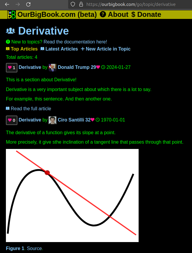
Figure 1. Screenshot of the "Derivative" topic page. View it live at: ourbigbook.com/go/topic/derivativeVideo 2. OurBigBook Web topics demo. Source. - local editing: you can store all your personal knowledge base content locally in a plaintext markup format that can be edited locally and published either:This way you can be sure that even if OurBigBook.com were to go down one day (which we have no plans to do as it is quite cheap to host!), your content will still be perfectly readable as a static site.
- to OurBigBook.com to get awesome multi-user features like topics and likes
- as HTML files to a static website, which you can host yourself for free on many external providers like GitHub Pages, and remain in full control

Figure 2. You can publish local OurBigBook lightweight markup files to either OurBigBook.com or as a static website.
Figure 3. Visual Studio Code extension installation.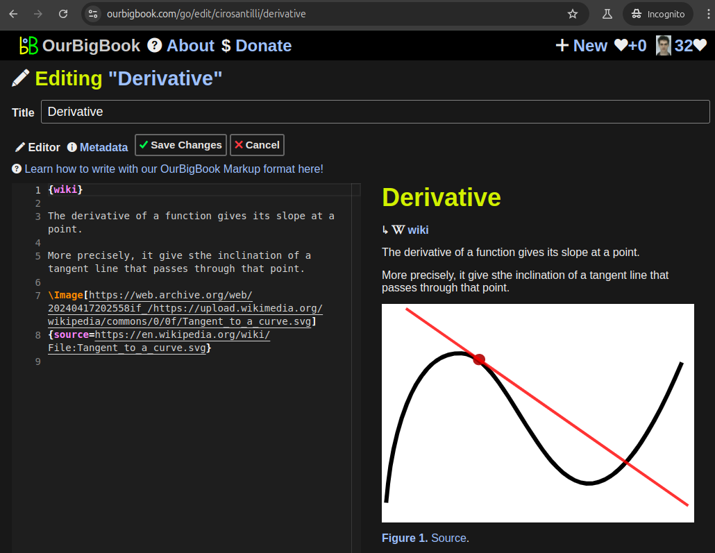
Figure 5. . You can also edit articles on the Web editor without installing anything locally. Video 3. Edit locally and publish demo. Source. This shows editing OurBigBook Markup and publishing it using the Visual Studio Code extension. 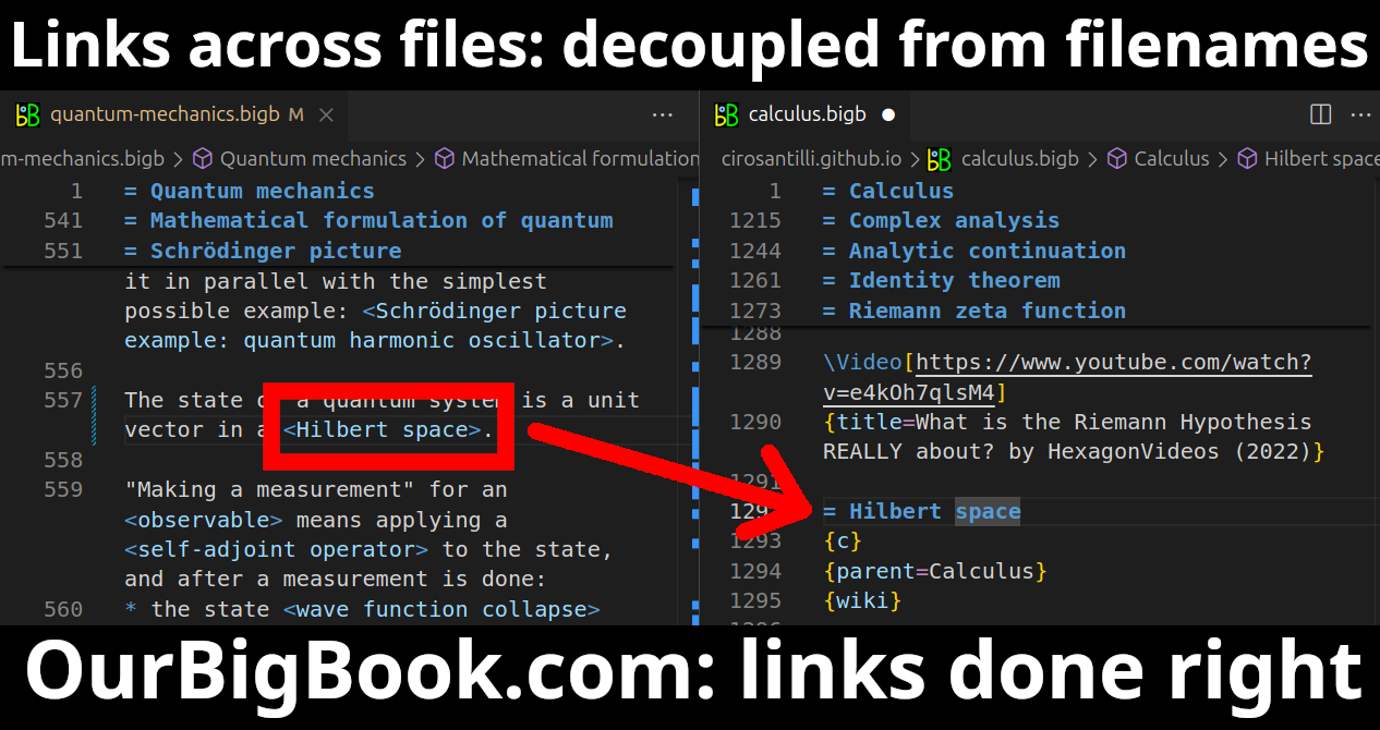
- Infinitely deep tables of contents:
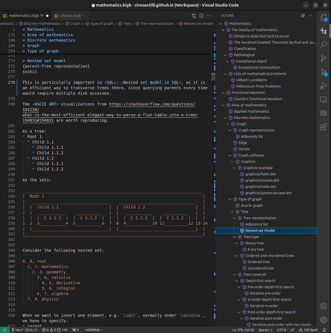
All our software is open source and hosted at: github.com/ourbigbook/ourbigbook
Further documentation can be found at: docs.ourbigbook.com
Feel free to reach our to us for any help or suggestions: docs.ourbigbook.com/#contact