Jesse Stombaugh has contributed to the geometric analysis of RNA structures. His 2002 paper with Neocles B. Leontis and Eric Westhof classifies noncanonical base pairs and organizes their geometric compatibility in RNA isostericity matrices. The paper's byline associates Stombaugh with Bowling Green State University's chemistry and biomolecular-sciences research.[ref]
The non-Watson-Crick base pairs and their associated isostericity matrices by Neocles B. Leontis and others. Title-page byline and affiliation; isostericity matrices, Tables 3–5
The underlying problem is that nucleotide sequence alone does not specify which edges of two nucleobases contact or how their attached backbones are arranged. A replacement can preserve a contact's geometry while changing its chemical composition. Stombaugh's coauthored classification provides a framework for testing such replacements, rather than treating every base pair with the same two letters as the same structure.
Stombaugh is first author of the 2009 study Frequency and isostericity of RNA base pairs by Jesse Stombaugh and others, with Craig L. Zirbel, Eric Westhof and Neocles B. Leontis. It introduces the RNA IsoDiscrepancy Index and compares geometrically aligned ribosomal RNA structures with sequence variation.[ref]
Frequency and isostericity of RNA base pairs by Jesse Stombaugh and others. Abstract and Results, definition and application of the index
An original comparison illustrates the question. The standard cis Watson–Crick A–U and G–C contacts are geometrically compatible, even though they have two and three hydrogen bonds, respectively. Replacing A–U by G–C can therefore preserve the approximate backbone scaffold without preserving every contribution to free energy or recognition by a protein. Conversely, simply reversing the order of a wobble pair need not preserve its geometry. Geometry, contact chemistry and local sequence context must be assessed separately.
These studies combine structural coordinates with comparative analysis; they do not infer a unique three-dimensional fold from a short sequence alone. A correlated substitution can support a structural hypothesis, but actual contacts and model errors still need checking against experimental structures.
Neil T. Hunt is a physical chemist who uses ultrafast infrared spectroscopy to study molecular structure and dynamics. His York profile describes applications of two-dimensional infrared spectroscopy to proteins, DNA and their interactions, together with instrument development at the Central Laser Facility.[ref]
In two-dimensional infrared spectroscopy, a sequence of pulses distributes the response over two frequency axes. Diagonal and off-diagonal features reveal different information; correlations between vibrational modes can help distinguish structures whose ordinary absorption bands overlap. Hunt's account discusses this approach for proteins in water, where solvent absorption complicates observation of the protein bands.[ref]
Hunt also coauthored the2022 RNA hairpin study using a rapid temperature jump followed by sufficiently fast cooling to observe refolding directly.[ref]
The distinction is experimentally useful: an equilibrium spectrum records the population-weighted response after relaxation, while a time-resolved measurement follows how that response changes after a perturbation. Measuring the dynamics requires a known time reference and a sufficiently fast instrumental response, not merely a spectrum with narrow frequency bins.
Anthony W. Parker, professionally known as Tony Parker, works on time-resolved spectroscopy of chemical and biological systems at the Central Laser Facility. His institutional profile describes Raman spectroscopy, infrared spectroscopy, two-dimensional infrared spectroscopy and development of spatially offset Raman spectroscopy.[ref]
One aim of time-resolved spectroscopy is to identify short-lived reaction intermediates rather than observe only the final products. A structural assignment comes from the spectrum; its changing amplitude can then constrain a kinetic model. Those two kinds of information support each other but are not interchangeable.
For an original idealized example, suppose an intermediate disappears in a first-order reaction and its baseline-corrected band amplitude is proportional to its concentration. ThenAt delay , the amplitude is , about36.8% of its starting value. A series of delayed measurements can estimate , whereas a single steady-state spectrum cannot determine that decay time. The model assumes a fixed band strength and a sufficiently fast measurement response.
Parker also coauthored the2022 study of RNA hairpin melting and refolding using a temperature jump and time-resolved infrared observation.[ref]
Measuring RNA UNCG Tetraloop Refolding Dynamics Using Temperature-Jump/Drop Infrared Spectroscopy by C. P. Howe and others. Byline and Abstract
This illustrates how a spectroscopic technique developed for transient chemistry can be applied to structural dynamics of nucleic acids.Barbara Procacci is a chemist whose work includes time-resolved nuclear magnetic resonance and infrared spectroscopy of photochemical reactions. Her University of York profile identifies mechanistic studies of light-induced transformations in metal complexes and development of NMR methods for following fast reactions.[ref]
The experimental strategy couples an initiating event to a later structural probe. A light pulse can perturb a molecular system; a measurement at a controlled delay then asks which products or intermediates are present. Repeating the measurement over delays connects spectral assignments with a reaction time course. A single final spectrum would lose that ordering information.
Procacci coauthored the2022 RNA hairpin refolding paper, which combines an infrared temperature jump with delayed spectroscopic observations during melting and cooling.[ref]
Measuring RNA UNCG Tetraloop Refolding Dynamics Using Temperature-Jump/Drop Infrared Spectroscopy by C. P. Howe and others. Title-page byline; Abstract; experimental method
Her contribution thus sits at the interface between a physical perturbation and a chemical interpretation of the response. An observed transient band must be related to a molecular change and to the measurement's timing; its occurrence after a pulse alone does not identify a unique reaction mechanism.
Gregory M. Greetham, known professionally as Greg Greetham, develops ultrafast spectroscopic instrumentation at the Central Laser Facility. His institutional profile describes work on the Ultra laser laboratories and their control systems, including two-dimensional infrared spectroscopy and femtosecond stimulated Raman spectroscopy.[ref]
Greg Greetham profile at the Central Laser Facility. Professional background and Ultra development
This instrumentation makes controlled pump-probe spectroscopy possible: one pulse initiates a change, and another probes the sample after a chosen delay. Pulse generation, timing, sample handling and acquisition all affect which molecular dynamics can be resolved. A faster probe alone cannot compensate for an initiating perturbation whose timing is poorly defined.
Greetham coauthored the2022 RNA hairpin refolding study, in which thin sample geometry enabled fast cooling as well as an initial temperature jump.[ref]
Measuring RNA UNCG Tetraloop Refolding Dynamics Using Temperature-Jump/Drop Infrared Spectroscopy by C. P. Howe and others. Byline; experimental description of sample geometry and cooling
The study illustrates the scientific role of instrument design. Following recovery toward the starting structure requires the thermal perturbation to relax on a useful timescale; a setup that only heats quickly can obscure the later folding dynamics.
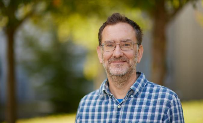
Greg Greetham
. Externally hosted profile photograph linked from the Central Laser Facility's named staff page.Clement Howe studied the melting and refolding dynamics of RNA hairpins and related DNA structures using infrared spectroscopy. His2023 University of York thesis identifies N. T. Hunt as supervisor and examines the influence of sequence and structure on these dynamics.[ref]
Howe was the first author of the2022 study combining a rapid temperature jump with a thin sample's rapid cooling to observe both melting and refolding of a UNCG tetraloop RNA hairpin and its DNA analogue.[ref]
A numerical comparison from that study separates forward and recovery dynamics. At70degrees Celsius, the reported melting times were microseconds for the RNA hairpin and microseconds for the DNA analogue, a ratio of about7.5. Their refolding occurred on similar timescales of roughly200microseconds. Similar recovery times therefore did not require similar melting times.[ref]
The thesis extends the question by changing the stem sequence and using two-dimensional infrared spectroscopy to follow relationships between vibrational modes. Sequence changes can therefore be tested through both their spectral consequences and their time-dependent structural response.[ref]
Eric Westhof is a structural biochemist whose research concerns the architecture and molecular interactions of RNA. His institutionally hosted CV identifies work at Strasbourg's IBMC and a professorship in structural biochemistry.[ref]
Eric Westhof curriculum vitae at IBMC. Professional cursus and institutional affiliation
With N. B. Leontis, Westhof developed a geometric nomenclature for RNA base-pair geometry. The classification records the interacting RNA base-pair edges and the relative orientations of the two glycosidic bonds, allowing noncanonical base pairs to be described systematically.[ref]
Geometric nomenclature and classification of RNA base pairs by N. B. Leontis and E. Westhof. Abstract, edge-based classification and cis/trans orientations
An important distinction is between two kinds of orientation. The glycosidic torsion angle describes how one base rotates relative to its own sugar, giving syn glycosidic conformation or anti glycosidic conformation. The pair's cis/trans descriptor instead relates the two glycosidic bonds across the base-pair interaction. Reporting one descriptor does not automatically report the other.
A geometric family also leaves room for different base identities. The question of RNA base-pair isostericity is whether substitutions can preserve the relevant relative placement and shape sufficiently to support a similar structural role. This makes geometry useful when comparing homologous RNA structures even when their sequences differ.[ref]
Geometric nomenclature and classification of RNA base pairs by N. B. Leontis and E. Westhof. Abstract, isosteric relationships and recurrent three-dimensional motifs
Westhof's contribution helped connect structural observations with a reusable language for comparing RNA folds, rather than restricting descriptions to the familiar sequence letters or canonical base pairs.
Neocles B. Leontis studied RNA structure and molecular recognition at Bowling Green State University. His institutional profile identifies him as a chemistry professor and biology adjunct working on nucleic acids.[ref]
Neocles Leontis lifetime-achievement profile at BGSU. Research description and professional affiliation
With E. Westhof, Leontis proposed a systematic classification of RNA base-pair geometry using three possible interacting RNA base-pair edges and two relative glycosidic-bond orientations.[ref]
A small counting argument explains the12basic families. Write for the Watson–Crick, Hoogsteen and Sugar edges. Treating interchange of the two bases as the same edge-family givesThere are6unordered choices with repetition. Each can have cis or trans relative glycosidic-bond orientation, giving . This counts geometric families, not nucleotide sequences or the number of hydrogen bonds in every individual interaction.
The classification provides a way to annotate noncanonical base pairs and compare structural motifs. The2001 paper also explains how geometric families can reveal RNA base-pair isostericity, connecting observed structures to plausible substitutions in related sequences.[ref]
Geometric nomenclature and classification of RNA base pairs by N. B. Leontis and E. Westhof. Abstract, automated annotation and isosteric relationships

Neocles Leontis
. Externally hosted photograph supplied with Bowling Green State University's named research profile.Roger M. Wartell is a biophysicist whose research connects nucleic-acid structure with physical measurements of folding and association. His Georgia Tech profile traces his training through physics degrees at Stevens and Rochester and identifies research on DNA melting, RNA-binding proteins and small RNA molecules in ice.[ref]
Roger Wartell profile at Georgia Tech. Biographical summary and research interests; linked CV, education and fields of interest
With Xueguang Sun and J. Michael Li, Wartell investigated a stable RNA hairpin that could form a metastable RNA dimer in frozen solution. Their study combined gel electrophoresis, sedimentation equilibrium and temperature-dependent absorbance to distinguish structures and their interconversion.[ref]
The scientific issue is broader than the existence of two gel bands. A band's electrophoretic mobility depends on molecular shape and experimental conditions as well as size. Independent measurements of association and thermal response help test whether the bands represent two folds of one strand or complexes containing different numbers of strands. Wartell's work illustrates how complementary physical measurements can constrain an otherwise ambiguous structural interpretation.
J. Michael Li is a coauthor, with Xueguang Sun and Roger M. Wartell, of the2007 study of frozen-solution conversion between an RNA hairpin and a metastable RNA dimer. The paper identifies the authors with Georgia Tech's School of Biology and gives Li's byline in this form.[ref]
Conversion of stable RNA hairpin to a metastable dimer in frozen solution by Xueguang Sun and others. Title-page byline and affiliation
The study examined a79-nucleotide RNA with two bands in native gel electrophoresis but one in denaturing conditions. Sedimentation equilibrium supported monomer and dimer assignments. Experiments on shorter segments implicated the first21nucleotides in dimer formation.[ref]
Conversion of stable RNA hairpin to a metastable dimer in frozen solution by Xueguang Sun and others. Abstract and Results, monomer/dimer identification and21-nucleotide segment
A useful distinction follows: two native gel bands alone do not count the number of strands in each structure. Changing the folding state can change electrophoretic mobility without changing strand mass. Combining a mobility observation with an independent association measurement addresses that ambiguity, which is central to interpreting this contribution.
Xueguang Sun studied the structure and RNA-binding properties of the bacterial protein Hfq. His Georgia Tech doctoral thesis, dated May2006, identifies Roger M. Wartell as his committee chair and documents structural analysis, binding experiments and studies of RNA association.[ref]
Characterization of E coli Hfq structure and its RNA binding properties by Xueguang Sun. Title and approval pages; chaptersII–IV
The thesis combines predicted protein structure with experiments probing RNA binding, including gel electrophoresis and fluorescence anisotropy. That combination tests a structural hypothesis against binding behavior rather than treating a predicted surface as sufficient evidence of its function.[ref]
Characterization of E coli Hfq structure and its RNA binding properties by Xueguang Sun. ChapterIII, sections on gel mobility shift, fluorescence anisotropy and mutant binding
Sun later coauthored the2007 frozen-solution study with J. Michael Li and Wartell, showing that a stable RNA hairpin could convert efficiently into a metastable RNA dimer under freezing conditions.[ref]
These projects connect molecular recognition with competing structures: a protein can present alternative binding surfaces, and an RNA strand can pair internally or associate with another strand. Experiments are needed to distinguish those possibilities; a sequence or a mobility band alone does not specify the resulting complex.
Paul Doty was a biochemist and biophysicist who studied the physical properties of proteins and nucleic acids. Harvard's memorial identifies DNA melting and reassociation of separated strands among his scientific contributions. His research helped make DNA structure accessible through measurable physical transitions rather than chemical composition alone.[ref]
Paul Doty memorial at Harvard. Departmental memorial, scientific contributions
With J. Marmur, Doty studied how DNA thermal-denaturation temperature relates to GC content. Their1962 paper followed260nm absorbance through the hyperchromic effect accompanying the transition and compared melting measurements with independently known base compositions.[ref]
An original calculation using the paper's empirical relation illustrates its quantitative purpose. Under the stated solvent condition of about0.2molar sodium ions, the reported fit iswith expressed numerically in degrees Celsius and the percentage GC content. A measured midpoint of89.8degrees Celsius corresponds toThis inference depends on the solution conditions and the empirical fit; it is not a universal temperature-to-composition conversion for every DNA sample.[ref]
Determination of the base composition of deoxyribonucleic acid from its thermal denaturation temperature by J. Marmur and P. Doty. Abstract; Results, dependence on ionic strength and base composition

Paul Doty at the Belfer Center in2005
. The Harvard Gazette identifies Doty standing beside his founder's photograph. Courtesy of the Belfer Center for Science and International Affairs.Julius Marmur was a biochemist whose work included DNA isolation, strand reassociation and DNA hybridization. His institutional biography at Albert Einstein College of Medicine identifies his collaboration with P. Doty at Harvard and his later research in yeast genetics.[ref]
Julius Marmur profile at Albert Einstein College of Medicine. About Dr.Julius Marmur
Marmur's contribution linked preparing intact DNA with testing its physical behavior. Purification makes it possible to compare changes in the nucleic acid itself rather than changes caused by a heterogeneous cell extract. DNA melting and subsequent pairing experiments then ask whether separated complementary strands can recover an associated structure. Isolation, strand separation and sequence-dependent reassociation are distinct experimental questions.
In the1962 thermal-denaturation study with Doty, the apparatus measured absorbance as a function of sample temperature. The authors used the midpoint of the hyperchromic effect and compared it with GC content determined independently. They corrected for thermal expansion of the solution, an example of separating a molecular absorption change from the changing concentration during heating.[ref]
Determination of the base composition of deoxyribonucleic acid from its thermal denaturation temperature by J. Marmur and P. Doty. Printedpp.109–110, abstract and Materials and Methods
The resulting approach made bulk optical measurements informative about DNA composition and structural transitions. It did not directly read the order of bases along a strand: composition, pairing behavior and nucleic acid sequence remain different descriptions.
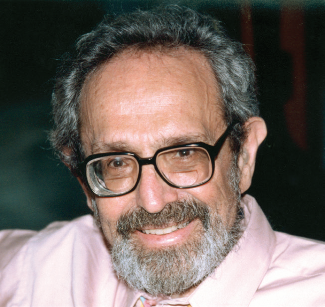
Julius Marmur
. Externally hosted portrait explicitly identified in Albert Einstein College of Medicine's institutional biography.Researcher in molecular biophysics. Hugel and collaborators tested connector rotation during DNA packaging by bacteriophage phi29, connecting a proposed mechanical model with a direct experimental observable.[ref]
Researcher in quantitative biology. With Naama Barkai, Leibler studied robustness in simple biochemical networks, connecting a system’s organization with its ability to maintain behavior despite changes in component parameters.[ref]
Robustness in simple biochemical networks by Naama Barkai and Stanislas Leibler. Author list and described study
Researcher in systems biology. With Stanislas Leibler, Barkai investigated robustness in biochemical networks, connecting the structure of regulatory interactions with reliable function under molecular variation.[ref]
Robustness in simple biochemical networks by Naama Barkai and Stanislas Leibler. Author list and described study
A physicist whose publications include the cold-atom Bloch oscillation experiment. His own publication list identifies the 1996 paper whose published byline uses Maxime Ben Dahan.
Publication list: www.phys.ens.fr/~dahan/Publications.html
Biophysicist whose work includes counting subunits of membrane-bound proteins through fluorescence measurements. With collaborators, Ulbrich connected observed signal changes with the number of labeled molecular components, making labeling efficiency part of the interpretation.[ref]
Subunit counting in membrane-bound proteins by Maximilian H. Ulbrich and Ehud Y. Isacoff. Method and interpretation
Biophysicist who used total internal reflection fluorescence microscopy to illuminate cell–surface contacts. Excitation near an interface connected optical confinement with selective observation of material close to a supporting surface.[ref]
Researcher in molecular imaging. Jungmann and collaborators used transient binding on DNA origami for single-molecule kinetics and super-resolution microscopy, connecting reversible molecular recognition with repeated localization measurements.[ref]
Researcher in cellular imaging. Manley and collaborators developed high-density mapping of single-molecule trajectories using photoactivated localization microscopy, connecting positions over time with spatially resolved molecular mobility.[ref]

Portrait of Suliana Manley
. EPFL; CC BY 4.0, via Wikimedia Commons. Source.Researcher in tumor biophysics. Stylianopoulos and collaborators investigated growth-induced solid stress in tumors, connecting mechanical compression with changes in the tissue environment and possible ways to relieve that stress.[ref]
Researcher in tissue mechanics. Mongera and collaborators investigated a fluid-to-solid jamming transition during vertebrate body-axis elongation, connecting collective physical behavior with embryonic tissue organization.[ref]
Researcher in cellular signaling. Irannejad and collaborators used conformational biosensors to investigate GPCR signaling from endosomes, connecting receptor activity with an intracellular location after internalization.[ref]
Conformational biosensors reveal GPCR signalling from endosomes by Roshanak Irannejad and others. Author list and described study
Researcher in membrane biophysics. Lorent and collaborators investigated differences between the two sides of plasma membranes in lipid unsaturation, packing and protein shape, connecting leaflet asymmetry with molecular organization.[ref]
Researcher in membrane biophysics. With Sarah L. Veatch, Keller studied liquid-phase separation in vesicles containing phospholipids and cholesterol, connecting composition with coexistence of distinct membrane regions.[ref]
Researcher in membrane biophysics. With Sarah L. Keller, Veatch investigated liquid-phase separation in giant vesicles, connecting observable membrane domains with physical interactions among lipids.[ref]
Researcher in membrane structural biology. Eicher and collaborators investigated access and deep binding pockets in AcrB, connecting a moving protein loop with the route taken by transported drugs.[ref]
Researcher in membrane structural biology. Murakami and collaborators determined multidrug-transporter structures supporting a functionally rotating mechanism, connecting different molecular conformations with stages of a transport cycle.[ref]
Researcher in cellular signaling. Saini and collaborators investigated receptor-induced movement of G-protein beta-gamma complexes and changes in Golgi organization, connecting signaling-component location with secretion.[ref]
Researcher in membrane signaling. Yen and collaborators investigated how PIP2 stabilizes active GPCR states and changes coupling selectivity, connecting membrane lipids with receptor behavior.[ref]
Researcher in molecular signaling. Mazhab-Jafari and collaborators investigated how K-Ras variants alter membrane-dependent occlusion of an effector-binding surface, connecting mutation with access to signaling partners.[ref]
Researcher in computational structural biology. Scheres developed RELION, connecting Bayesian inference with reconstruction of molecular structures from cryo-electron microscopy images.[ref]
Researcher in structural biology. Yip and collaborators investigated atomic-resolution protein structure determination by cryo-electron microscopy, connecting improved imaging performance with the visibility of fine molecular detail.[ref]
Atomic-resolution protein structure determination by cryo-EM by Ka Man Yip and others. Author list and described study
Researcher in structural biology. Nakane and collaborators demonstrated single-particle cryo-electron microscopy at atomic resolution, connecting instrumental and analytical improvements with more detailed molecular interpretation.[ref]
Single-particle cryo-EM at atomic resolution by Takanori Nakane and others. Author list and described study
Researcher in cryo-electron microscopy analysis. Zheng and collaborators developed MotionCor2 to correct beam-induced sample motion, connecting image alignment with preservation of high-resolution structural information.[ref]
Researcher in genetic recombination. Yang and collaborators investigated RecA–DNA synaptic and D-loop structures, connecting atomic organization with strand exchange between DNA molecules.[ref]
Mechanism of strand exchange from RecA–DNA synaptic and D-loop structures by Haijuan Yang and others. Author list and described study
Researcher in genetic recombination. Xu and collaborators studied human RAD51 filaments during strand exchange, connecting structures at different catalytic stages with the mechanism of homologous recombination.[ref]
Researcher in genetic recombination. Chen and collaborators investigated RecA bound to single- and double-stranded DNA, connecting molecular structures with the exchange of homologous DNA strands.[ref]
Mechanism of homologous recombination from the RecA–ssDNA/dsDNA structures by Zhucheng Chen and others. Author list and described study
Researcher in receptor biophysics. Manglik and collaborators investigated the dynamic process of beta-2-adrenergic receptor signaling, connecting structural states with transitions that a single static receptor model cannot fully describe.[ref]
Coauthor of experiments on inositol-trisphosphate effects in isolated pancreatic-cell fractions. The study connected a candidate intracellular messenger with responses in selected cellular components; the published initials are retained without an unverified identity expansion.[ref]
Researcher in microbial genetics. With Salvador Luria, Delbrück analyzed variation in virus resistance among bacterial cultures, connecting fluctuations in resistant-cell numbers with mutations arising before selection.[ref]
Mutations of Bacteria from Virus Sensitivity to Virus Resistance by Salvador Luria and Max Delbrück. Author list and described study

Portrait of Max Delbrück
. Jonathan Delbruck, source and rights-releasing party named in the File; original photographer not separately established. Public-domain release by the party identified on Commons as author/copyright holder; worldwide fallback permission for any purpose; VRT ticket 2008120310001036., via Wikimedia Commons. Source.Researcher in molecular motors. Noji and collaborators directly observed rotation of the F1 sector of ATP synthase, connecting enzymatic activity with mechanical motion at the single-molecule scale.[ref]
Direct observation of the rotation of F1-ATPase by Hiroyuki Noji and others. Author list and described study
Researcher in molecular biophysics and coauthor of direct observation of F1-ATPase rotation. The experiment connected ATP synthase activity with an observable rotary motion rather than an inferred mechanical analogy.[ref]
Direct observation of the rotation of F1-ATPase by Hiroyuki Noji and others. Author list and described study
Researcher in structural biology. Guo and collaborators determined bacterial ATP synthase structures, connecting subunit organization with the coupling of a rotary mechanism to ATP production.[ref]
Structure of a bacterial ATP synthase by Hui Guo and others. Author list and described study
Researcher in structural biology. Rubinstein coauthored studies of bacterial ATP synthase, connecting cryo-electron microscopy structures with the mechanics and regulation of a rotary energy-converting complex.[ref]
Structure of a bacterial ATP synthase by Hui Guo and others. Author list and described study
Researcher in molecular motors. Watanabe analyzed rotary catalysis of ATP synthase, connecting rotation, chemical steps and coupling between the enzyme’s membrane and soluble sectors.[ref]
Rotary catalysis of FoF1-ATP synthase by Rikiya Watanabe. Author list and described study

Rikiya Watanabe. Portrait from the institutional profile.
Researcher in ATP-synthase biophysics. Nakamoto coauthored experiments on temperature-dependent single-molecule rotation, connecting interactions between subunits with the catalytic dwell of the motor.[ref]
Articles by others on the same topic



A list of biophysicists would typically include individuals who have made significant contributions to the field of biophysics, which is an interdisciplinary science that combines principles of biology and physics to understand biological systems. While there are many notable biophysicists, here are some key figures known for their work in this field: 1. **Max Delbrück** - A pioneer in molecular biology who contributed to the understanding of bacteriophages.