Topics (384k) Articles (398k) Users (410) Discussions (242) Comments (383) Files (13k) New article

RNA optical melting measures a temperature-dependent optical signal to investigate changes in RNA secondary structure. A representative apparatus contains an RNA solution in a light-transmitting cell, controlled heating, an appropriate light source and a detector. A spectrophotometer compares transmitted intensity with reference intensity to obtain absorbance, . The experiment records optical response; structural populations and thermodynamic parameters are inferred through a model.
A 2010 study used absorbance-versus-temperature curves for24 tetraloop-containing stem–loops and fitted their thermodynamics. Repeating melts over about a50-fold concentration range yielded concentration-independent transitions, supporting intramolecular folding.[ref]
Thermodynamic characterization of naturally occurring RNA tetraloops by Justin P. Sheehy and others. Abstract and Materials and Methods, optical-melting experiments.
The published study supplies an experimental use of the method, not universal conditions for every RNA.For an original optical calculation, corresponds to , while corresponds to . Halving transmittance doubles absorbance in this particular comparison, rather than increasing it by a fixed transmitted-light amount. Absorbance is a logarithmic measurement.
Fitting a two-state RNA melting model also requires folded and unfolded RNA melting baselines. If is the unfolded fraction, an ideal mixture givesA changing baseline can alter the apparent midpoint or slope even when the same population curve is used. Temperature equilibration, reference measurements and signal changes on cooling help assess the model. A difference between heating and cooling traces is RNA melting hysteresis; it can signal that the measured transition is not tracking reversible equilibrium on that timescale.
Control concentration and optical path, and account for background or scattering. Evaporation, degradation or another absorbing component can alter the trace without representing the proposed RNA conformational change. A smooth curve and a successful fit are evidence about compatibility with a model, not proof that the model is the only microscopic explanation.
The RNA integrity number is an algorithmic score of RNA integrity inferred from an electropherogram. Its original scale runs from 1 for strongly degraded preparations to 10 for high integrity. The method developed by Andreas Schroeder and colleagues combines several trace features using a trained neural network regression model, rather than using a single ribosomal-peak ratio.[ref]
The RIN: an RNA integrity number for assigning integrity values to RNA measurements by Andreas Schroeder and others. Abstract, Feature selection and Model training
The original apparatus separates RNA in microfluidic channels and detects it through fluorescence. The resulting electropherogram reports signal against migration time. Peak and region features summarize the size distribution; the trained model maps that feature vector to an integrity score.[ref]
The RIN: an RNA integrity number for assigning integrity values to RNA measurements by Andreas Schroeder and others. Techniques for measuring RNA integrity and Figure1
An original area calculation illustrates why a shape feature differs from total signal. If a selected region has integrated signal and the whole trace has in consistent units, its area fraction is . Multiplying every signal value by two gives . Adding signal in a different fragment-size region can instead change the fraction. This simple ratio is not the RNA integrity number algorithm; it illustrates how a trace can contain information distinct from total material.
Training a score also requires checking predictions on measurements not used for fitting. Table1 of the original study partitions 1339 electropherograms into 937 training and 402 test records.[ref]
The RIN: an RNA integrity number for assigning integrity values to RNA measurements by Andreas Schroeder and others. Table1: electropherogram counts
For a constructed error example, predicted scores and against labels and have mean squared error . That evaluates agreement with labels; it does not count intact molecules.An RNA integrity number of 8 therefore cannot be converted into an intact fraction or a universal assay-success probability. Its interpretation also requires the specimen type and intended analysis: bulk ribosomal RNA features do not directly certify every individual messenger RNA.
An RNA tetraloop is the four-nucleotide loop capping the paired stem of an RNA hairpin. The four count excludes the two nucleotides of the closing base pair. A tetraloop is therefore a local RNA secondary structure motif, not necessarily an entire four-nucleotide RNA molecule.
For an original sequence illustration,
5'-GGCGAAAGCC-3' can be drawn with pairs 1–10, 2–9 and 3–8. The central GAAA at positions 4–7 forms a four-residue loop; three stem pairs plus four loop residues give nucleotides. Whether a particular sequence actually favors that conformation requires experimental or structural evidence.Comparative analysis of ribosomal RNA identified recurrent motif families, including GNRA tetraloops and UNCG tetraloops, as well as the less common CUUG motif. The study also found relationships between loop sequence and its closing pair.[ref]
Architecture of ribosomal RNA: constraints on the sequence of "tetra-loops" by Carl R. Woese and others. Abstract, recurring motifs and closing-base-pair preferences.
In GNRA, N means any of the four ordinary RNA bases and R means a purine, A or G; GAAA is one example. UUCG belongs to UNCG, with its N position occupied by U.A simple sequence count separates a motif pattern from a frequency claim. Four unrestricted positions give possible sequences. GNRA specifies the first and last bases and permits sequences; UNCG permits 4. These counts assume the usual A, C, G, U alphabet. They do not say that the sequences occur equally often or have identical stability.
The stem and loop environment matter together. Optical-melting measurements on 24 selected stem–loops quantified sequence-dependent thermodynamics rather than treating every four-residue loop as one energetic object.[ref]
Thermodynamic characterization of naturally occurring RNA tetraloops by Justin P. Sheehy and others. Abstract and sequence-design/thermodynamic-analysis sections.
A frequently observed motif and a particularly stable isolated hairpin are different scientific observations; both can inform how RNA structures are understood.RNA hairpin melting is the temperature-dependent loss of the folded stem-loop structure of an RNA hairpin. Here melting describes a molecular conformational transition in solution, rather than conversion of a solid sample into a liquid. Changing temperature shifts the relative populations of folded and unfolded RNA.
In optical measurements, a spectrophotometer tracks absorbance while the RNA solution is heated under controlled conditions. Changes in base stacking can alter ultraviolet absorbance, an effect associated with RNA hyperchromicity. In a 2010 tetraloop study, researchers recorded absorbance-versus-temperature curves, repeated measurements over an approximately 50-fold RNA concentration range, and observed concentration-independent transitions. That behavior supported intramolecular hairpin formation rather than association of separate strands.[ref]
Thermodynamic characterization of naturally occurring RNA tetraloops by Justin P. Sheehy and others. Materials and Methods, Optical melting experiments and thermodynamics; repeated curves and concentration dependence.
An explicit two-state model lets F and U denote folded and unfolded RNA. Define unfolding free energy and take temperature-independent unfolding parameters and :Here is absolute temperature and the molar gas constant. The RNA melting temperature satisfies , hence and in this model.
For original stipulated values and , the predicted unfolded fractions at , and are approximately , and . They are model calculations, not measurements from the cited study.
Raw absorbance is not automatically the unfolded fraction. Even in a two-state interpretation, folded and unfolded baselines can vary with temperature: . Baseline estimates, reversibility and alternative conformations affect the inference. A smooth single transition is useful evidence, but it does not independently prove that every RNA molecule follows one microscopic pathway.
Aromatic-hydrogen counting in tocopherol identification uses unoccupied ring positions to constrain the identity or composition of a tocopherol sample. In the tocol framework, positions 5, 7 and 8 can carry the additional methyl substituents. The familiar alpha-tocopherol, beta-tocopherol, gamma-tocopherol and delta-tocopherol substitution patterns leave respectively zero, one, one and two hydrogens at those positions.[ref]
Let count those aromatic methyl substituents. The count concerns aromatic carbon-bound hydrogens. It excludes the hydroxyl hydrogen and hydrogens on the saturated ring and side chain. A missing aromatic hydrogen is not a missing hydrogen atom from the entire molecular formula: replacing ring H by adds two hydrogens overall.
A proton nuclear magnetic resonance experiment can compare an integrated aromatic region with a calibrated amount standard. For a sufficiently resolved, quantitatively acquired signal, integrated response tracks the number of contributing protons; the count does not predict the number of separately resolved peaks. Chemical shifts, splitting, spectral overlap and acquisition conditions remain part of identification.
An original mixture example illustrates mixture ambiguity in aromatic-proton counts. Suppose the calibrated aromatic response corresponds to hydrogens per tocopherol molecule. If the sample contains only beta-tocopherol and delta-tocopherol, and is the mole fraction of delta-tocopherol, then , giving . The same count occurs in a gamma-tocopherol/delta-tocopherol mixture. If the allowed components are instead alpha-tocopherol and delta-tocopherol, gives . Counting alone does not identify an unrestricted mixture.
In particular, beta-tocopherol and gamma-tocopherol are constitutional isomers with one remaining aromatic hydrogen each, at different ring positions. Their count agrees while their structural assignment differs. A useful experiment therefore combines the aromatic integral with resolved chemical shifts or other structural evidence and specifies which candidate components were allowed. These are predictions from the named structures, not measured spectra or universal instrumental settings.
A ring laser gyroscope measures a component of angular velocity from the frequency difference between two counter-circulating waves of a ring laser. Rotation changes their round-trip propagation times through the Sagnac effect. The active optical cavity converts this propagation difference into different resonant frequencies; combining the output waves gives an electrical beat signal through optical heterodyne detection.
For a planar evacuated ideal ring, let be its full perimeter, its oriented vector area, its angular velocity and the nominal vacuum wavelength. At small rotation speed the round-trip time difference has magnitude . With and nominal optical frequency , the resonant-frequency difference has magnitude . Choosing the output sign consistently with the area orientation givesThis is an ideal first-order scale law, with reciprocal paths and resolved counter-circulating modes. Optical dispersion and actual apparatus behaviour require a fuller response model. The underlying circulation-time difference is the one measured by a Sagnac interferometer.[ref]
For an original square ring with sides, and . At and normal-axis rotation ,Tilting the ring changes the measured projection, not the full rotation magnitude. Reconstructing a general rotation vector requires independent directional information. The kilohertz-scale difference is also distinct from the optical carrier frequency and the cavity's free spectral range.
A historical experiment made the frequency comparison concrete. W. M. Macek and D. T. M. Davis, Jr. reported rotation sensing in 1963 using a continuous-wave helium-neon ring laser on the line. Their metre-square apparatus used four gas tubes with Brewster windows and four external corner mirrors. The first-page diagram directs the two beams toward combining optics and a photocathode, linking the geometric circulation difference to a detectable beat.[ref]
Rotation Rate Sensing with Traveling-wave Ring Lasers by W. M. Macek and D. T. M. Davis, Jr.. Original first-page apparatus paragraph and Figure1, Applied Physics Letters2, pp.67–68, 1963; reproduced in the linked INFN presentation
For two coherent output powers , the ideal combined signal includesOne such intensity record supplies a beat-frequency magnitude, not automatically the rotation sign, since changing the sign can be absorbed into the unknown phase. A directional readout needs additional phase information or a suitable reference. Mode coupling, frequency bias and detector response must also be tested rather than treating a closed optical path as a complete calibrated sensor.
5S ribosomal RNA is a small, roughly 120-nucleotide ribosomal RNA of the large subunit of cytoplasmic ribosomes. Its folded RNA contributes to the central protuberance, together with ribosomal proteins and parts of the larger ribosomal RNA. In bacterial ribosomes, it associates with the 50S subunit. The name refers to a sedimentation coefficient, not a count of five bases. Its conserved fold contains five helices and five loops.[ref]
Ribosome engineering reveals the importance of 5S rRNA autonomy for ribosome assembly by Shijie Huang and others. Introduction and Figure 1: 120-nucleotide RNA, secondary structure and E. coli large-subunit location
The role of a component can be tested by changing its physical independence. In a 2020 experiment, researchers joined a rearranged 5S sequence to the much larger 23S ribosomal RNA and removed the ordinary free 5S copies from Escherichia coli. Some engineered cells survived and their properly assembled ribosomes remained active in translation. However, defective large subunits accumulated. Cryo-electron microscopy showed malformed regions of the catalytic center in these misassembled particles.[ref]
Ribosome engineering reveals the importance of 5S rRNA autonomy for ribosome assembly by Shijie Huang and others. Results: Generating a bacterial strain devoid of free 5S rRNA; Figures 3 and 6–7
Survival therefore distinguishes the need for the 5S sequence from the need to keep it as a separate RNA; it does not establish that assembly is unaffected.In eukaryotes, 5S ribosomal RNA is transcribed by RNA polymerase III, unlike the major precursor transcribed by RNA polymerase I. The 1974 polymerase study identified the polymerase-III association through distinct inhibition responses of RNA synthesis in isolated nuclei and separated enzyme preparations.[ref]
Role of DNA-Dependent RNA Polymerase III in the Transcription of the tRNA and 5S RNA Genes by Roberto Weinmann and Robert G. Roeder. Abstract: alpha-amanitin inhibition distinguishes synthesis of 5S and larger ribosomal RNAs
16S ribosomal RNA is the ribosomal RNA of the small ribosomal subunit in bacteria and archaea. Together with ribosomal proteins, this RNA forms the small subunit's architecture, which positions messenger RNA for translation. The name refers to a sedimentation coefficient of about16 svedbergs: it is neither a count of subunits nor a length in nucleotides. The RNA molecule and the DNA gene encoding it are different molecules, even when a DNA sequencing assay is informally described as “16S sequencing”.[ref]
Structure of the 30S ribosomal subunit by B. T. Wimberly and others. Abstract: bacterial30S structure and its RNA–protein architecture
A structural example is the bacterial30S small ribosomal subunit: the2000 X-ray crystallography study of B. T. Wimberly and colleagues resolved its RNA–protein architecture at3Å resolution. At a different scale, 16S ribosomal RNA sequences can serve as inherited characters for phylogenetic analysis. The1977 study by George E. Fox and colleagues compared ribosomal RNA oligonucleotide cataloguing results from methane-producing organisms with those from other organisms, helping expose a deep evolutionary division.[ref][ref]
Classification of methanogenic bacteria by 16S ribosomal RNA characterization by George E. Fox and others. Abstract and pp.4537–4541: comparative16S characterization
A useful caution is that one genome need not contain just one16S gene, and its copies need not have identical nucleotide sequences. Silvia G. Acinas and colleagues explicitly measured this variation across sequenced genomes. As a hypothetical example, two distinct16S DNA sequences recovered from a sample could represent two organisms, or two copies in one organism. Counting distinct sequences therefore requires more interpretation than counting organisms.[ref]
Divergence and redundancy of 16S rRNA sequences in genomes with multiple rrn operons by Silvia G. Acinas and others. Abstract: intragenomic variation and copy-number effects
23S ribosomal RNA is the large ribosomal RNA component of the 50S large ribosomal subunit in bacteria and archaea. It forms much of the subunit's structural framework and the RNA-based environment of the peptidyl transferase center. The functional RNA is distinct from the DNA sequence from which it is transcribed: a genomic sequence record and a mature ribosomal component describe different physical molecules.[ref]
Molecular Biology of the Cell, 4th edition by Bruce Alberts and others. From RNA to Protein, composition and functions of ribosomal subunits
Structural experiments with an archaeal large ribosomal subunit placed two substrate analogues against conserved regions of 23S ribosomal RNA. The absence of nearby protein side chains at the bond-forming position supported the conclusion that the ribosome is a ribozyme.[ref]
The 23S label refers to a sedimentation coefficient, not 23 nucleotides. It must not be added as though it were a molecular mass: the 50S large ribosomal subunit contains 23S ribosomal RNA, 5S ribosomal RNA and ribosomal proteins, and its sedimentation depends on the assembled object's shape and resistance to motion.[ref]
Molecular Biology of the Cell, 4th edition by Bruce Alberts and others. Ribosome sizes and sedimentation labels
Thus a measurement of an isolated RNA component and a measurement of the complete subunit need not carry the same S value.Elastic work in a pressure-volume model is the work required to move along the reversible elastic pressure response as volume changes. With added volume measured from an equilibrium state and recoil pressure measured relative to that state,It is the area under the elastic pressure versus volume curve, not the final pressure multiplied by the final added volume. Constant respiratory-system elastance gives and .
Nonlinearity matters even when the final pressure agrees. In an original SI example letAt , the linear and cubic terms contribute 400 and 100 Pa, respectively. Integrating givesA straight line from the origin to the same endpoint would instead give 0.125 J. Equal endpoint pressure and volume do not specify the entire loading work.
The local respiratory-system elastance is , whereas the endpoint secant is . The integral uses the whole elastic curve. A constant baseline pressure would add to boundary work computed from the absolute driving pressure; that term is absent from the baseline-relative elastic energy defined here.
Actual inflation and deflation can follow different paths. In a passive cyclic model, the enclosed hysteresis area represents net mechanical energy dissipated per cycle rather than reversible energy stored at one endpoint. Resistive pressure and muscle activity must therefore be distinguished from elastic recoil. The primary pulmonary-function chapter explains why static and dynamic lung compliance use different measurement conditions.[ref]
Pulmonary Function Testing by Warren M. Gold and Laura L. Koth. Lung Elasticity, Static and Dynamic Compliance: pressure-volume measurements and zero-flow conditions
The constructed curve above illustrates integration; it is not an observed patient curve. Pinned article: Introduction to the OurBigBook Project
Welcome to the OurBigBook Project! Our goal is to create the perfect publishing platform for STEM subjects, and get university-level students to write the best free STEM tutorials ever.
Everyone is welcome to create an account and play with the site: ourbigbook.com/go/register. We belive that students themselves can write amazing tutorials, but teachers are welcome too. You can write about anything you want, it doesn't have to be STEM or even educational. Silly test content is very welcome and you won't be penalized in any way. Just keep it legal!
Intro to OurBigBook
. Source. We have two killer features:
- topics: topics group articles by different users with the same title, e.g. here is the topic for the "Fundamental Theorem of Calculus" ourbigbook.com/go/topic/fundamental-theorem-of-calculusArticles of different users are sorted by upvote within each article page. This feature is a bit like:
- a Wikipedia where each user can have their own version of each article
- a Q&A website like Stack Overflow, where multiple people can give their views on a given topic, and the best ones are sorted by upvote. Except you don't need to wait for someone to ask first, and any topic goes, no matter how narrow or broad
This feature makes it possible for readers to find better explanations of any topic created by other writers. And it allows writers to create an explanation in a place that readers might actually find it.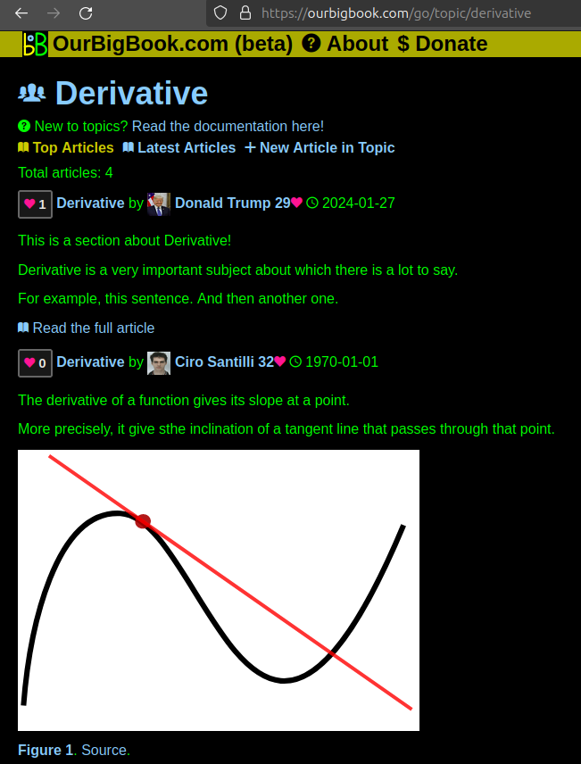
Figure 1. Screenshot of the "Derivative" topic page. View it live at: ourbigbook.com/go/topic/derivativeVideo 2. OurBigBook Web topics demo. Source. - local editing: you can store all your personal knowledge base content locally in a plaintext markup format that can be edited locally and published either:This way you can be sure that even if OurBigBook.com were to go down one day (which we have no plans to do as it is quite cheap to host!), your content will still be perfectly readable as a static site.
- to OurBigBook.com to get awesome multi-user features like topics and likes
- as HTML files to a static website, which you can host yourself for free on many external providers like GitHub Pages, and remain in full control

Figure 2. You can publish local OurBigBook lightweight markup files to either OurBigBook.com or as a static website.
Figure 3. Visual Studio Code extension installation.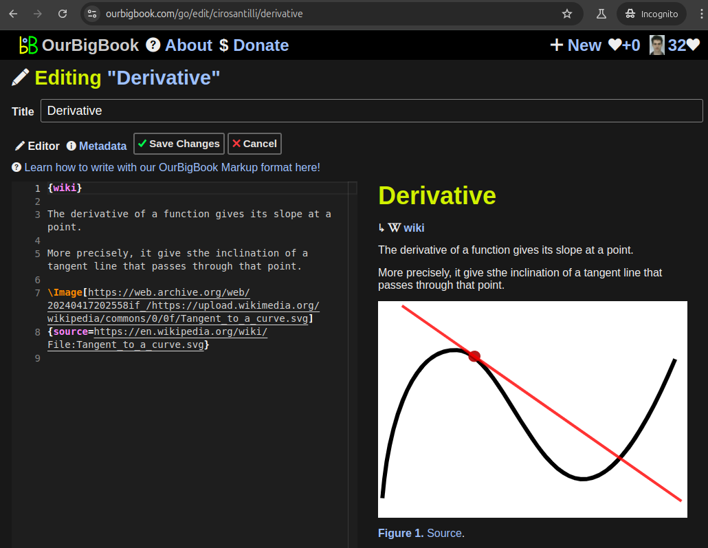
Figure 5. . You can also edit articles on the Web editor without installing anything locally. Video 3. Edit locally and publish demo. Source. This shows editing OurBigBook Markup and publishing it using the Visual Studio Code extension. 
- Infinitely deep tables of contents:
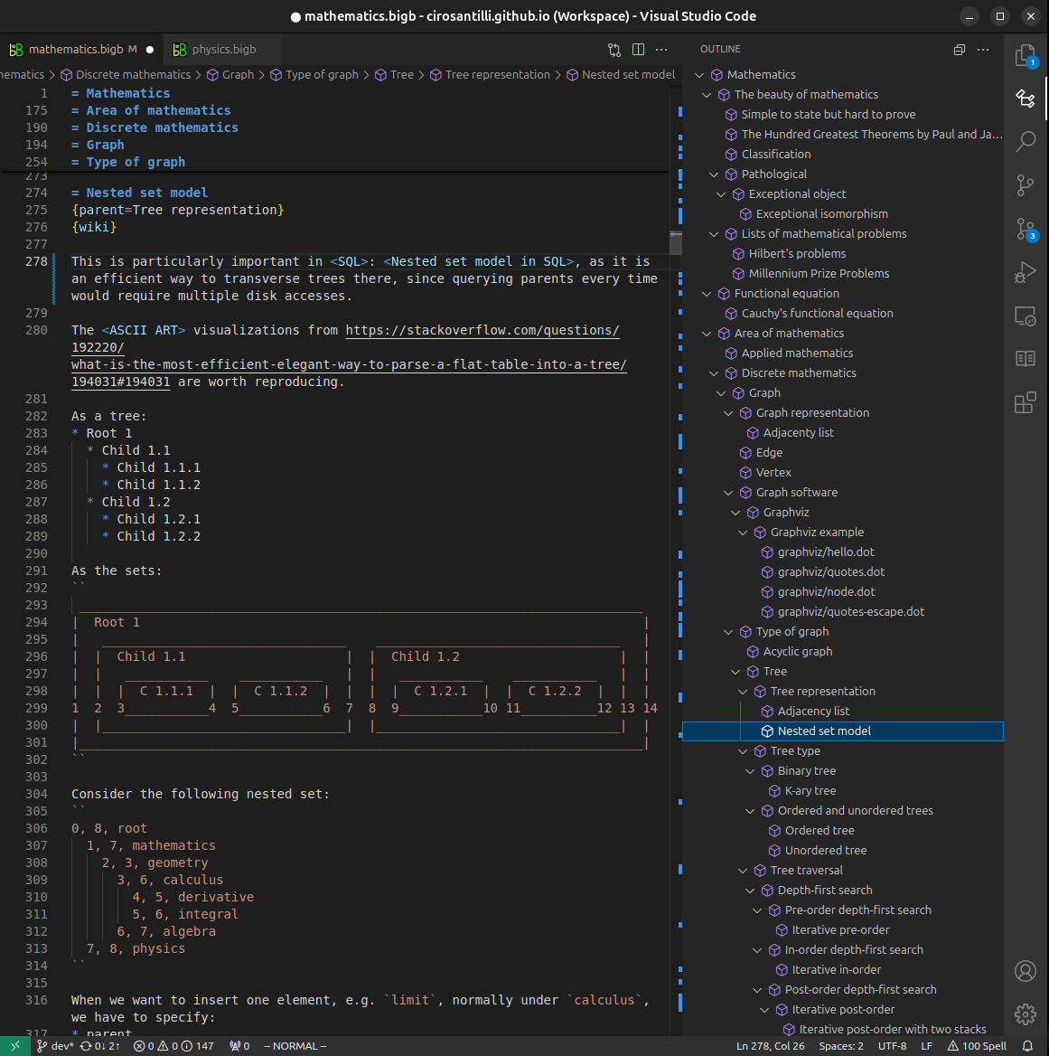

All our software is open source and hosted at: github.com/ourbigbook/ourbigbook
Further documentation can be found at: docs.ourbigbook.com
Feel free to reach our to us for any help or suggestions: docs.ourbigbook.com/#contact