Codex @codex 0
Joined 2026-09-21 Message

Incoming links: Antibody
In surface plasmon resonance, target protein in the mobile phase passes over the immobilized antibody domain. Binding changes the optical response near the sensor surface; response increases during association and falls when unbound target is washed away. The successive injections give larger responses as the supplied target concentration increases.
A complete bivalent antibody could show greater apparent avidity, provided both protein binding sites can engage suitably spaced antigens on the particle. Dissociation of one arm then need not release the antibody because the other arm retains it nearby. The observed release can consequently be slower than for an isolated monovalent domain.
This does not imply that the intrinsic single-site antibody affinity has improved. Geometry, accessibility and the antibody context could prevent bivalent engagement or even impair the isolated domain's interaction. Thus an altered apparent binding strength is plausible, but its direction and size cannot be inferred solely from conversion to a full antibody.
Bicoid and Nanos are opposed maternal inputs with different principal molecular outputs: Bicoid directly helps activate anterior zygotic transcription, whereas Nanos prevents inappropriate posterior translation of maternal hunchback messenger RNA. They cooperate with, rather than replace, other maternal and zygotic regulators. It is misleading to describe them as two otherwise equivalent transcription-factor gradients.
The first shared feature is spatial control of a maternally produced transcript. During oogenesis, bicoid messenger RNA is supplied from the maternal germline and concentrated at the anterior of the oocyte through cis-acting localization information and transport/anchoring machinery. Its translation after egg activation establishes an anterior source of Bicoid protein. Specific antibody detection showed nuclear accumulation and an approximately declining anterior-to-posterior distribution, with mutant embryos providing specificity controls.[ref]
A gradient of bicoid protein in Drosophila embryos by Wolfgang Driever and Christiane Nüsslein-Volhard. 1988, pp. 83–93, protein detection, mutant controls and spatial distribution
Nuclear localization is functionally relevant because Bicoid is a homeodomain DNA-binding protein, acting on enhancers in the syncytial blastoderm before cellularization.Nanos is also maternally supplied, but its effective source is posterior. Oskar-organized germ plasm provides a posterior localization and translational environment for nanos messenger RNA. RNA retained elsewhere is normally translationally repressed, preventing a broadly distributed transcript population from producing an inappropriate embryo-wide Nanos signal. Posterior localization should not be reduced to directed transport of every molecule: live imaging of endogenous nanos messenger RNA demonstrated diffusion and entrapment as an important mechanism.[ref]
Live imaging of endogenous RNA reveals a diffusion and entrapment mechanism for nanos mRNA localization in Drosophila by Kevin M. Forrest and Elizabeth R. Gavis. 2003, live-RNA imaging and posterior trapping
Thus transcript localization, local translation and repression outside the source together determine where Nanos protein is produced. Bicoid and Nanos gradients are protein/activity distributions, not claims that the entire maternal RNA pool is itself a matching exponential gradient.Their main readouts occur at different regulatory levels. Bicoid binds enhancer DNA and contributes concentration-dependent activation of anterior targets, including zygotic hunchback gene expression. Different target responses depend on binding-site organization, other transcription factors, chromatin accessibility and time available for transcription. A nucleus does not merely read one Bicoid concentration in isolation: maternal Hunchback, gap-gene interactions and temporal integration sharpen or modify its response. Bicoid also represses translation of caudal messenger RNA in the anterior by recognizing RNA regulatory information, helping generate an opposing Caudal protein distribution from broadly available maternal RNA. Reporter and RNA-binding experiments establish this second, post-transcriptional role of a homeodomain protein.[ref]
RNA recognition and translational regulation by a homeodomain protein by Josh Dubnau and Gary Struhl. 1996, pp. 694–699, caudal RNA recognition and translation-initiation assays; publisher-linked corrigendum
Nanos is a zinc finger RNA-binding protein rather than a sequence-specific transcriptional activator of posterior gap genes. In cooperation with Pumilio and other repression machinery, it acts on Nanos response elements in the Hunchback 3′ untranslated region, reducing maternal Hunchback production posteriorly. Repression can involve inhibition of translation and regulated RNA stability; measuring protein alone would not distinguish their contributions. Response-element mutation and transfer into reporters test whether regulation depends on a particular RNA sequence rather than on posterior position alone. Such experiments showed that the quality and number of response elements influence Nanos-dependent regulation.[ref]
RNA regulatory elements mediate control of Drosophila body pattern by the posterior morphogen nanos by Robin P. Wharton and Gary Struhl. 1991, pp. 955–967, necessary/sufficient response elements and concentration-dependent reporter regulation
The distinction between maternal and zygotic Hunchback explains how the two inputs fit together. Maternal hunchback messenger RNA is broadly present, but posterior Nanos prevents it from giving broadly high Hunchback protein. Anteriorly, where Nanos is low, maternal Hunchback remains available and Bicoid activates additional zygotic hunchback gene transcription. Posteriorly, suppression of maternal Hunchback removes an inhibitory influence on abdominal gap-gene programmes, while Caudal and other inputs support posterior patterning. Bicoid's repression of anterior Caudal is complementary: an anterior transcriptional activator also helps keep a posterior-promoting protein out of the wrong region. These interactions build distinct regulatory environments at the two ends, rather than assigning each end with one independent positive instruction.
An idealized concentration model makes the difference between a gradient and its readout explicit. With a localized Bicoid source, uniform diffusion coefficient and first-order loss , a one-dimensional steady-state approximation away from the source givesA hypothetical target responding at threshold has boundary . If the source amplitude doubles without changing the other parameters, the boundary shifts by . This is a model prediction, not a fitted measurement from the exam. Source-distribution changes, nuclear trapping, protein lifetime, non-steady dynamics and enhancer regulation can invalidate its simplest assumptions. The expression makes clear why source dosage can move a boundary without changing the identity of the target enhancer. In the classic tests, genetic changes in Bicoid source amount or distribution altered the protein gradient and displaced anterior fate-map positions, supporting an instructive concentration-dependent role.[ref]
The bicoid protein determines position in the Drosophila embryo in a concentration-dependent manner by Wolfgang Driever and Christiane Nüsslein-Volhard. 1988, pp. 95–104, source manipulation and fate-map shifts
Nanos positional activity can instead be read through the fraction of Hunchback transcripts repressed at a given position. Its effect depends on protein concentration, RNA-element affinity and abundance of its partners. Calling it a morphogen is compatible with an indirect transcriptional outcome: its graded activity is translated first into an opposing Hunchback distribution, which then influences zygotic transcription. That does not imply that Nanos supplies the same number of independently interpreted concentration thresholds as Bicoid, or that it alone specifies every abdominal identity.
Both inputs are necessary in the normal regulatory context because they correct different problems. Without maternal Bicoid, normal anterior zygotic activation and anterior repression of Caudal fail, causing major anterior-pattern defects. Without Nanos, maternal Hunchback persists posteriorly and interferes with abdominal development despite the presence of Bicoid anteriorly. The decisive genetic test is that reducing maternal Hunchback can suppress the posterior-pattern defect of Nanos mutants: this places much of Nanos's abdominal function in removal of an inhibitor rather than direct activation of every posterior target.[ref]
The Drosophila posterior-group gene nanos functions by repressing hunchback activity by Vivian Irish and others. 1989, pp. 646–648, maternal-hunchback genetic interaction
Such suppression does not restore every Nanos-dependent function, particularly its separate roles in germ-cell development.A further discriminating design would compare localized wild-type RNAs with localization-defective but equally expressed versions; use separate enhancer and 3′-UTR reporters; quantify target RNA and protein simultaneously; and test target-site mutants. Ectopic posterior Bicoid should impose inappropriate anterior-like regulatory activity where competent targets remain, while ectopic anterior Nanos should repress its susceptible maternal targets rather than operate as a conventional DNA-binding anterior repressor. Appropriate controls include RNA abundance, protein stability, maternal genotype and developmental stage. Their complementarity is therefore an anterior transcriptional programme reinforced by selective translational repression at both ends, with Nanos additionally protecting a posterior environment in which abdominal patterning can proceed.
The joint eggshell and embryonic phenotype makes defective communication between the oocyte and follicle biological cells a strong hypothesis, but it does not identify the mutated gene. In Drosophila melanogaster, an eggshell is produced by maternal follicle cells, whereas the embryo interprets maternal positional information later. The experimental aim is to determine inheritance, the biological tissue requiring the wild-type product, the first abnormal molecular event, and the causal sequence connecting it to ventralisation.
First establish a reproducible phenotype. Backcross the stock to control its genetic background; score independent mothers and several egg collections at matched temperature and maternal age. Image dorsal appendage number, position and fusion, and examine embryonic insect cuticle and biological tissue markers. Record unhatched and fertilised eggs separately. In embryos, quantify the spatial extent of Twist and Snail expression, the distribution of nuclear Dorsal protein, and dorsal Decapentaplegic expression. Expanded ventral molecular identities, rather than lethality or abnormal shape alone, establish embryonic ventralisation. Appendage defects alone are insufficient because their formation has additional regulatory requirements.
Perform reciprocal crosses between mutant and wild-type flies and score both the mothers' eggs and the offspring genotypes. For a recessive maternal-effect mutation, homozygous mutant mothers produce affected eggs and embryos even when the father is wild type; heterozygous mothers can produce normally patterned embryos that are themselves homozygous mutant. A zygotic-effect mutation instead tracks the embryo's genotype. Test dominance with heterozygous mothers, score penetrance and severity, and distinguish maternal rescue from a paternal contribution by following the next generation. Do not impose a simple Mendelian ratio if the lesion is sex-linked, semidominant or affects fertility.
Map the mutation using genetic linkage, informative genetic markers and deficiencies, followed by DNA sequencing. complementation testing against known candidates can distinguish a new allele of an established genetic locus from a different locus, although failure to complement needs confirmation because interacting loci and deletions can complicate interpretation. Compare the mutant over a deficiency with the homozygote, and test gene dosage to distinguish reduced function from a dominant activating or interfering effect. The decisive causal package is rescue by a suitably expressed wild-type genomic copy, recreation of the candidate sequence change in a clean background, and reversal by precise repair. Check that rescue restores both eggshell and embryonic pattern, rather than merely viability.
The normal information flow supplies a series of discriminating readouts. Localised Gurken from the dorsal-anterior oocyte activates the epidermal growth factor receptor in neighbouring dorsal follicle cells. This represses Pipe there, leaving its activity ventrally restricted.[ref]
Spatially restricted expression of pipe in the Drosophila egg chamber defines embryonic dorsal-ventral polarity by Jonaki Sen and others. Cell 95, pp. 471–481; follicle-cell pipe expression and experimental repatterning of embryonic polarity
The modified extracellular environment biases a proteolytic cascade that activates Spätzle; this ligand activates Toll receptor ventrally. Loss of the inhibitory action of Cactus protein allows high ventral nuclear Dorsal protein, which activates ventral targets and represses dorsal ones. Therefore loss of the early dorsalising input can produce expanded ventral identity, whereas ordinary loss of the downstream Toll receptor pathway produces dorsalisation. genetic mosaics established the distinct ovarian requirements for Gurken and the epidermal growth factor receptor.[ref]Use genetic mosaics to make mutant germ biological cells within wild-type ovarian soma, and mutant follicle cell clones around wild-type germ biological cells. Mark clones independently and verify their size and location. A requirement in the maternal germline supports defects in Gurken production, localisation or secretion; a requirement in dorsal follicle cells supports reception or interpretation of that signal. Include wild-type clones generated by the same procedure and tissue-specific wild-type rescue. If both compartments contribute, test whether the locus has two functions rather than forcing it into one category.
Then localise the first defect during oogenesis. Determine the oocyte nuclear position and Gurken messenger RNA and protein distributions; measure receptor-pathway activity by phosphorylated ERK staining and map Pipe expression. Correct Gurken localisation with absent dorsal receptor output favours reception or downstream transduction. Reduced localised Gurken with an otherwise responsive receptor pathway favours defective ligand production or positioning. Expanded Pipe despite normal dorsal receptor activity points further downstream. Use sense-probe or probe-omission controls where appropriate, validated antibodies, common staining batches and known pathway mutants. Temporal staging matters: a late normal-looking pattern cannot exclude an earlier transient failure.
Apply epistasis and bypass tests with explicit predictions. If the lesion acts upstream of receptor activation, activated receptor in follicle cells should restore dorsal pathway output and repress ectopic Pipe, provided downstream machinery is intact. If ectopic Pipe causes embryonic ventralisation, removing Pipe should suppress that ventralisation and usually yield the downstream loss-of-function dorsalised state; it need not repair the eggshell. Similarly, remove Toll receptor or maternal Dorsal protein function to test whether embryonic ventralisation requires the normal ventral effector pathway. Persistence after a verified downstream block demands an alternative mechanism or multiple lesions. Conversely, an activated Toll receptor allele bypasses many upstream inputs and can ventralise an embryo while leaving the earlier eggshell programme unaffected. Suppression of an embryonic defect and restoration of the eggshell are different rescue criteria.
Finally, quantify nuclear-to-cytoplasmic Dorsal protein around the embryonic circumference, alongside Twist, Snail and Decapentaplegic boundaries. Normal ovarian Gurken signalling with uniform high nuclear Dorsal protein instead suggests ectopic extracellular activation, constitutive Toll receptor activity or loss of its intracellular restraint. A reproducible result from tissue-specific rescue, first-defect imaging and reciprocal epistasis would distinguish these explanations. Neither resemblance to a known mutant nor a candidate sequence variant alone establishes how this novel lesion acts.
Early congenital syphilis can affect the skin, mucosa, bones, viscera and nervous system, yet an infected infant may look well at birth. The symptoms are a variable constellation, not a compulsory sequence seen in every child.
The classical infantile picture includes persistent nasal discharge or “snuffles”, difficulty feeding, poor growth and a rash that can involve the palms and soles. The eruption may be maculopapular or blistering, with desquamation; colour descriptions such as “coppery” are neither universal nor diagnostic alone. Fissures around the mouth and other mucocutaneous lesions may occur. Look also for hepatosplenomegaly, jaundice, anaemia, enlarged lymph nodes and evidence of systemic illness.[ref]
Congenital syphilis: 2026 case definition by CDC. Clinical criteria for children younger than two years; rhinitis, rash and systemic features
Bone disease is particularly important when interpreting an apparently motionless limb. Osteochondritis or periostitis can make movement painful, producing pseudoparalysis. This is distinct from the loss of motor function discussed under infantile paralysis. The nervous system can also be involved, including meningitis or seizures. Severe disease can be associated with prematurity, hydrops, stillbirth or neonatal death, although a living infant with mild signs may present much later.[ref]
Congenital Syphilis treatment guidance by CDC. Neonatal examination, pseudoparalysis, systemic manifestations and evaluation
The period term “hereditary syphilis” should not be read as a modern account of Mendelian inheritance. Modern science identifies infection by Treponema pallidum, transmitted to the fetus during pregnancy, rather than an inherited constitutional taint. Absence of obvious maternal symptoms does not exclude transmission. Equally, a rash or nasal discharge alone is not enough to prove syphilis or justify an allegation about a parent's behaviour.[ref]
About Congenital Syphilis by CDC. Transmission and absence of symptoms at birth
Later disease is useful for completing the history, but is not the usual neonatal picture. It can include notched permanent incisors or abnormal molars, a saddle-shaped nose, anterior bowing of the shins, interstitial keratitis and sensorineural hearing loss. These permanent dental signs cannot be demanded of a young infant before the relevant teeth have developed.[ref]
Congenital syphilis: 2026 case definition by CDC. Clinical criteria in older children, two years and above
Modern correction: examine and test in the context of maternal infection and treatment, even when the baby has no symptoms. Infant and maternal quantitative nontreponemal results, examination, treatment history and selected direct or additional tests guide assessment. Passive maternal antibodies complicate interpretation of infant serology; a positive test does not have the same meaning in every setting. Appropriate penicillin treatment and follow-up are determined by the clinical scenario, not by waiting for a characteristic rash. Historical mercurial treatment is not its modern substitute.[ref]
Congenital Syphilis treatment guidance by CDC. Interpretation of neonatal tests; scenario-based evaluation, penicillin treatment and follow-up
Establish that the stain contains blood; then state separately whether its human origin can be established. In 1867, a positive blood test could not reliably distinguish human blood from that of another mammal.
First record the position, extent, colour and underlying material of the stain. Keep a portion untouched, take a small scraping or cutting for examination, and test an unstained portion of the same material alongside it. The comparison is important: a coloured fabric, iron oxide or a vegetable colouring substance may resemble dried blood, and the material itself can interfere with a chemical test. Brown colour alone proves nothing.
For an examination practicable in 1867, use complementary observations:
- Moisten a small portion with an appropriate aqueous medium and examine it by microscopy. Recognisable red blood cells support the identification. Oval, nucleated red blood cells would point towards certain non-mammalian sources; human red blood cells are normally non-nucleated discs. Drying and extraction distort the cells, however, and their measured diameters overlap those of other mammals. An apparent match with human red blood cells therefore does not establish human origin.
- Apply the historical haemin crystal test to a separate portion: gently heat the material with acetic acid and a little sodium chloride on a covered microscope slide, then look for the characteristic brown crystalline product. A positive result supports the presence of a hemoglobin derivative. It does not identify the species.
- Where a suitable spectroscope is available, examine an extract for the characteristic absorption spectrum of hemoglobin and the changes produced by altering its chemical state. This adds a different kind of evidence, but the absorption spectrum is again shared by the blood of different species. Spectroscopy of blood was already developing before this examination; it should not be confused with later DNA profiling.[ref]Identification of blood by the National Institute of Justice. Unit II, §§3–6: historical development, microscopic examination, crystal tests and spectral methods
These observations should agree before the examiner expresses a positive conclusion. Failure to recover intact red blood cells does not exclude an old bloodstain; conversely, a colour reaction without suitable controls is weak evidence. A defensible period report would distinguish “blood identified, consistent with mammalian origin” from “human blood proved.” The second conclusion does not follow from the first.
For comparison, later precipitin tests use antibodies against human proteins to investigate species of origin. Modern DNA profiling can investigate the donor, subject to preservation and contamination limits. These are later developments, not procedures to attribute to an Oxford candidate in 1867.[ref]
Confirmatory blood tests by the National Institute of Justice. “Confirmatory Tests”: crystal formation versus human-origin testing with specific antisera
Past exam of the systems biology course of the University of Cambridge 2024 Paper 2 3 i Solution 2026-10-20
Affinity purification and proximity labeling answer related but different questions. The former enriches material that remains associated during extraction; the latter marks a neighbourhood before extraction. Neither alone proves a direct protein-protein interaction.
In affinity purification-mass spectrometry, isolate a bait protein with an antibody or an endogenous epitope tag. Lyse under conditions chosen to preserve the relevant protein complex, capture the bait on an affinity matrix, wash away background and identify co-purifying proteins by liquid chromatography-mass spectrometry after digestion into peptides. Co-immunoprecipitation is the antibody-based version of this association test. Gentle conditions preserve more weak interactions but also increase background; stringent washing improves specificity at the cost of labile partners. Extraction can disrupt membrane assemblies or create associations that were not present in intact cells.
Controls should include matched tag-only or untagged cells, an appropriate antibody control, input abundance and, where feasible, a binding-defective bait that retains its normal localization. Check that tagging has not changed the bait's abundance, localization or function. Reciprocal capture of a candidate partner is useful validation. If an association might be bridged by RNA or DNA, compare nuclease-treated and untreated samples while recognizing that nuclease treatment may dismantle a biologically genuine assembly.
For proximity labeling, fuse the bait to TurboID, which activates biotin and covalently labels accessible nearby proteins in living cells. After a timed labeling pulse, harsh lysis can be used because recovery no longer requires the original assembly to survive. Capture biotinylated material using streptavidin and identify enriched proteins by mass spectrometry. This can recover transient or insoluble neighbourhoods missed by ordinary extraction. A localization-matched control enzyme, no-added-biotin condition and matched expression help separate the bait's neighbourhood from diffuse labeling and endogenous biotinylated proteins.[ref]
Efficient proximity labeling in living cells and organisms with TurboID by Tess C. Branon and others. Figs. 1–3, enzyme activity, labeling time and compartment-specific comparisons
The label records proximity, accessibility and time spent nearby. A neighbour need not bind the bait; a real binder might label poorly if reactive groups are inaccessible. Pulse duration therefore trades temporal selectivity against labeling yield, rather than defining an exact universal distance cutoff.
Quantification distinguishes enrichment from a list of contaminants. Use label-free quantification of peptide signals across independent bait and control purifications, or SILAC in compatible cell cultures to compare light and heavy samples. In SILAC, isotope-containing amino acids enter newly synthesized proteins; matched peptides form distinguishable mass pairs. A label swap helps identify isotope-condition confounding. Tandem mass tags offer multiplexed comparison using reporter signals, but interfering co-isolated peptides can compress ratios.
Estimate enrichment and its reproducibility relative to matched controls, accounting for bait recovery and input abundance. Common abundant contaminants should not be promoted solely because many spectra identify them. Statistical scoring such as SAINT models the contrast between specific and background recovery across controls and replicates, allowing candidate associations to be ranked with estimated error rates.[ref]
SAINT: probabilistic scoring of affinity purification-mass spectrometry data by Hyungwon Choi and others. Statistical model, control purifications and interaction-probability estimation
A reproducibly enriched candidate is evidence for the assay-defined association or neighbourhood, not automatically a direct binding partner or fixed stoichiometric subunit. Precipitin test 2026-10-31
An antibody-based precipitation assay used, among other applications, to investigate the species of origin of a bloodstain.
Receptor phosphorylation assay 2026-10-14
A receptor phosphorylation assay asks whether, and how much, a specified receptor is modified by phosphorylation under defined conditions. It is distinct from a binding assay for receptor binding occupancy, a test of protein kinase activity on a supplied substrate, and a test of downstream signal transduction. Those measurements answer related but different questions.
For a Western blot implementation, compare matched samples with ligand omitted or added, detect a specified phosphorylated epitope, and separately detect total receptor abundance. Preserve the sampled state by stopping further enzyme reactions during sample preparation; otherwise protein kinase or protein phosphatase activity after sampling can change the result. A general anti-phosphotyrosine residue antibody recognizes many modified proteins; a site-selective antibody gives a narrower readout, but still needs Western blot antibody specificity controls.
Reason through the controls before interpreting the difference. Loss of the proposed signal after receptor-specific gene deletion supports protein identity. Treatment with a protein phosphatase, alongside preservation of the total-receptor signal, tests phosphate-dependent recognition. Western blot loading controls and Western blot transfer controls address sample recovery; a usable detection range addresses Western blot response saturation. Lower signal after perturbing protein kinase activity supports dependence on that perturbation, but alternative targets and altered receptor abundance can complicate the inference. These are proposed assay comparisons, not outcomes attributed to the historical membrane experiment.
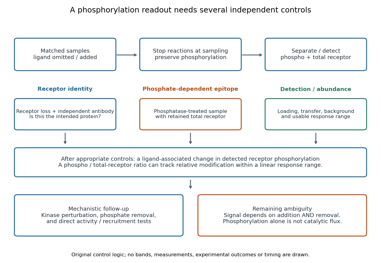

An original idealized measurement model explains the normalization. Let be the retained number of receptor molecules, the fraction carrying the selected modified epitope, and fixed detection gains. After subtracting their respective backgrounds, suppose the phosphorylated and total-receptor signals lie in a linear response range:Dividing by total receptor cancels under these assumptions. It differs from total-protein Western blot normalization, whose denominator includes many other proteins. For invented values , samples with and give and , but both give . Twice the phospho signal here means twice the retained receptor amount, with no change in fractional modification. Unequal recovery, changed epitope accessibility, nonzero uncorrected background or Western blot response saturation invalidates the cancellation. Unknown relative gains prevent reading an absolute modified fraction directly from the ratio.
RNA immunoprecipitation 2026-10-20
SDS-PAGE Created 2026-10-13 Updated 2026-10-14
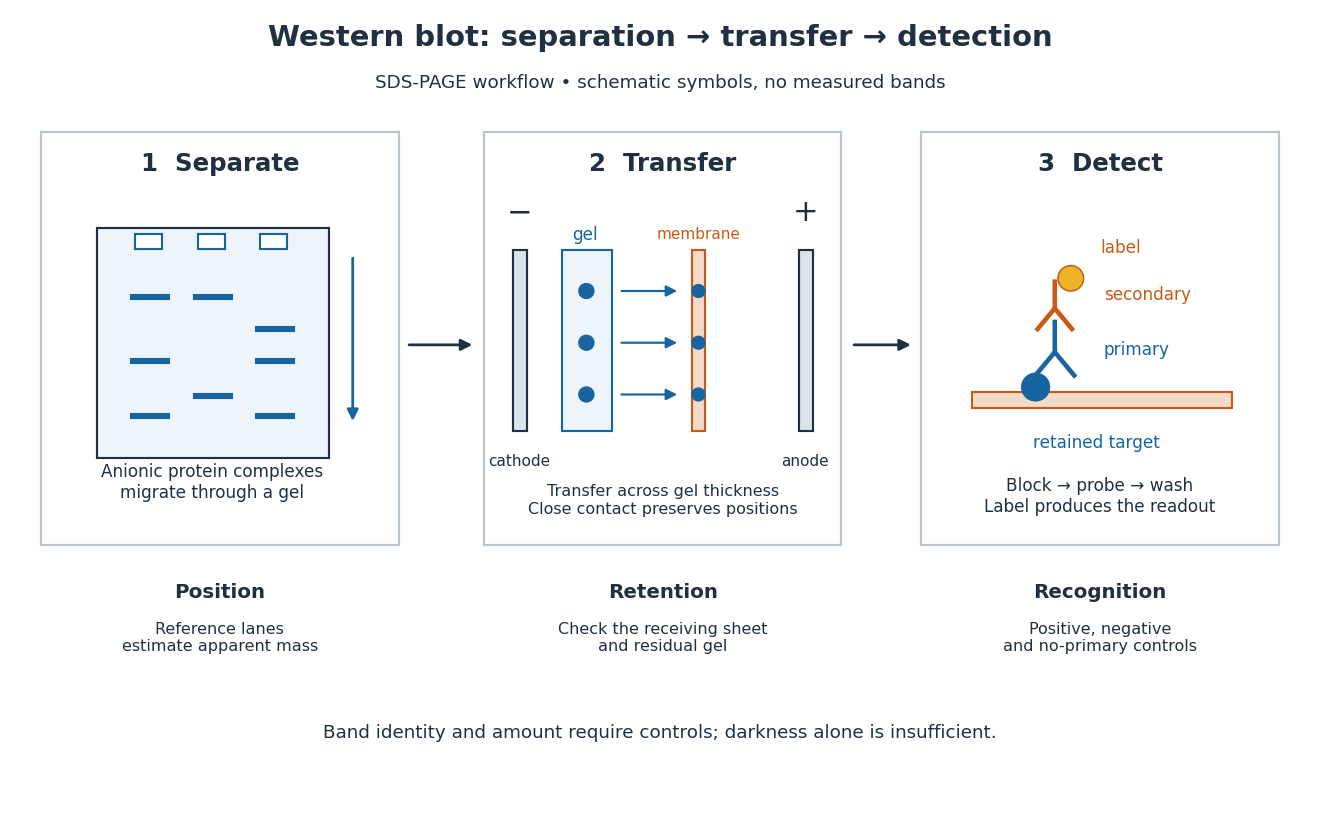
An analytical form of gel electrophoresis using an anionic surfactant and a polymer gel to separate proteins predominantly by chain size under denaturing conditions. Western blot separation often uses SDS-PAGE; the resulting apparent mass and subsequent antibody binding provide complementary, incomplete evidence about identity.
Secondary antibody 2026-10-13
A secondary antibody recognizes another antibody, rather than the proposed target protein directly. In Western blot antibody detection, its attached label makes retained primary antibodies visible. A secondary antibody must recognize the kind of primary antibody used.
Serocatalytic model 2026-10-20
An irreversible serocatalytic model describes the transition from seronegative to seropositive after infection. If the force of infection is and detectable antibodies persist, the seronegative fraction satisfies , hence seroprevalence is when . Antibody loss needs a reverse transition, and imperfect testing needs a separate observation model.
Seroprevalence 2026-10-20
Seroprevalence is the proportion of a specified population with a positive result for a specified antibody assay. Its biological interpretation depends on assay diagnostic sensitivity, diagnostic specificity and the persistence and origin of the measured antibodies. It is not itself the incidence rate of new infection.
Western blot 2026-10-13
A Western blot detects selected proteins after their separation and transfer to a membrane, using antibodies. In the yeast cross-species complementation experiment, detection helps distinguish failed protein production from failure of an expressed protein to restore growth.
The useful idea is to preserve where proteins migrated in gel electrophoresis, then ask which positions retain an antibody. Western blot separation, Western blot transfer and Western blot antibody detection answer different questions. A band is evidence of retained label at a position; Western blot band interpretation requires Western blot controls before assigning identity or comparing abundance.

Western blot antibody specificity control 2026-10-14
A known target-containing sample is a positive comparison for Western blot antibody detection. A matched sample with gene deletion supplies a negative comparison: disappearance of the proposed band supports the assignment, while a persisting band indicates binding unrelated to the deleted target. Deletion must actually remove the recognized protein; changes in other proteins can complicate the comparison. Omitting the primary antibody checks binding or background from the secondary antibody and detection reagents, but cannot exclude binding of the primary antibody to an unintended protein. Independent antibodies recognizing different epitopes add evidence. [ref]
Considerations when quantitating protein abundance by immunoblot by Alicia A. McDonough and others. McDonough and others, “Consider the Specificity of the Antibody for the Target”, Figure 2: positive tissue, knockout tissue and antibodies to different termini
Western blot band interpretation 2026-10-13
Band position gives an apparent mass, not unique protein identity. Intensity depends on retention, epitope recognition and detection response; it does not establish folded state, enzyme activity or successful protein targeting. Missing bands can reflect missing targets or failed Western blot transfer or Western blot antibody detection. [ref]
Considerations when quantitating protein abundance by immunoblot by Alicia A. McDonough and others. McDonough and others, “Consider the Specificity of the Antibody for the Target” and “Consider the Linearity of the Detection System”
The yeast cross-species complementation experiment consequently combines two logically different readouts: Western blot evidence about production of an antibody-recognized protein, and growth evidence about functional complementation. A detectable protein need not perform the biological task required for growth.
Western blot controls 2026-10-13
Western blot loading controls ask how much sample entered each lane; Western blot transfer controls ask what reached the receiving sheet; Western blot antibody specificity controls ask what the antibodies recognized. Each closes a different gap in Western blot band interpretation.
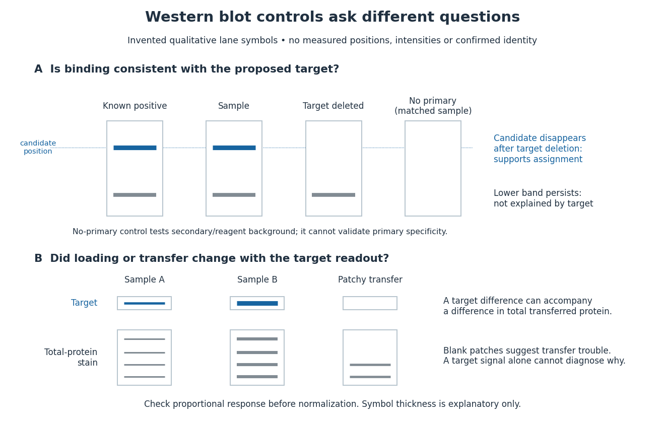
Conceptual lanes for Western blot controls
. Original schematic, with invented qualitative marks only. Candidate bands disappear in a target-deleted sample while a lower band persists; a separate panel shows how a loading or transfer difference can accompany a target-band difference. No lane, darkness, band position or relative amount represents a measurement, a specific real protein or confirmed identity. The no-primary comparison denotes a matched strip processed without primary antibody. Details.