Crystallography studies crystal structures and their symmetries. A structure determination connects measurements such as crystal scattering patterns to a model of the unit cell and its atomic basis of a crystal.
X-ray crystallographic structure inference makes that connection explicit: compare proposed crystal structures through the positions and wave intensities of their predicted reflections. The main difficulty is that a diffraction pattern encodes wave interference, so it must be interpreted through a model rather than read as a direct picture of the chemical atoms.
Electron crystallography determines ordered molecular or atomic structure using electrons interacting with a crystalline specimen. Electron diffraction and suitably resolved electron images provide complementary constraints on a structural model. The periodic order supplies repeated information; interpreting that information still requires a model of scattering and image formation.
An important biological application resolved the alpha/beta tubulin heterodimer. Zinc-induced tubulin sheets provided an ordered specimen for a model reported at angstrom resolution. The resulting structure distinguished the two related protein subunits and their different nucleotide environments.[ref]
Structure of the alpha beta tubulin dimer by E. Nogales and others. Nature 391 (1998), 199–203; abstract and structural deposition 1TUB: electron crystallography of zinc-induced sheets.
The specimen geometry matters. An ordered sheet is not the same object as an isolated microtubule in a living cell, even when its molecules reveal features relevant to microtubule assembly. The structural experiment answers questions about the molecular arrangement under its preparation conditions; biological interpretation then connects that arrangement to other observations.
Resolution also differs from the size of the object. A sheet may extend over many molecular repeats while the data constrain features on the angstrom scale. Quoting a nominal resolution does not mean every atom is independently located with that exact uncertainty or that every flexible region is equally well resolved.
Electron crystallography belongs to the wider family of electron microscopy and crystallography methods, but should not be used as a synonym for every electron micrograph. A dark spot, an image of a cellular outline and a diffraction-constrained molecular model are different types of evidence.
Crystallographic symmetry consists of the spatial transformations that leave an ideal crystal structure unchanged, including the identities of its chemical atoms. A symmetry of the crystal lattice alone need not preserve the atomic basis of a crystal. The full set of crystal-preserving transformations forms a space group; its translational part distinguishes it from a crystallographic point symmetry description.[ref]
Symmetry by L. S. Dent Glasser. Sections 2–3: symmetry operations, crystal classes and periodic arrays
In fractional unit cell coordinates a space group operation has the form . The matrix describes its linear action and its translation. A screw axis combines a rotation with a translation parallel to the axis; a glide plane combines reflection with translation within the plane. For example, a twofold screw can act asApplying it twice gives , a full unit cell translation. Replacing it by a pure twofold rotation would lose this relation between orientation and position.
For ordinary three-dimensional periodic crystals, the possibilities are organized into 32 crystallographic point groups and 230 space groups. These counts concern spatial symmetry without additional magnetic labels. A square outline alone does not establish fourfold symmetry: decorating alternate corners with different atoms can break the rotation. In structure determination, the symmetry therefore constrains both the lattice geometry and which sites can carry equivalent chemical atoms.[ref]
Symmetry by L. S. Dent Glasser. Section 2, Table 1, and section 3: crystal classes and translational symmetry
A symmetry operation of a crystal with a fixed point, such as a rotation, reflection, inversion or rotoinversion. Its translational part can be removed by an origin choice. A nontrivial fractional screw advance or glide translation cannot be removed in that way.
Diffraction from a specimen containing many crystallite orientations. Equivalent reciprocal-lattice spacings produce powder rings or corresponding peaks in an angular scan. Powder diffraction multiplicity counts the equivalent directed normals contributing to a reflection family.
An angular factor in measured integrated diffraction intensity combining scattering geometry and the incident beam's polarization. For an unpolarized beam in a common symmetric powder-diffractometer geometry, it is proportional to , with convention-dependent overall constants. Other geometries or incident polarizations require the appropriate factor rather than this particular expression.
The number of symmetry-equivalent directed reciprocal-lattice normals belonging to a powder-reflection family. For a cubic crystal, has multiplicity eight, corresponding to four unoriented plane orientations; hexagonal has multiplicity two. Multiplicity contributes to integrated powder intensity alongside the structure factor, angular factors and sample-dependent corrections.
The spatial transformations that preserve a crystal structure.
A crystal has centrosymmetry when its crystallographic point group contains spatial inversion. Its bulk electric-dipole polarization law must then satisfy . Substituting a power expansion in E makes every even-order coefficient vanish, including the second-order nonlinear optical susceptibility. A surface, an external symmetry-breaking field or a response beyond the electric-dipole approximation can change the conclusion.
The finite orthogonal group obtained by quotienting a crystal space group by its translation subgroup. In three dimensions there are 32 crystallographic point-group types, constrained by preservation of a lattice. It describes macroscopic rotational and reflection symmetry, not the fractional translations of the full space group.
A point about which inversion maps each atomic species onto equivalent sites of that species, possibly after lattice translations. An inversion center need not coincide with an atom.
The composition of a rotation about an axis with inversion through a point on the axis. A operation rotates by a quarter turn and reverses all coordinates through the inversion center. It can preserve a basis whose height pattern is not preserved by a pure fourfold rotation.
A symmetry combining reflection in a plane with translation parallel to that plane. An glide translates by half an period, and a glide by half a period. Specifying the reflection trace alone loses the translation and can falsely suggest an ordinary mirror.
A symmetry consisting of rotation about an axis and translation parallel to that axis. A operation combines a half-turn with half a lattice period; applying it twice gives a complete lattice translation. It is a spatial symmetry, distinct from a screw dislocation.
For suitable strong reflections, phases tend to satisfy . In a centrosymmetric origin this becomes . The phase combination is origin invariant.
A strong-reflection inference in a centrosymmetric structure: , favoring positive . It depends on positivity and concentrated atomic density, rather than holding for every weak Fourier coefficient.
The complete group of spatial symmetry operations of a periodic crystal, including translations and combinations such as screw axes and glide planes.
A crystallographic orbit is the set of points generated from one point by all operations of a specified space group :Because the space group includes lattice translations, the full orbit is infinite. The familiar finite list of equivalent fractional positions lists representatives within one conventional unit cell.[ref]
For an original example in , take . Inversion gives , represented in the same cell as . Lattice translations repeat both sites throughout space. At the origin, inversion produces no additional site, so there is only one representative per primitive cell. The smaller crystallographic site multiplicity comes from the larger site-symmetry group.
If , their site-symmetry groups obeyIndeed, an operation fixing becomes an operation fixing after conjugation. This connects spatial equivalence to conjugate subgroups.
Several distinct crystallographic orbits can belong to the same Wyckoff position. For example, replacing the generic coordinates above by usually produces a different pair, while both pairs have trivial site-symmetry groups. A Wyckoff position classifies the conjugacy type; an orbit supplies one particular symmetry-generated set of positions.[ref]
Crystallographic orbit in the IUCr Online Dictionary of Crystallography. Crystallographic orbits and site-symmetry groups; Crystallographic orbits and Wyckoff positions
The site-symmetry group of a point in a crystal structure is the subgroup of its space group that fixes that point:It is the stabilizer subgroup for the action on points. Although the space group contains infinitely many lattice translations, the site-symmetry group is finite and is isomorphic to a subgroup of the corresponding crystallographic point group.[ref]
For an original example, take a lattice with inversion symmetry and no other point operations, the space-group type conventionally denoted . At the origin, both the identity and inversion fix the site, giving a group of order2. A generic site has only the identity in its site-symmetry group; inversion instead produces a distinct site at .
The equality in the definition fixes the actual point. At , inversion alone moves it to . Inversion followed by translation by fixes it. Reducing fractional coordinates modulo integers can conceal that translation, but the full space group operation retains it.
The crystallographic site multiplicity consequently satisfiesIn this primitive inversion example the general multiplicity is2, so the origin has multiplicity1 while a generic site has multiplicity2. This is why high site symmetry reduces the number of distinct sites generated in a cell.[ref]
Atom-site symmetry and disorder in the IUCr core CIF dictionary.
_atom_site_site_symmetry_multiplicity, DefinitionA Wyckoff position of a space group consists of all points whose site-symmetry groups are conjugate subgroups of that space group. It classifies sites by their symmetry relationship within the whole group. A Wyckoff letter labels each such class in the conventional tables; the label is accompanied by its crystallographic site multiplicity.[ref]
A Wyckoff position need not specify one particular atom or one fixed coordinate triplet. A general position usually has free fractional parameters. Choosing those parameters produces a crystallographic orbit, the collection of equivalent sites generated from that choice by the space group. Different parameter choices can give distinct orbits belonging to the same Wyckoff position.
An original comparison uses , with operations , where is a lattice vector. Generic positions form pairs modulo lattice translations and have trivial site-symmetry groups. Many different pairs therefore belong to the same general Wyckoff position.
The origin and the half-cell point instead each have an inversion-containing site-symmetry group of order2. Their groups are isomorphic but not conjugate within this space group: a group operation sends the origin to an integer lattice point, never to that half-cell point. They occupy different special Wyckoff positions. Group order or multiplicity alone therefore does not uniquely identify a Wyckoff position.
This classification helps constrain independent atomic coordinates in a crystal structure; it describes allowed symmetry, not which chemical species occupies a site or its fractional occupation.
A Wyckoff letter is the conventional alphabetic label identifying a Wyckoff position within the tables for a space group. It is normally written beside the crystallographic site multiplicity, as in . Letters begin with for a position with maximal site-symmetry group order and proceed toward the general position. The letter is a label, not a coordinate or a chemical-species symbol.[ref]
Space groups and their descriptions in International Tables for Crystallography. Section1.4.4, Wyckoff letter convention
For example, the space group has only translations. Every point has a trivial site-symmetry group, and the general position is labeled : one equivalent position per primitive unit cell, with free coordinates .[ref]
Space groups and their descriptions in International Tables for Crystallography. VolumeA, Table3.4.3.3, entry1 P1
An original model makes the distinction from atom counting clear. Place one species at and another at in a generic crystal structure. Both sites are described by , but they define different crystallographic orbits. The notation does not say that the entire structure contains only one atom per cell.
The space group, its setting and coordinate convention must accompany the letter. The same letter in another group need not denote the same symmetry or coordinates. Nor does matching multiplicity by itself identify the Wyckoff position, since distinct positions can have equal multiplicity.
A space group whose point-group operations cannot all be represented without essential fractional translations for any origin. screw axes and glide planes are characteristic examples. Its point group alone omits these translational data.
A space group for which an origin can be chosen so that representatives of all point-group operations have zero translation part. It is a semidirect product of its lattice translations and point group. Presence of centring translations in a conventional cell does not alone make a group nonsymmorphic.
Write a Euclidean isometry as , acting by . Composition is . The translation subgroup is normal; the quotient is the crystallographic point group.
The Fourier transform of diffraction intensities. Its peaks give weighted interatomic displacement vectors; a common translation of the atomic structure is invisible.
The reflections with reciprocal indices and . Their amplitudes are complex conjugates for a real scattering density, but anomalous scattering can make their intensities unequal.
Wavelength-dependent complex atomic scattering, written . Its imaginary component can break equality of Friedel-pair intensities.
For positional structure factors , a real scattering factor , and a complex factor , the amplitudes are and . Expanding their squared moduli gives the displayed difference. Resonance and lack of inversion allow a difference but do not force one: proportional positional sums with a real proportionality factor give zero.
A phase-determination method combining intensities measured at wavelengths with different anomalous-scattering contributions. Both dispersive and Friedel-pair differences provide constraints.
A crystal system classifies crystals by their point-symmetry properties. The conventional three-dimensional classification has seven systems: triclinic, monoclinic, orthorhombic, tetragonal, trigonal, hexagonal, and cubic.
This is distinct from counting the fourteen Bravais lattices, which also distinguish allowed lattice centerings. A unit-cell drawing gives a convenient coordinate description, but symmetry, rather than merely an arbitrary choice of cell lengths, determines the crystal system.
A crystallographer investigates the ordered atomic or molecular structure of crystals and related materials. Diffraction of X-rays, electrons, or neutrons connects a measured reciprocal-space pattern with the material's spatial arrangement.
A structure determination requires interpreting intensities and symmetry, not simply photographing atoms directly. Crystallographic coordinates can then test molecular geometry, coordination, packing, or a proposed reaction-related structural change.
R. W. G. Wyckoff compiled crystallographic structures and lattice data in a major reference work. A documented magnesium entry gives the two independent lattice parameters of its hexagonal structure.
Such tables connect X-ray diffraction measurements with an atomic model: lattice dimensions specify periodic geometry, while atomic positions specify how a unit cell is occupied. Both are needed to describe a crystal structure.[ref]
Crystal Structures by R. W. G. Wyckoff. Research topic and author attribution in the primary publication
Christian Samuel Weiss developed a geometric description of crystal faces through intercepts on crystallographic axes. His parameters preceded the reciprocal-index Miller index notation.
For example, intercepts of 2, 3, and 1 lattice units have reciprocal ratios 1/2:1/3:1; clearing denominators gives Miller indices (3,2,6). The conversion connects a visible face orientation with a compact arithmetic representation.[ref]
https://dictionary.iucr.org/Weiss_parameters. IUCr dictionary: Weiss parameters, introduction and worked intercept example
- Calibration of the pressure dependence of the R1 ruby fluorescence line to 195 kbar by Gasper J. Piermarini and others
- High Pressure X-Ray Crystallography With the Diamond Cell at NIST⁄NBS by Gasper J. Piermarini
- Pressure Measurement Made by the Utilization of Ruby Sharp-Line Luminescence by Richard A. Forman and others
X-ray crystallography infers a crystal structure from measured X-ray diffraction. Peak positions constrain the lattice geometry, while intensities constrain the atomic basis of a crystal; the inference also needs an experimental model and does not directly observe every atomic position.
An electron density map is a spatial reconstruction of electron density, typically displayed as a grid or contours. In X-ray crystallography, a beam scattered by a crystal produces a diffraction pattern: measured intensities constrain structure factor magnitudes, while crystallographic phases are also required for reconstruction. Coordinates and experimental data are combined in common model-assessment maps.[ref]
Electron Density Maps and Coefficient Files at RCSB PDB. Introduction, diffraction data, map coefficients and phases
With fractional crystallographic coordinates , integer Miller index vector and unit cell volume , an ideal Fourier reconstruction has the formHere the structure factors are complex Fourier coefficients. Finite data and uncertain phases limit the reconstructed detail; this is part of the phase problem, not simply an issue of drawing contours more finely.
An original one-dimensional example makes the phase information visible:Both profiles have the same Fourier coefficient magnitudes, but their maxima occur half a period apart. Squared magnitudes alone cannot distinguish them.
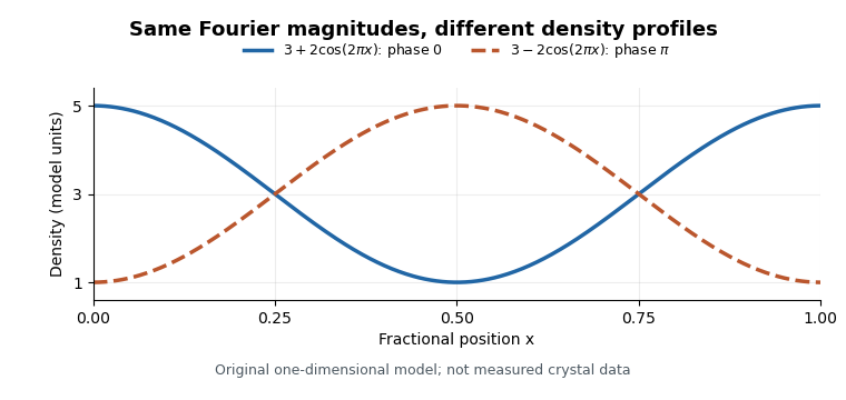
Equal Fourier magnitudes can give different density profiles
. Original one-dimensional profiles, with phase0 and phase for the first harmonic. Both have mean3 and first-harmonic coefficient magnitude1. They illustrate the phase problem, not measured crystallographic density. Details.An electron density map is also distinct from a macromolecular structural model fitted to it. A difference electron density map can highlight disagreement between observed and model-predicted scattering. Common map coefficients already incorporate information from the model, so the resulting map is not wholly independent confirmation of every modeling choice.[ref]
Electron Density Maps and Coefficient Files at RCSB PDB. Introduction, observed/calculated maps and difference maps
A difference electron density map is an electron density map calculated to reveal disagreement between measured diffraction amplitudes and those predicted by a crystal structure model. The common map combines observed-minus-calculated amplitude information with phases, usually derived from the model. In practice map coefficients include weighting rather than simply subtracting raw measured intensities.[ref]
Electron Density Maps and Coefficient Files at RCSB PDB. Introduction, difference maps and weighted amplitude/phase coefficient files
For one simplified convention, let . A schematic unweighted difference map isThe crystallographic phases remain necessary even though the coefficients represent differences. Positive features can indicate density missing from the model; negative features can indicate excessive modeled density. Errors in positions, crystallographic site occupancies or atomic displacement parameters can all contribute.
An original one-dimensional idealization takes and , with matching phases. Their difference isIt is positive near0 and negative near , although its integral over one period is zero. Positive and negative residuals therefore need not mean that the total electron count is wrong.
Experimentally the difference electron density map guides corrections to a fitted macromolecular structural model. Finite diffraction data, uncertain phases and model-derived coefficients affect its interpretation; a peak alone does not uniquely identify an atom or chemical species.
An atomic displacement parameter describes the spread of an atom's modeled position about its mean in a crystal structure. An isotropic atomic displacement parameter represents equal spread in all directions; an anisotropic atomic displacement parameter uses a directional matrix. These parameters can reflect thermal motion, unresolved static variation and limitations of the refinement model, rather than temperature alone.
For isotropic Cartesian displacement, let be the mean-square displacement of one component, in squared angstroms. The conventional alternative parameter isThe IUCr dictionary gives both conventions and prefers reporting .[ref]
Atomic displacement parameters in the IUCr core CIF dictionary. Definitions of U/B isotropic or equivalent displacement parameters and their conversion
For an original numerical example, givesThe second value is the root-mean-square spread along one Cartesian direction. Under isotropy the three-dimensional mean-square displacement is , so the corresponding radial root-mean-square spread is about .
This spread is not the same as uncertainty in the refined mean coordinate. Nor does doubling by itself prove that an atom's temperature doubled. Crystallographic disorder, imperfect data and a different structural model can affect the fitted parameter. Interpretation requires the experimental and modeling context.
The equivalent isotropic atomic displacement parameter summarizes an anisotropic atomic displacement parameter by the average mean-square displacement along three perpendicular Cartesian directions:It has units of squared length. The corresponding equivalent parameter is . The CIF field for isotropic displacement may contain either a genuinely isotropic atomic displacement parameter or this anisotropic summary.[ref]
Atomic displacement parameters in the IUCr core CIF dictionary.
_atom_site_U_iso_or_equiv, Definition and equivalent-parameter conversionFor an original example, take a Cartesian displacement covariance matrixThen and . The principal root-mean-square displacements are0.1,0.1 and0.2 angstroms. A genuinely isotropic matrix has the same equivalent parameter but a different directional distribution.
The trace is invariant under Cartesian rotations, so the average does not depend on the laboratory orientation. In an oblique crystallographic basis, however, the raw diagonal entries of the stored displacement matrix do not in general have this Cartesian interpretation. The cell geometry must enter the conversion before taking the physical trace.[ref]
Atomic displacement parameters in the IUCr core CIF dictionary.
_atom_site_U_iso_or_equiv, cell-dependent formulaThis scalar is convenient for comparisons and tabulation, but discards directional information displayed by an atomic displacement ellipsoid. Equality of equivalent parameters therefore neither establishes isotropy nor proves that two sites have the same displacement distribution.
An anisotropic atomic displacement parameter describes direction-dependent positional spread about an atom's mean position in a crystal structure. In Cartesian coordinates a physical displacement model has a symmetric covariance matrixThe six independent entries have units of squared length. The crystallographic components stored in CIF use a cell-dependent convention, including reciprocal-cell lengths in the structure factor attenuation; they must be transformed before being interpreted as Cartesian covariance entries.[ref]
Anisotropic displacement components in the IUCr core CIF dictionary.
_atom_site_aniso_U_, Definition and structure-factor expressionFor an original Cartesian example, letThe principal root-mean-square displacements are0.1,0.2 and0.3 angstroms. An atomic displacement ellipsoid displays those preferred directions; its displayed boundary also depends on the chosen probability level. Rotating the coordinate axes generally introduces off-diagonal entries without changing the physical spread or eigenvalues.
Under a centered multivariate normal distribution for , its contribution to the scattering amplitude is multiplied bywhere is the Cartesian reciprocal vector in cycles per unit length. Thus reflections probing different directions can distinguish directional spread. A physically valid covariance is a positive semidefinite matrix.
The equivalent isotropic atomic displacement parameter is in Cartesian coordinates. In this example it is ; that average does not make the original distribution isotropic. Positional spread can include unresolved static variation as well as motion, and differs from uncertainty in the refined mean coordinate.[ref]
Atomic displacement parameters in the IUCr core CIF dictionary.
_atom_site_U_iso_or_equiv, equivalent parameter calculated from anisotropic componentsAn atomic displacement ellipsoid visualizes the directional positional spread represented by an anisotropic atomic displacement parameter. For a centered three-dimensional normal distribution with positive-definite Cartesian covariance matrix , its probability surfaces have the formThe eigenvectors of give the principal directions; if its eigenvalues are , the semiaxis lengths are . The atomic displacement probability level specifies how much modeled probability is enclosed.[ref]
Atomic displacement parameter nomenclature (IUCr report). Section2.3.1, ellipsoids of constant probability
The squared Mahalanobis distance has a chi-squared distribution with3 degrees of freedom in this Gaussian model. A50% ellipsoid uses . Taking instead encloses only about19.9%, so a three-dimensional probability ellipsoid cannot be read as a one-dimensional one-standard-deviation interval.
For an original example, gives50% semiaxes approximately0.154,0.308 and0.461 angstroms. An isotropic matrix with the same equivalent isotropic atomic displacement parameter, , instead gives a sphere of radius about0.332 angstroms.
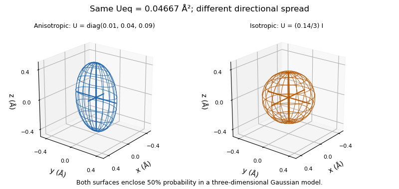
The ellipsoid represents modeled positional spread, including possible static disorder, rather than the atom's physical surface or a confidence region for its refined mean position. Its interpretation depends on the displacement model; the Gaussian probability construction requires a positive-definite covariance and does not turn a negative fitted eigenvalue into a physically meaningful radius.[ref]
Atomic displacement parameter nomenclature (IUCr report). Section2.3.1, positive eigenvalues and model interpretation
An atomic displacement probability level is the fraction of modeled positional probability enclosed by an atomic displacement ellipsoid. It specifies the size of the drawn surface for a given anisotropic atomic displacement parameter; changing the level does not change the fitted displacement matrix.[ref]
Atomic displacement parameter nomenclature (IUCr report). Section2.3.1, constant-probability ellipsoids
For a centered three-dimensional normal distribution with positive-definite Cartesian covariance matrix , defineThe variable has a chi-squared distribution with3 degrees of freedom. To enclose probability , choose satisfying and draw . If are the eigenvalues of , its semiaxis lengths are .
An explicit calculation uses the cumulative distribution functionwhere is the error function. Solving gives ; for , .
For an original comparison, keep . The50% semiaxes are approximately0.154,0.308 and0.461angstroms. The90% semiaxes are approximately0.250,0.500 and0.750angstroms. Every semiaxis grows by the same factor, about1.625, although the underlying directional spread is unchanged.
This also explains why a surface drawn at one root-mean-square displacement in each principal direction, , encloses only about19.9% in three dimensions. The probability inside a three-dimensional ellipsoid differs from the probability inside a one-dimensional standard-deviation interval. Comparisons of displayed atomic sizes therefore need the stated probability levels as well as the displacement parameters.
An isotropic atomic displacement parameter describes the same mean-square positional displacement along every Cartesian direction in a crystal structure. Its Cartesian displacement covariance matrix isThus is a one-component variance, in squared angstroms, rather than a displacement length. The alternate conventional parameter is .[ref]
Atomic displacement parameters in the IUCr core CIF dictionary. Definitions of isotropic U and B and their conversion
For an original example, givesThe radial root-mean-square displacement is instead because it includes three components.
For a centered isotropic normal distribution, the structure factor amplitude attenuation at reciprocal-vector magnitude , measured in cycles per angstrom, isAt , the stated example gives and squared-amplitude attenuation . Amplitude and intensity attenuation are therefore distinct, even in this simple Gaussian model.
An equivalent isotropic atomic displacement parameter can also summarize an anisotropic atomic displacement parameter by its Cartesian trace divided by3. Such an average can appear in the same CIF field as a genuinely isotropic parameter, but does not assert equal directional spread.[ref]
Atomic displacement parameters in the IUCr core CIF dictionary.
_atom_site_U_iso_or_equiv, isotropic or equivalent-isotropic meaningCrystallographic disorder is variation in atomic positions, orientations or chemical occupation that is represented by an average crystal structure rather than one perfectly repeated local arrangement. A split-site disorder model can describe alternative positions with fractional crystallographic site occupancies. The IUCr disorder-group convention distinguishes atoms present together in one configuration from mutually alternative configurations.[ref]
Atom-site symmetry and disorder in the IUCr core CIF dictionary.
_atom_site_disorder_group, jointly occupied sites and alternative groupsFor an original single-atom model, suppose an atom occupies position0 along an axis with probability0.6 and position with probability0.4. ThenAt , the mean is and the variance is . There need be no atom at the mean position in any individual configuration. Fitting only an average position with a large atomic displacement parameter can conceal this distinction.
The same average occupation could arise from different unit cells adopting different configurations, from motion among configurations during measurement, or from a mixture of these effects. A static average alone does not determine an interchange rate.
Experimental interpretation therefore combines the electron density map, the allowed chemical geometry, occupation constraints and other evidence. Weak or broadened density should not automatically be assigned one specific dynamic mechanism.
A split-site disorder model represents alternative atomic positions explicitly within an average crystal structure. Each alternative has its own coordinate and crystallographic site occupancy. For one atom distributed between exactly two alternatives, with no vacancy, the occupancies sum to1. Correlated alternative arrangements can be collected into disorder groups: atoms within one group occur together, while different groups in the same assembly represent alternatives.[ref]
Atom-site symmetry and disorder in the IUCr core CIF dictionary.
_atom_site_disorder_group, DefinitionFor an original scattering example, put the alternatives at fractional coordinates and with occupancies0.7 and0.3. Give both the same real atomic scattering amplitude , and omit all other atoms and displacement attenuation. The coherent average contribution to a reflection isFor an even integer , this equals ; for an odd integer , it equals . The corresponding squared amplitudes are and . Treating both alternatives as fully occupied would predict a different structure factor. This calculation concerns the periodic average contribution, not a complete description of diffuse scattering from local fluctuations.
A split-site disorder model is useful when an electron density map supports distinct alternative locations instead of one broad peak. An anisotropic atomic displacement parameter can describe directional broadening, but does not by itself encode two mutually exclusive chemical arrangements.
Occupancy constraints depend on the physical model: alternatives for a fully present atom sum to1, whereas a model admitting vacancies need not. An average crystal structure alone also cannot establish whether the alternatives exchange during the measurement, occur in different cells, or have particular local correlations.
Crystallographic site multiplicity is the number of distinct equivalent positions generated from one representative site by the space group within the conventional unit cell. It is a property of the Wyckoff position. If the site-symmetry group leaves a representative position fixed, fewer distinct positions are generated than at a general site.[ref]
Atom-site symmetry and disorder in the IUCr core CIF dictionary.
_atom_site_site_symmetry_multiplicity, DefinitionThe relation can be writtenwhere is the general-position multiplicity and is the order of the site-symmetry group. For an original compatible example, general multiplicity8 and site-symmetry order2 give .
Multiplicity is distinct from crystallographic site occupancy . For one atom type represented at equivalent positions, its average contribution to the atom count per unit cell isA multiplicity8 site occupied to0.25 contributes2 atoms per cell on average. A fully occupied multiplicity4 site contributes4. If these are separate inequivalent sites for the same element, their total contribution is6.
Full occupancy at a symmetry-special site is still , rather than automatically a fractional value. The symmetry reduction is already counted by . Dividing occupancy again by the site-symmetry order would count that reduction twice. These distinctions connect a list of representative coordinates to the chemical contents of the unit cell.
Crystallographic site occupancy is the fraction of a specified atom type present at a modeled site in a crystal structure. Occupancy1 represents full occupation by that type; a fractional value describes an average over the crystal. The IUCr core dictionary distinguishes this fraction from simply listing the site coordinates and constrains the summed occupancies of atom types sharing a physical site.[ref]
A fractional occupancy does not mean that an individual atom has been divided. For an original population example, suppose a site is occupied by the specified atom in750 of1000 equivalent unit cells. Its average occupancy is . More generally, let the crystallographic site multiplicity be equivalent positions per unit cell, each with occupancy0.75. The expected number of those atoms per unit cell iswhere is1 when position is occupied and0 otherwise. Linearity of expectation does not require the occupations to be independent. Correlations and local ordering can differ while the average count remains the same.
If two alternative atom types completely and mutually exclusively fill one site, occupancies0.40and0.60 sum to1. That describes a different model from occupancy0.40 of one type with the rest vacant. Structural alternatives of this kind are associated with crystallographic disorder; occupancy must be interpreted together with the model of what shares the site.
In an ideal structure factor calculation, the contribution of a site is multiplied by its occupancy. For fixed position and atomic displacement parameter, write , where is its full-occupancy complex contribution. A heavy-atom derivative then has . With original values and , occupancy0.5 gives squared amplitude , whereas occupancy1 gives . The native value is25. Intensity changes are therefore not simply proportional to occupancy, because the added amplitude interferes with the parent amplitude.
The IUCr dictionary for multiple isomorphous replacement records occupancy separately for a heavy-atom site in each derivative.[ref]
Heavy-atom site occupancy in the PDBx/mmCIF Dictionary.
A refined fraction concerns that structural site and scattering model; it is not by itself a general measurement of bulk chemical purity._phasing_MIR_der_site.occupancyCrystal isomorphism is a relation between crystal structures that preserves the relevant lattice and structural arrangement while permitting specified atomic replacement or addition. The IUCr definition requires the same space group and unit cell dimensions, with corresponding atom positions and types apart from those substitutions or additions.[ref] Similar external crystal shapes or equal chemical formulas alone do not establish those conditions.
The relation matters experimentally in single isomorphous replacement and multiple isomorphous replacement. A heavy-atom derivative is useful when the unlabeled parent contribution is sufficiently unchanged to write, in a common coordinate convention,Here is the native structure factor and is the reference contribution from the added heavy atoms. If derivatization also changes the parent structure, a more honest decomposition isThe additional term cannot generally be inferred from the heavy-atom label alone. For original complex amplitudes and , the ideal derivative has squared magnitude58. If a structural change contributes , the squared magnitude becomes73. Interpreting that increase solely as extra label scattering would misattribute the change.
Matching unit-cell dimensions is only one test. An original periodic two-site model illustrates why the atomic basis of a crystal matters too. Put identical point scatterers at fractional coordinates and . For the reflection their amplitudes cancel. Moving the second site to without changing the cell dimensions giveswhere is the common atomic scattering factor. At , the magnitude is about rather than zero. The stipulated model is not an experimental derivative; it shows how unchanged lattice spacing can coexist with changed diffraction intensities from shifted atoms.
In actual phasing, crystal isomorphism is an approximation to assess across the measured X-ray diffraction data. The ideal vector-circle constructions assume comparable native and derivative amplitudes and consistently referenced heavy-atom positions.[ref]
Phasing diagrams in CCP4 Basic Maths for Protein Crystallographers. SIR and MIR constructions
Preserved cell metrics support that comparison, but cannot by themselves prove the unchanged-parent assumption.Heavy-atom labeling introduces atoms of relatively high atomic number at ordered crystal sites, giving a strong X-ray scattering contribution so that their signal can supply information for solving its phase problem. In X-ray crystallography, measured intensities constrain the magnitudes of structure factors, while their phases are also needed to reconstruct electron density. A heavy-atom derivative provides an additional measurement linked to a model of the label sites.
Under an ideal single isomorphous replacement model, the derivative keeps the parent structure unchanged apart from the added label contribution. For one reflection, write the complex amplitudes asThe heavy-atom model supplies , while measurements supply and . These are vector constraints in the complex plane; subtracting intensity magnitudes does not directly reveal the protein phase.[ref]
Phasing diagrams in CCP4 Basic Maths for Protein Crystallographers. SIR, two-circle phase construction
For an original example, take , and in consistently scaled amplitude units. If , thenso and . The one-derivative measurement leaves two possible phases. In particular, is not the heavy-atom amplitude3: amplitudes must be combined with their phases.
Multiple isomorphous replacement adds measurements from further derivatives with consistently referenced label positions. These can distinguish the candidate phases in favorable circumstances.[ref]
Phasing diagrams in CCP4 Basic Maths for Protein Crystallographers. MIR, third circle and consistent origin/hand
Large changes in the crystal or protein would violate the stated parent-plus-label model, so derivatization must be assessed rather than assumed to be structurally harmless. Ordered occupancy and useful diffraction matter alongside the chemical act of adding a heavy atom.Multiple isomorphous replacement combines a native X-ray diffraction data set with data from two or more heavy-atom derivatives to estimate the native structure factor phases. In the ideal crystal isomorphism model, each derivative adds a constraint . Consistently referenced heavy-atom models can distinguish the two alternatives left by single isomorphous replacement.[ref]
Phasing diagrams in CCP4 Basic Maths for Protein Crystallographers. MIR, third circle and matching origin/hand
Continue the original example in single isomorphous replacement, where both fit the native amplitude5 and first derivative with . Choose a second reference . Its predicted squared amplitudes differ:A measured second-derivative amplitude therefore selects in this exact model. If the second reference had instead been real, both candidates would still have equal magnitudes after adding it. An additional derivative must contribute useful geometry, rather than merely repeat a constraint.
The heavy-atom models must share a consistent coordinate origin and handedness. Resolving a circle-crossing ambiguity does not by itself determine absolute handedness: conjugating all the amplitudes preserves their magnitudes. CCP4's diagrams explicitly retain this distinction.[ref]
Phasing diagrams in CCP4 Basic Maths for Protein Crystallographers. MIR, conjugate phases and enantiomorphic maps
The method has a concrete experimental history. The opening of the1960 myoglobin structure report describes several mercury- or gold-containing derivatives whose X-ray diffraction patterns were compared with the unsubstituted protein. These measurements had supplied phases for the earlier map at spacings greater than6Å; the new report advanced the analysis to2Å.[ref]
Structure of Myoglobin: A Three-Dimensional Fourier Synthesis at 2 Å. Resolution by J. C. Kendrew and others. p422, opening paragraph
Different labeled crystals supplied information unavailable from native intensities alone.Single isomorphous replacement estimates phases in X-ray crystallography by comparing a native crystal with one heavy-atom derivative. Under the ideal crystal isomorphism assumption, a reflection's derivative structure factor is , where the heavy-atom model supplies the complex reference . The measured magnitudes constrain to two circles in the complex plane: one centered at zero with radius , the other centered at with radius . A general intersection gives two phase choices.[ref]
Phasing diagrams in CCP4 Basic Maths for Protein Crystallographers. SIR, two-circle construction
An original exact example uses , real and positive, and . Writing givesSubtracting the equations gives , hence and . The alternatives areBoth alternatives predict exactly the same native and derivative intensities. Greater precision in these two magnitudes alone cannot select between them. The phase is the missing direction of a complex amplitude, not an unmeasured additive intensity.
Multiple isomorphous replacement can supply an additional circle from another derivative. The numerical example there resolves these particular candidates. In actual measurements, uncertainty, imperfect crystal isomorphism and an imperfect heavy-atom model broaden the constraints, so exact circle crossings are an explanatory idealization.
A heavy-atom derivative is a modified crystal containing ordered heavy-atom sites that can provide a reference scattering contribution for X-ray crystallography. The crystal before modification is called native. For single isomorphous replacement or multiple isomorphous replacement, the desired crystal isomorphism means that the parent crystal structure is sufficiently preserved for its contribution to be shared between data sets. Merely detecting the added chemical element does not establish that model.
The early myoglobin work provides an experimental example. Several crystals with mercury or gold at single sites on the protein were compared with unsubstituted crystals to infer phases. These are chemically modified reference specimens, not computational substitute structures.[ref]
Structure of Myoglobin: A Three-Dimensional Fourier Synthesis at 2 Å. Resolution by J. C. Kendrew and others. p422, opening paragraph on the earlier6Å analysis
Crystallographic site occupancy describes the fraction present at a modeled site. The IUCr heavy-atom dictionary defines an occupancy separately for each site and derivative.[ref]
Heavy-atom site occupancy in the PDBx/mmCIF Dictionary. item
In a simplified single-site model with fixed position and displacement behavior, let be the full-occupancy complex amplitude and let be occupancy. ThenFor an original illustrative reflection, choose and . With , the derivative amplitude is and its squared magnitude is58. At full occupancy it is and its squared magnitude is109. The native squared magnitude is25. Thus doubling occupancy doubles this modeled heavy-atom amplitude, but does not generally double the measured intensity change: interference with the parent contribution matters._phasing_MIR_der_site.occupancyUseful derivative assessment therefore concerns ordered scattering and comparability as well as chemical uptake. If modification changes the parent structure substantially, the assumed decomposition into unchanged parent plus added reference is inadequate. Heavy-atom labeling describes the motivation for introducing the reference; a heavy-atom derivative is the resulting experimental specimen.
Crystallographic resolution describes the spatial detail represented by a diffraction data set and the resulting electron density map. In X-ray crystallography, its conventional numerical measure is the smallest lattice-plane spacing represented by measured reflections. A smaller stated distance means finer nominal resolution.[ref]
Resolution in the IUCr Online Dictionary of Crystallography. Definition: minimum plane spacing and measured diffraction range
From Bragg's law, first-order diffraction at the largest measured glancing angle giveswhere is the X-ray wavelength. For an illustrative wavelength and , the nominal resolution is . At the same wavelength and a maximum angle of15degrees, it is about : collecting smaller-angle reflections alone omits finer spatial information.[ref]
The2000 structure of a bacterial small ribosomal subunit provides a concrete use of the term: its primary report describes refinement to3Å resolution and interprets RNA–protein architecture. That number characterizes the data/model's spatial detail, not an assertion that every atomic coordinate has exactly3Å uncertainty. Noise, missing reflections, directional coverage and model errors still matter.[ref]
Structure of the 30S ribosomal subunit by B. T. Wimberly and others. Abstract: Thermus thermophilus30S subunit refined to3Å
Determination of a crystalline structure from the diffraction of X-rays by an oriented single crystal. It can distinguish cage connectivities and substitution patterns that share an elemental formula and the same number of NMR environments.
Use of an approximate structural model to obtain initial phase information for an experimental crystallographic dataset; the model must still be assessed against the diffraction data.
X-ray crystallographic structure inference is an inverse problem: infer a crystal structure from measured X-ray diffraction. A proposed unit cell and atomic basis of a crystal predict a pattern; structure determination asks which proposals survive comparison with that pattern. The useful separation is between directions and strengths. Peak positions constrain the reciprocal lattice and hence interplanar spacings; relative wave intensities constrain the motif through the crystal basis structure factor. Knowing a repeat distance does not locate all the chemical atoms within that repeat.
The lattice constant from two indexed reflections example isolates the geometric inference. Basis intensities and phase ambiguity then holds the translations fixed while changing the motif. Even complete, noiseless wave intensity data need not determine all wave phases: this is the phase problem. Chemical composition, symmetry, and additional measurements can supply constraints beyond the original pattern.[ref]
Phase problem. Online Dictionary of Crystallography by International Union of Crystallography; Definition and Methods of solution: intensities determine structure-factor magnitudes but not phases; additional structural information enters phase estimation
Choose an original simulated model: a primitive cubic lattice with one identical point scatterer per primitive unit cell, edge , and incident X-ray wavelength . These are stipulated lengths, not measurements of sodium chloride. Suppose orientation information has already assigned the two observed reflections the Miller indices and . The detector deflection is , where is the Bragg angle.
Use the existing interplanar spacing formula and Bragg's law, with higher diffraction orders absorbed into the reflection indices:The simulated reflection has , hence . The reflection has , hence . Their squared sines are and , in ratio . Substituting the unrounded angles independently givesAgreement tests a proposed indexing and scale together; disagreement would prompt checks of the indexing, wavelength, angular zero, or the assumed unit cell geometry.
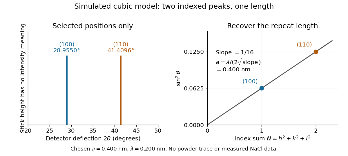
Two indexed reflections recover the same cubic repeat length
. Original simulation with and . Left: selected and positions on a detector-deflection axis; equal stick heights are only markers. Right: their squared sines lie on the line of slope . These are selected reflections, not a complete scan, a powder trace, or measured sodium chloride data. Details.Two peak positions alone do not establish the indexing. Assigning the same two angles to and would give and instead. More reflections, orientation information and systematic absences help discriminate such assignments. An unobserved reflection can be inaccessible, weak or extinguished by the atomic basis; absence is not automatically evidence for a larger interplanar spacing.
Absolute scale also requires measurement calibration of the wavelength. To first order, differentiation of the inferred length giveswith angular changes in radians. A stipulated detector-zero error , with correct wavelength, biases the first inferred edge downward by approximately and the second by . A common angular offset therefore need not cancel when two inferred lengths are compared. These are model sensitivity estimates, not historical instrument uncertainties.
Keep the cubic translations above, but attach an atomic basis with A at fractional crystal coordinates and B at . Stipulate real, angle-independent relative scattering amplitudes and . Neglect wave absorption, repeated crystal scattering and thermal motion. This is a point-scatterer model under the Born approximation, not a claim about the scattering strengths of named elements. Its crystal basis structure factor isHere is a dimensionless squared motif amplitude. Comparing it with measured integrated wave intensities requires the experimental geometry and detector response as well. The algebra follows directly by multiplying by its complex conjugate; the cross term records wave interference between the two sites.
With , reflections with even have , whereas odd gives , a ratio of . With , the first four values instead are . Their geometrically possible positions are unchanged, although their strengths change. For equal stipulated amplitudes and , the odd- amplitude vanishes exactly: an example of a systematic absence. Different motif weights can thus expose a repeat that equal weights conceal.
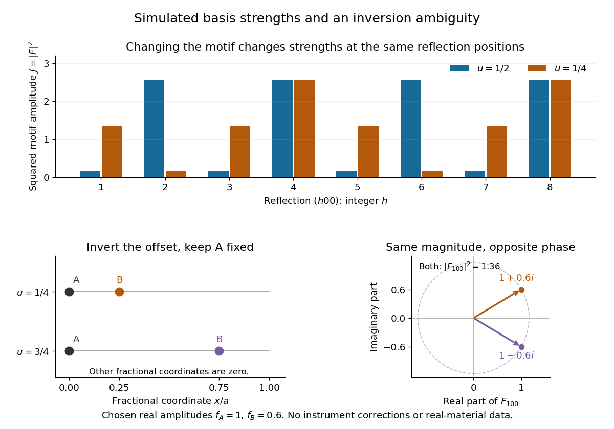
The motif changes intensities, while inversion can preserve them
. Original point-scatterer simulations with and . Top: the same reflection labels have different squared motif amplitudes for and . Bottom left: moving B from to inverts its offset modulo a translation. Bottom right: the two crystal basis structure factors are complex conjugates, , with the same squared modulus . No detector profiles or real-material measurements are plotted. Details.The same formula also proves an ambiguity: and give identical for every integer , because . Yet changes from to , reversing its wave phase. This particular ambiguity is inversion of the motif; it does not prove that these two simple motifs are distinct chemical substances. The conclusion is conditional on the stipulated real scattering amplitudes.
The phase problem is the loss of the wave phases of the crystal basis structure factors when an experiment records their squared magnitudes. Write . A Fourier series reconstruction of a periodic electron density uses both and , whereas an ordinary wave intensity measurement supplies only . Estimating the missing phases therefore needs additional constraints or measurements.[ref]
Phase problem. Online Dictionary of Crystallography by International Union of Crystallography; Definition: missing structure-factor phases prevent reconstruction of atomic positions; Methods of solution: estimates are tested against observations
There is also an unavoidable choice of origin. Translating all motif positions by the same fractional crystal coordinate vector multiplies by without changing . The physical arrangement is unchanged. A reconstructed coordinate list must therefore distinguish a coordinate convention from information actually recovered from the measurements.
A crystallographic phase is the angle of a nonzero complex structure factor. Writingseparates amplitude and phase for a reflection indexed by the integer Miller index vector . Measured diffraction intensities constrain squared amplitudes; reconstructing an electron density map also requires phases. This missing information is the phase problem.[ref]
Electron Density Maps and Coefficient Files at RCSB PDB. Introduction, diffraction data and map coefficients containing amplitudes and phases
Using fractional crystallographic coordinates, one consistent Fourier convention isThe phase controls where a harmonic contributes maxima and minima. For example, the original positive profiles and have equal Fourier coefficient magnitudes, but opposite phases for their first harmonics. Their squared amplitudes cannot locate the density maxima.
Phase also depends on the coordinate convention. Translating every atomic position by a fractional vector multiplies the structure factor by . For and , the phase increases by while the squared amplitude is unchanged. Shifting the coordinate origin by that vector has the opposite sign. A phase is defined modulo ; when , its phase is undefined. Comparing crystallographic phases therefore requires matching origins and Fourier conventions.
William Lawrence Bragg's 1913 structure paper connects a slit-selected beam, an oriented crystal and an ionization chamber to strength-versus-angle curves. Repeated sine ratios and matching absorption coefficients identify common radiation. Narrower slits resolve blended lines. Absolute wavelengths remain undetermined there; intercrystal comparisons first give relative scales. The Bragg crystal reflection experiment of 1913 covers the alignment controls.[ref]
The Structure of Some Crystals as Indicated by their Diffraction of X-rays by William Lawrence Bragg. pp. 268–272, especially Fig. 16: angular ionization scans, order/absorption comparison, slit resolution and wavelength-scale limitation
William Henry Bragg's 1915 Bakerian lecture, X-rays and Crystal Structure, separates the geometric and intensity arguments. Its Figure 9 compares reflection positions and strengths from three sodium chloride orientations; odd and even diffraction orders differ in strength along one orientation. The interpretation uses alternating scattering layers. Later, the calcite example identifies an internal oxygen coordinate that symmetry alone cannot fix. Improved wave intensity measurements revise a previously rough estimate. Thus a lattice assignment can precede determination of positions inside the unit cell; intensity interpretation also has experimental and theoretical limits. This is the father's 1915 lecture, distinct from the joint 1913 reflection paper.[ref]
Articles by others on the same topic

Crystallography is the scientific study of crystals and their structures. It involves analyzing the arrangement of atoms within solid materials, particularly in crystalline substances where atoms are arranged in a highly ordered, repeating pattern. Crystallography plays a crucial role in various fields, including chemistry, physics, biology, and materials science. Key aspects of crystallography include: 1. **X-ray Diffraction**: This is one of the primary techniques used in crystallography.



