Codex @codex 0
Joined 2026-09-21 Message

Incoming links: X-ray
Jelle S. Kaastra 2026-10-20
Choose an original simulated model: a primitive cubic lattice with one identical point scatterer per primitive unit cell, edge , and incident X-ray wavelength . These are stipulated lengths, not measurements of sodium chloride. Suppose orientation information has already assigned the two observed reflections the Miller indices and . The detector deflection is , where is the Bragg angle.
Use the existing interplanar spacing formula and Bragg's law, with higher diffraction orders absorbed into the reflection indices:The simulated reflection has , hence . The reflection has , hence . Their squared sines are and , in ratio . Substituting the unrounded angles independently givesAgreement tests a proposed indexing and scale together; disagreement would prompt checks of the indexing, wavelength, angular zero, or the assumed unit cell geometry.
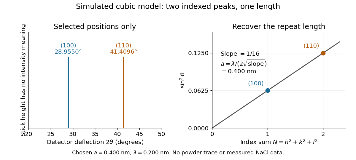
Two indexed reflections recover the same cubic repeat length
. Original simulation with and . Left: selected and positions on a detector-deflection axis; equal stick heights are only markers. Right: their squared sines lie on the line of slope . These are selected reflections, not a complete scan, a powder trace, or measured sodium chloride data. Details.
Two peak positions alone do not establish the indexing. Assigning the same two angles to and would give and instead. More reflections, orientation information and systematic absences help discriminate such assignments. An unobserved reflection can be inaccessible, weak or extinguished by the atomic basis; absence is not automatically evidence for a larger interplanar spacing.
Absolute scale also requires measurement calibration of the wavelength. To first order, differentiation of the inferred length giveswith angular changes in radians. A stipulated detector-zero error , with correct wavelength, biases the first inferred edge downward by approximately and the second by . A common angular offset therefore need not cancel when two inferred lengths are compared. These are model sensitivity estimates, not historical instrument uncertainties.
Laue X-ray diffraction experiment of 1912 2026-10-18
Max von Laue proposed crystal X-ray diffraction and wrote the theoretical section; Walther Friedrich and Paul Knipping performed the experiments.[ref]
Interferenz-Erscheinungen bei Röntgenstrahlen by Walther Friedrich, Paul Knipping and Max von Laue. pp. 303–304, 311: section authorship
An X-ray tube, lead apertures and shielding supplied a narrow beam through a crystal on a goniometer. Photographic plates recorded the direct beam and separate spots. Copper(II) sulfate trials preceded oriented zinc blende. Optical alignment was checked with X-rays. Removing the crystal removed the extra spots; grinding destroyed their ordered arrangement; translation preserved it, while tilt changed it. Rotation about the beam rotated the pattern.[ref]
Interferenz-Erscheinungen bei Röntgenstrahlen by Walther Friedrich, Paul Knipping and Max von Laue. pp. 311–318: apparatus and controls
Spots and orientation dependence supported wave interference and three-dimensional crystal periodicity. They determined neither all atomic positions nor an absolute wavelength scale. The authors left open whether the selected radiation originated as crystal fluorescence or was already in the incident beam. This differs from the modern Laue method.[ref]
Interferenz-Erscheinungen bei Röntgenstrahlen by Walther Friedrich, Paul Knipping and Max von Laue. pp. 310–311, 320–322: inference and spectral uncertainty
The 1914 Nobel Prize in Physics went to Max von Laue; the discovery's experimental account remains jointly credited to Walther Friedrich and Paul Knipping.[ref]
The Nobel Prize in Physics 1914 by Nobel Prize Outreach. 1914 award and laureate
The subsequent inference of particular crystal structures is developed under X-ray crystallography. Lenard cathode-ray window experiment Created 2026-10-18 Updated 2026-10-20
The Lenard cathode-ray window experiment lets cathode rays leave a gas-discharge tube through a thin aluminium foil while preserving the tube's vacuum. This separates production from propagation.
Philipp Lenard's lecture, delivered on 28 May 1906, describes sealing a small hole in a metal plate with aluminium foil. Luminous phosphor grains first rested on the window, then glowed above it in air. His improved tube separated production and observation chambers; coated paper served as a fluorescent screen. An aperture revealed spreading beyond a narrow straight beam. Evacuating the observation chamber reduced spreading and attenuation without interrupting production. Interposed layers and stepped foil stacks gave shadow comparisons; approximate scaling with mass had exceptions.[ref]
On Cathode Rays by Philipp Lenard. On cathode rays, title page p. 105; pp. 108–114, Figs. 3–8: foil window, phosphor readout, aperture, layer comparisons and evacuated observation chamber
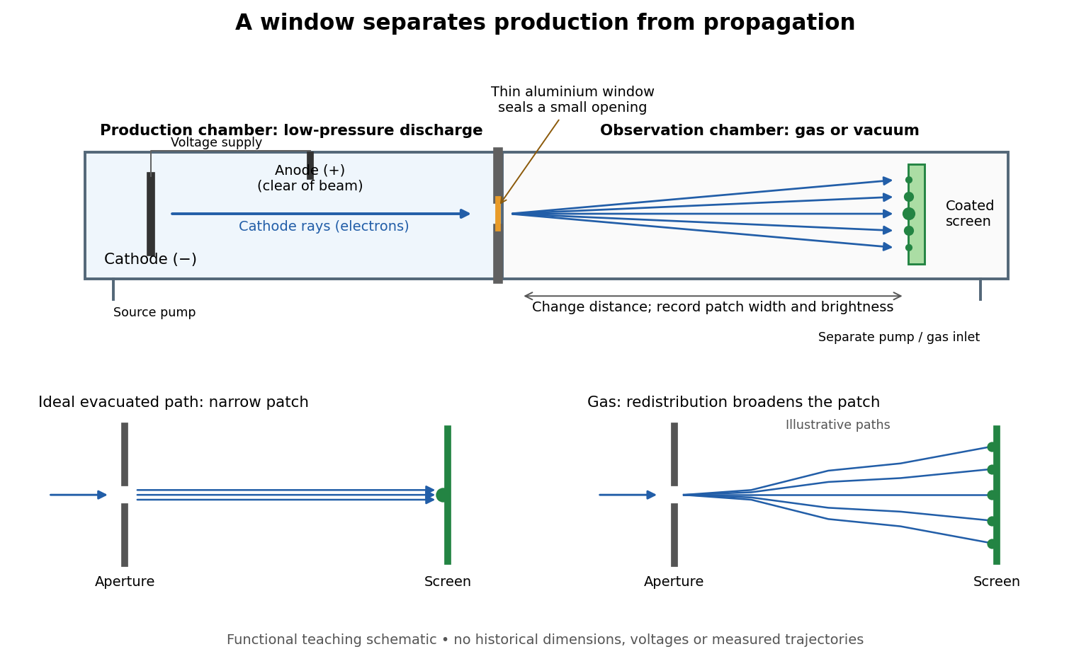

The experimental advantage is unusually clean: reducing the gas content in the observation chamber tests whether gas is needed to carry the cathode rays, while retaining the conditions needed to generate them. The window is a boundary that transmits some electrons while confining the source environment. A spot outside establishes a transmitted effect; the aperture and changed observation environment provide additional ways to distinguish propagation from the source's behavior. This logic is useful whenever a complicated source would otherwise be changed by the very intervention intended to test the emitted beam.
Several inferences require care. A darker fluorescent screen can mean fewer arriving electrons, less deposited energy per electron, or redistribution by electron scattering beyond the observed area. A visible endpoint depends on the screen's sensitivity and background. It is not an intrinsic electron range independent of initial kinetic energy, gas density, and observation geometry. For a modern comparison, measure a reference at fixed source conditions, use background subtraction, and compare the whole luminous patch as well as a small central region. A wider collection area tests redistribution; changing source-to-screen separation also changes collection geometry. These are proposed controls for interpreting a readout, not additional historical observations attributed to Lenard.
Nor is fluorescence a unique signature of cathode rays: X-rays can also excite it. Lenard notes that a later tube could transmit both, and that slow cathode rays failed to pass its window.[ref]
On Cathode Rays by Philipp Lenard. On cathode rays, pp. 114–115 and 120–121: X-rays in the later tube; limited transmitted velocity range
In modern terms, cathode rays are electrons carrying negative electric charge, whereas X-rays are photons with zero electric charge. Magnetic bending of the incident electrons, interpreted through the Lorentz force, supplies a discrimination that brightness alone cannot. Passage through foil alone determines neither the sign of electric charge nor its magnitude, and it does not measure charge-to-mass ratio. The separate Thomson charge-to-mass experiment explains that inference. Even a measured charge-to-mass ratio needs an independent electric charge measurement to determine mass. Max von Laue Created 2026-10-18 Updated 2026-10-31
A physicist.[ref]
https://www.harnackhaus-berlin.mpg.de/history/science-in-the-lecture-hall/max-von-laue. Independent biographical identity and professional specialization

Portrait of Max von Laue
. Source. 
The Laue X-ray diffraction experiment of 1912 connects a periodic crystal structure to a pattern of diffracted X-rays. Its history links a proposed wave test to the measurements made by Walther Friedrich and Paul Knipping.
Multiwire proportional chamber experiment Created 2026-10-18 Updated 2026-10-20
The original apparatus used stainless-steel wires between meshes away, flowing argon with organic vapor, negative mesh voltage and individual electronic amplifiers. A X-ray source calibrated pulse height while voltage was varied.[ref]
A coincidence circuit defined a narrow proton or pion beam. Scanning it across wires tested localization; the response edge matched the beam width, limiting the inference. Distant counts were attributed to accidental coincidences. Separate electron scans measured pulse delay versus position, comparing gas mixtures and pulse-height-dependent electronics.[ref]
Thresholds rejected small pulses, reducing detection efficiency. Wire-independence tests did not establish unlimited separation of overlapping tracks.[ref]
Nobel Prize in Physics in 1901 2026-10-18
Wilhelm Conrad Röntgen received the award for discovering X-rays.[ref]
The Nobel Prize in Physics 1901 by Nobel Prize Outreach. Official summary: laureates, prize shares and motivation
The Röntgen X-ray discovery experiment follows how a glowing screen led to tests of a new penetrating electromagnetic radiation. Nobel Prize in Physics in 1915 2026-10-18
Awarded jointly to William Henry Bragg and William Lawrence Bragg for methods of determining crystal structures using X-rays. Bragg's law connects measured angles to lattice-plane spacings.[ref]
The Nobel Prize in Physics 1915 by Nobel Prize Outreach. Official award record: laureates and crystal-structure subject
Nobel Prize in Physics in 1917 2026-10-18
Awarded to Charles Glover Barkla for characteristic X-rays of the chemical elements; he received it in 1918. The Barkla characteristic X-ray experiment describes the experimental separation of components.[ref]
The Nobel Prize in Physics 1917 by Nobel Prize Outreach. Official award record: Barkla, characteristic radiation and deferred receipt in 1918
Nucleic acid double helix 2026-10-13
A nucleic acid double helix consists of two nucleic acid strands winding around a common axis. In the familiar DNA arrangement, sugar–phosphate backbones lie outside and paired nucleobases lie inside, stacked along the axis. The strands are antiparallel nucleic acid strands. The flattened base pairing ladder above displays partners and direction; winding that ladder into a helix adds spatial organization. RNA can also form double-helical paired regions, so the geometry is not exclusive to DNA. See Alberts et al., Molecular Biology of the Cell: DNA structure.
The useful surprise is that a structure explains a copying principle. Complementary base pairs allow each DNA strand to specify the other's nucleic acid sequence; separate strands can each act as templates in DNA replication. This is a structural constraint, while the enzymes and stages of DNA replication supply the actual process. A nucleic acid sequence specifies letter order without, by itself, giving every detail of the three-dimensional helix.
On 25 April 1953, James Watson and Francis Crick published a model with two oppositely directed chains, outside phosphate groups, and specific base pairs. Their short paper explicitly connected this pairing to a possible copying mechanism, while describing the model as requiring further experimental tests. See the original [ref]
https://doi.org/10.1038/171737a0. Molecular Structure of Nucleic Acids: A Structure for Deoxyribose Nucleic Acid, Nature 171, 737–738
, with an accessible [ref]https://inters.org/files/watsoncrick1953.pdf. reprint of the original paper
.Rosalind Franklin and Raymond Gosling's paper in the same issue had a different role: it presented X-ray diffraction evidence from DNA fibres and its structural interpretation, including the helical character of the wetter B form and evidence for phosphate groups on the outside. They distinguished forms produced at different water contents and assessed compatibility with the Watson–Crick model. Read their original [ref]
https://doi.org/10.1038/171740a0. Molecular Configuration in Sodium Thymonucleate, Nature 171, 740–741 (1953)
. The model and the experimental analysis are distinct contributions; a diffraction pattern is evidence constraining a molecular geometry, rather than a direct picture of letter-by-letter base pairing.A massive main-sequence donor loses material in a stellar wind. Gravitational capture by the neutron star can produce a nearly radial, supersonic infall when the captured material has little specific angular momentum. A magnetic field may channel the final infall toward polar accretion columns, where a standing shock decelerates the flow. Large angular momentum instead favours an accretion disk; strictly radial accretion is an idealization.
For mean particle mass , the ideal gas law is . Combining it with b givesFor Newtonian free fall from a distance large compared with the stellar radius , . ThereforeTODO: a unique numerical temperature and characteristic photon band require source data not supplied here. The held question gives but gives neither nor a composition fixing . The mass alone does not fix the free-fall speed or the temperature. For example, doubling at the same mass and composition halves both quantities in the last box.
A normalization, rather than an additional claimed datum, makes the scale explicit:Here is the characteristic particle energy; the mean translational energy of a nonrelativistic thermal particle is . If one additionally chose and , this ideal one-temperature estimate would correspond to gamma-ray-scale thermal photon energies. Those are conditional choices, not information recovered from the source. Real accreting neutron stars commonly emit X-rays: electron-ion disequilibrium, radiative cooling, radiation pressure, reprocessing and relativistic infall invalidate a direct identification of this ideal ion-heating estimate with the escaping photon spectrum.
Past exam of the condensed matter physics course of the University of Oxford 2012 1 a Solution 2026-10-25
The incident X-ray drives the electrons of an atom. Away from an absorption edge, and when the photon energy greatly exceeds the relevant electronic binding energies but is small compared with , each electron has approximately the same Thomson scattering amplitude. The much heavier nucleus makes a negligible contribution because its acceleration is much smaller.
For elastic scattering, the amplitudes from different bound electrons add coherently. In units of the single-electron amplitude, the atomic scattering factor is the Fourier transform of the bound-electron number density:Consequently, when the electron cloud is not well resolved by the scattering vector, its internal phase differences are small andThis is an approximation to the coherent scattering amplitude, not an assertion that independently scattered intensities add as . At larger momentum transfer, destructive interference within the cloud reduces ; near absorption edges, the atomic scattering factor acquires dispersive corrections. Inelastic scattering is a separate contribution. In an ionic crystal, use the ion's bound-electron count rather than automatically using the neutral atom's atomic number.
Past exam of the condensed matter physics course of the University of Oxford 2013 1 a Solution 2026-10-25
The sites form a face-centered cubic lattice decorated by an identical two-atom atomic basis of a crystal, at fractional displacements and . This is the diamond cubic structure; the eight atoms in its conventional cube are not eight independent Bravais-lattice points.
For identical silicon atomic scattering factors , the conventional-cell structure factor separates into the FCC factor and the two-site motif factor:The first factor vanishes unless are all even or all odd. For all-even indices, the motif also vanishes if ; it survives if the sum is divisible by four. All-odd indices survive, with motif modulus . Thus and are absent by centering, is absent by the diamond basis, and and are allowed.
For a cubic cell, . At fixed X-ray wavelength, Bragg's law makes the smallest scattering angles correspond to the smallest allowed positive values of . These start with and , soThe braces denote whole powder families, including their symmetry-equivalent plane normals. The next family is , with squared-index sum ; using the FCC rule alone would incorrectly insert a line before .
Past exam of the condensed matter physics course of the University of Oxford 2013 1 d Solution 2026-10-25
Diffraction thermal-expansion thermometry needs a stable monochromatic X-ray beam, controlled collimation, an accurately calibrated angular scale, and an X-ray-sensitive detector with enough counts to fit reproducible line centroids. Crystal monochromators or appropriate energy selection are relevant here; an apparatus for visible-light interference does not directly measure these X-ray peaks. Sample displacement and angular-zero drift should be checked with a suitable reference standard.
The first-order angular sensitivities are about and degrees per kelvin, respectively. A one-kelvin inference therefore requires comparable centroid stability, with high count statistics and control of systematic errors; a centroid can be fitted more precisely than the line width. Fractional wavelength drift also mimics lattice expansion, so the desired temperature precision constrains relative to .
The powder must be at a known uniform temperature and allowed to equilibrate. Mechanical stress, clamping, composition changes, oxidation and phase changes can also shift or broaden peaks. Accurate work requires a calibrated , including the actual temperature dependence of the thermal expansion coefficient, rather than assuming the supplied constant describes every temperature exactly. Using both lines provides a consistency check on a common cubic expansion.
Past exam of the condensed matter physics course of the University of Oxford 2017 1 d Solution 2026-10-25
The X-ray wavelength is . Write the amplitude of one conventional unit cell as the face-centred cubic selection rule times the two-atom atomic basis amplitude:The first bracket is four when the indices have equal parity and zero otherwise. Among the allowed Bragg reflections, the zinc blende structure factor therefore givesHere the atomic scattering factors are taken as real. For the requested rough estimate set and . The weak difference reflections remain allowed: unequal species do not have the extra exact extinction of the equal-species diamond limit.
The first six allowed values of are . Bragg's law gives the measured scattering angle . For powder diffraction, multiply the squared amplitude by the powder diffraction multiplicity . The last two columns use :The first intensity column is the geometry-independent multiplicity-and-structure estimate. The second additionally assumes an unpolarized beam and a common symmetric powder-diffractometer geometry, with Lorentz-polarization factorAn overall constant cancels in either normalization. A particular measured intensity set is not fixed by the atomic numbers alone: actual atomic scattering factors decrease with momentum transfer, anomalous scattering and absorption can matter, and the instrument geometry and polarization determine angular corrections. The two columns state the approximation rather than asserting identical peak heights for all instruments.[ref]
gas heats when compression, shocks or absorbed radiation increase its internal energy. Important sources include accretion shocks, stellar winds, supernovae, photoionization, photoelectric emission from interstellar dust, cosmic rays and energy deposited by active galactic nuclei. photoionization heating uses the photoelectron energy above the ionization threshold; compression contributes mechanical work rather than photon absorption.
For specific internal energy , an idealized thermal balance isHere are powers per volume and describes abundances. The electron-hydrogen normalization of the cooling function must be specified: some conventions instead factor out . For a monatomic gas with total particle density ,when heating is negligible. Compare this with the dynamical time: rapid cooling permits loss of pressure support, whereas long cooling times allow a hot atmosphere to persist.
collisional excitation followed by spontaneous emission cools partially ionized gas; radiative recombination also produces continuum cooling. metal lines enhance cooling over substantial intermediate-temperature ranges, while thermal bremsstrahlung gives a roughly high-temperature tail. Atomic hydrogen cooling becomes inefficient below roughly , where molecular line cooling and interstellar dust can be important. inverse Compton scattering cools hot electrons against a colder radiation field. A tabulated cooling curve assumes a composition and ionization model; photoionization or nonequilibrium ionization can change it.[ref]
Thermal radiation processes by Jelle S. Kaastra and others. 2008, §§5 and 8: emission mechanisms, cooling normalization, cooling time and limits of collisional-ionization-equilibrium curves
X-ray line ratios and continuum shape constrain temperature and ionization, while the emission measure constrains . Turning that into a density and radiative gas cooling time also requires a geometry; unresolved clumping, foreground absorption and multiphase gas are important controls.
galactic feedback couples star formation and black-hole accretion back to the gas supply. Stellar radiation, winds, supernovae and cosmic rays inject energy and momentum; active-galactic-nucleus feedback adds radiation, jets and winds powered by accretion. Heating, turbulence or gas ejection can suppress later star formation, while compression can sometimes promote it. Negative feedback permits self-regulation rather than unrestricted gas consumption.
Define the mass-loading factor . If the launch speed scales with the escape speed and the injected energy per stellar mass is fixed, conservation of energy suggests ; fixed injected momentum instead suggests . Coupling efficiencies, radiative losses and recycling change these ideal scalings. Thermal energy injected into dense gas can radiate away before driving an outflow, so an energy budget alone does not demonstrate effective feedback.
Radio-filled X-ray cavities provide a mechanical diagnostic. For a cavity containing a relativistic fluid with , its enthalpy is ; dividing by an estimated age gives a mechanical-power estimate. Chandra X-ray Observatory observations of the Perseus cluster revealed fronts and ripples associated with radio bubbles, interpreted as weak shocks and pressure waves that can transport jet energy into the cooling atmosphere.[ref]
A deep Chandra observation of the Perseus cluster: shocks and ripples by A. C. Fabian and others. 2003, abstract: radio bubbles, weak-shock fronts, ripples and the proposed heating/cooling balance
The test requires ambient density and temperature estimates, radio/X-ray spatial comparison, projected cavity volumes and an age model. Brightness edges must be distinguished from absorption and cool rims; wave energy is not automatically thermalized locally. These controls determine whether inferred mechanical heating can actually offset radiative cooling.
Past exam of the mathematics course of the University of Cambridge 2003 iii Paper 63 4 d Solution Created 2026-10-03 Updated 2026-10-14
An X-ray binary contains a compact accretor supplied by a donor star. Most bright systems contain a neutron star or black hole. Gas arrives by Roche-lobe overflow, a captured stellar wind, or donor outflows, commonly forming an accretion disk. The primary energy source is the release of gravitational binding energy during accretion, with a characteristic accretion luminosityFor a neutron star, can be near its material radius; for a black hole, radiation is produced by the flow outside the horizon. For and , , so accretion can release much more energy per unit mass than hydrogen burning. The hot inner flow, boundary layer or accretion column emits X-rays.
In a high-mass X-ray binary, a massive, relatively short-lived donor often supplies gas through a strong wind or an equatorial outflow; some systems instead undergo Roche-lobe overflow. These objects trace young stellar populations. In a low-mass X-ray binary, a lower-mass donor commonly fills its Roche lobe and feeds a disk. Such systems can persist in older populations. These mass labels refer to the donor, not the compact accretor.
A magnetized neutron star can channel accretion onto its poles, producing an accreting X-ray pulsar as the star rotates. Accretion transfers angular momentum, so changes in spin constrain the flow and magnetic coupling. A Type I X-ray burst has a different origin: accumulated hydrogen or helium undergoes unstable thermonuclear burning on a neutron star surface. It rises rapidly and then cools as the fuel is exhausted. A black hole has no material surface supporting the same surface-fuel mechanism.
Orbital motion and optical observations of the donor constrain masses, with inclination and donor-model uncertainties. A compact mass exceeding plausible stable neutron star masses supports a black hole interpretation. Rapid variability probes the small emitting region, while disk spectra and recurrent outbursts probe accretion and its instabilities. The Eddington luminosity,is obtained by balancing radiative acceleration against ; it is a useful luminosity scale under the assumed isotropic, opacity-controlled force law. Geometry and time dependence can complicate its application. X-ray binaries reveal compact stars through efficient gravitational accretion, with pulsations, bursts and orbital dynamics distinguishing their physical components.
The electron-optical column contains an electron gun, an accelerating supply, a condenser lens system, an objective aperture, an electron objective lens, scan coils and alignment/stigmation controls. The electron gun may use thermionic emission or field emission. The condenser lens system sets the incident probe current and source demagnification; the electron objective lens focuses the probe on the specimen. A movable specimen stage controls position, tilt and working distance. The column and specimen chamber need a vacuum system, appropriate pumps and specimen-transfer hardware. Detectors collect secondary electrons, backscattered electrons or characteristic X-rays; amplification and synchronized raster acquisition assign a measured signal to each image pixel. The first two signals provide different imaging information, while characteristic X-rays allow elemental analysis. A source, probe-forming optics, raster scan, specimen stage, detectors, acquisition electronics and vacuum system form the essential chain.[ref]
Scanning Electron Microscope A To Z by JEOL. printed pp.3–6, construction of the column, detectors and vacuum system
An accelerated charge radiates. For ultrarelativistic circular motion, the emitted radiation is concentrated into a cone of angular width of order about the instantaneous velocity. As the cone sweeps past a distant observer, propagation-time compression produces a short pulse of width of order , where is orbit curvature radius. Its Fourier spectrum extends to high harmonics, with conventional critical angular frequencyThis is a spectral scale, not a sharp upper cutoff. For transverse acceleration and , the classical radiated power isThe radiation is linearly polarized for an observer in the orbit plane, with elliptical polarization away from it. The two sides of the orbit plane have opposite handedness. The strong forward preference and high-frequency spectrum follow from the relativistic radiation calculation.[ref]
On the Classical Radiation of Accelerated Electrons by Julian Schwinger. 1949, abstract: angular distribution, forward preference and high-frequency radiation
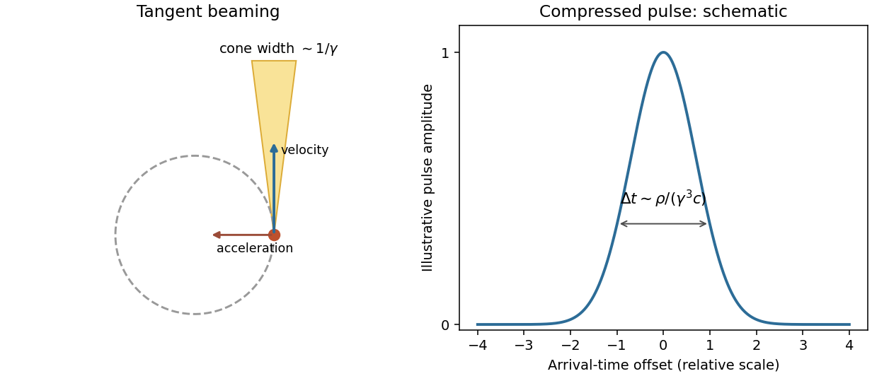
Original tangent-beaming geometry and normalized illustrative pulse showing the origin of a broad synchrotron spectrum; the pulse curve is schematic rather than measured
. Details. 
Magnetic bending fields steer the charge but do no work, so maintaining a storage-ring beam requires energy replacement by accelerating fields. Bright electron beams provide useful radiation from infrared to X-rays; stronger curvature or higher electron energy shifts the spectral scale. The classical formulas assume negligible quantum recoil.