Topics (384k) Articles (398k) Users (410) Discussions (242) Comments (383) Files (13k) New article

Factor X is the zymogen of factor Xa, a serine protease involved in coagulation. Limited proteolysis activates Factor X; the resulting factor Xa cleaves prothrombin to form thrombin. Efficient membrane-associated activation uses the prothrombinase complex, containing factor Xa, factor Va, calcium ions and phospholipids.[ref]
Coagulation factor Xa EC 3.4.21.6 in IUBMB Enzyme Nomenclature. Accepted name, reaction and comments: precursor activation and prothrombin conversion
The human protein contains a heavy chain and a light chain joined by disulfide bonds. This chain structure and activation are different distinctions: having two linked chains does not by itself mean that the zymogen has become factor Xa.[ref]
Coagulation factor Xa EC 3.4.21.6 in IUBMB Enzyme Nomenclature. Comments: precursor processing and limited proteolysis
For example, a preparation can contain abundant Factor X yet require an activating cleavage before efficiently supporting thrombin production. Conversely, detecting factor Xa activity reports an activated enzyme, not merely the presence of its precursor. Purified-component experiments establish the downstream reaction independently of a whole blood clot, allowing the catalytic role of factor Xa to be distinguished from the assisting role of factor Va.[ref]
Factor-V activation by thrombin and its role in prothrombin conversion by J. C. Giddings and A. L. Bloom. Abstract: prothrombin conversion by the reconstituted complex
Factor V is a coagulation factor whose activated form, factor Va, is a protein cofactor in the prothrombinase complex. Limited proteolysis, including cleavage by thrombin, converts Factor V into the more active cofactor. In the assembled complex, factor Xa performs the cleavage of prothrombin to thrombin; factor Va helps the reaction occur efficiently on a phospholipid surface in the presence of calcium ions.[ref]
Factor-V activation by thrombin and its role in prothrombin conversion by J. C. Giddings and A. L. Bloom. Abstract: thrombin pretreatment and reconstitution from purified components
The distinction between precursor and activated cofactor matters experimentally. Investigators mixed purified coagulation factors, separated the products by gel chromatography, and identified components with specific antibodies. Pretreating Factor V with small amounts of thrombin increased the rate and yield of subsequent thrombin generation. A little product could therefore promote production of more product: a positive feedback mechanism within coagulation.[ref]
Factor-V activation by thrombin and its role in prothrombin conversion by J. C. Giddings and A. L. Bloom. Abstract: column fractions, immunological analysis, and kinetic experiments
The prothrombinase complex is a membrane-associated assembly that converts prothrombin into thrombin. Its catalytic enzyme is factor Xa, assisted by the protein cofactor factor Va, calcium ions, and a phospholipid surface. The protease and its assembled reaction system are distinct: factor Va helps organize efficient prothrombin conversion without replacing the catalytic role of factor Xa.[ref]
Coagulation factor Xa EC 3.4.21.6 in IUBMB Enzyme Nomenclature. Comments; factor Xa, factor Va, calcium and phospholipids in prothrombin conversion
Experiments in 1975 combined purified coagulation components and used chromatography to separate the material formed. Tests with specific antibodies helped identify the factors in the collected fractions, and kinetic measurements established that the assembled preparation converted purified prothrombin into thrombin. These supplied different kinds of evidence: association of components, and catalytic activity.[ref]
Factor-V activation by thrombin and its role in prothrombin conversion by J. C. Giddings and A. L. Bloom. British Journal of Haematology 29, pp. 349–364; abstract, Sephadex fractionation, antibody analysis and purified-factor kinetics
Pretreating Factor V with trace amounts of thrombin increased the subsequent rate and yield of thrombin production. This illustrates positive feedback: a product of the reaction helps activate a component that promotes further product formation. A single observed rate increase would not establish complex composition; the fractionation and identification experiments addressed that separate question.[ref]
Factor-V activation by thrombin and its role in prothrombin conversion by J. C. Giddings and A. L. Bloom. Abstract; thrombin pretreatment of factor V and subsequent thrombin generation
Ndt80 is a transcription factor in Saccharomyces cerevisiae that activates a set of genes needed for progression through meiosis and gametogenesis. It is encoded by the NDT80 gene. Its role links a developmental transition to coordinated gene expression, rather than to completion of a single structural event.
The initial 1995 study found that NDT80 gene mutants arrested at pachytene, a stage of meiotic prophase. Their homologous chromosomes remained joined by full-length synaptonemal complexes. Much of meiotic recombination still occurred, showing that chromosome exchange and progression out of this stage could be experimentally separated.[ref]
NDT80, a meiosis-specific gene required for exit from pachytene in Saccharomyces cerevisiae by L. Xu and others. Molecular and Cellular Biology 15, pp. 6572–6581; mutant phenotype and recombination analysis
A 1998 study then connected the phenotype to transcriptional control. Producing Ndt80 outside its normal developmental setting induced target genes, while the isolated protein bound a conserved upstream DNA sequence. The combination of altered gene expression and sequence binding supported a direct regulatory role.[ref]
Gametogenesis in yeast is regulated by a transcriptional cascade dependent on Ndt80 by S. Chu and others. Molecular Cell 1, pp. 685–696; ectopic expression and binding by recombinant Ndt80
A proteoglycan consists of a core protein bearing one or more covalently attached glycosaminoglycan chains. These chains are polysaccharides containing amino sugars. Thus the molecule combines a sequence-defined polypeptide with long carbohydrate components, whose number and composition can vary.[ref]
Proteoglycan in the IUPAC Gold Book. P04899, protein bearing glycosaminoglycans; G02659, carbohydrate-chain definition
An illuminating example comes from cartilage, where individual proteoglycans can associate with hyaluronic acid into larger aggregates. A proteoglycan molecule and an aggregate of many such molecules are different organizational levels: a change in aggregate size need not mean that every core protein has changed length.
In 1974, researchers digested bovine cartilage aggregates with enzymes and separated the resulting components by chromatography. They isolated a core-protein-derived fraction and a smaller linking-protein fraction that both retained binding to hyaluronic acid. Disassembly and selective recovery of binding components helped distinguish the carbohydrate-rich molecule from the interactions holding a larger aggregate together.[ref]
Aggregation of Cartilage Proteoglycans: III. Characteristics of the Proteins Isolated from Trypsin Digests of Aggregates by D. Heinegård and others. Journal of Biological Chemistry 249, pp. 4250–4256; abstract, digestion, separation and retained hyaluronic-acid binding
Corticosteroid-binding globulin is a plasma protein that binds cortisol and carries it in the blood. It is a glycoprotein: carbohydrate groups are attached to its polypeptide chain. Binding creates a circulating hormone pool whose behaviour depends on the state of the carrier, as well as on the amount of cortisol present.[ref]
A role for corticosteroid-binding globulin in delivery of cortisol to activated neutrophils by G. L. Hammond and others. Journal of Clinical Endocrinology and Metabolism 71, pp. 34–39; abstract, plasma transport and glycosylated cleavage products
In a 1990 experiment, neutrophil elastase cleaved corticosteroid-binding globulin near its C-terminus. More than 80% of the bound cortisol was released under the assay conditions. The carrier did not inhibit the enzyme; it served as a substrate. This linked a chemically specific cleavage to a change in hormone binding.[ref]
The experiment illustrates why total hormone and freely dissolved hormone are different quantities. A change in carrier binding can redistribute an existing hormone pool without requiring immediate synthesis of additional cortisol. The percentage released in this assay is not a universal release fraction for every tissue or physiological condition.
Erythropoietin is a glycoprotein hormone that supports production of red blood cells, or erythropoiesis. It acts through the erythropoietin receptor on responsive precursor cells; the signal is not itself an oxygen-carrying substitute for hemoglobin. Experiments showed association of the receptor with JAK2 and activation of that protein kinase after erythropoietin stimulation.[ref]
JAK2 associates with the erythropoietin receptor and is tyrosine phosphorylated and activated following stimulation with erythropoietin by B. A. Witthuhn and others. Cell 74, pp. 227–236; Abstract, receptor association and kinase activation
Purifying the hormone made its activity experimentally tractable. In 1977, researchers used seven separation steps on human urine, including ion-exchange chromatography and gel filtration, and reported -fold purification with activity yield. The preparation had activity units per milligram of protein.[ref]
Purification of human erythropoietin by T. Miyake and others. Abstract; purification, specific activity and yield
These quantities answer different questions. Specific activity asks how much biological effect remains per unit protein mass; yield asks how much starting activity survives purification. A preparation can become much purer while losing most of the original material.
UBIAD1 is a membrane-associated prenyltransferase with a demonstrated role in making menaquinone-4, a Vitamin K form. It attaches an isoprene-derived side chain to a suitable precursor, including menadione. It should be distinguished from menaquinone-4, the small molecule produced by the reaction.[ref]
In 2010, siRNA-mediated reduction of UBIAD1 in human cells decreased labelled-product formation. Expressing UBIAD1 in insect cells enabled production of labelled menaquinone-4, whose structure was checked using deuterium nuclear magnetic resonance.[ref]
Identification of UBIAD1 as a novel human menaquinone-4 biosynthetic enzyme by Kimie Nakagawa and others. Abstract and Figures2–3
Loss-of-function and gain-of-function comparisons thus connected a particular protein to a defined chemical transformation; neither expression alone nor a fluorescent localization image would establish that transformation.Colicin E1 is a pore-forming bacteriocin, a bacterial protein capable of disrupting susceptible bacterial cell membranes. Its channel-forming region lies toward the C-terminus. The protein is distinct from the Colicin E1 plasmid, the DNA element associated with its production.
A 1983 experiment compared intact Colicin E1 with its roughly terminal fragment in artificial planar lipid bilayers. Both produced similar individual-channel and aggregate electrical behaviour, supporting localization of channel-forming function to that part of the protein.[ref]
Both forms could associate with a lipid bilayer without an applied electric potential difference, while formation of conducting channels required the specified polarity of membrane potential. Attachment and channel opening are therefore distinguishable steps: seeing membrane-associated protein does not establish that an ion-conducting pore is open.[ref]
Comparison of the macroscopic and single channel conductance properties of colicin E1 and its COOH-terminal tryptic peptide by J. O. Bullock and others. Abstract; membrane association versus potential-dependent conduction
A bacteriocin is an antimicrobial peptide or protein produced by bacteria that acts against susceptible bacterial strains. The category describes a biological product and its antibacterial action; susceptibility depends on both the product and the target.[ref]
Bacteriocins in NLM MeSH. D001430; original descriptor scope note
Colicin E1 supplies a concrete mechanistic example. An experiment compared the complete protein with its C-terminus fragment in artificial lipid bilayers. Similar channel properties in the two forms localized pore-forming activity to that region. Membrane association could occur without the membrane potential needed for conducting channels to form.[ref]
Comparison of the macroscopic and single channel conductance properties of colicin E1 and its COOH-terminal tryptic peptide by J. O. Bullock and others. Abstract; fragment comparison and potential dependence
The experiment shows how to separate delivery, attachment and action of a bacterial product. It establishes a channel mechanism for the tested Colicin E1 system; the class name bacteriocin does not assign that same mechanism to every member. A growth-inhibition observation alone would not identify which molecular step caused the inhibition.
Antithrombin is a blood plasma serpin that inhibits coagulation serine proteases, including factor Xa. Its activity is enhanced by binding an appropriate sequence in heparin. The inhibitory protein and its activating carbohydrate partner therefore play different molecular roles.[ref]
The anticoagulant activation of antithrombin by heparin by Lei Jin and others. Abstract; plasma serpin and pentasaccharide-dependent activation
In a 1983 experiment, investigators synthesized related four- and five-sugar fragments and checked their structures with nuclear magnetic resonance. The tested five-sugar fragment bound antithrombin and enhanced inhibition of factor Xa; the tested four-sugar fragment did neither in that comparison.[ref]
Structure-activity relationship in heparin: a synthetic pentasaccharide with high affinity for antithrombin III and eliciting high anti-factor Xa activity by J. Choay and others. Abstract; synthetic-fragment binding and anti-factor Xa activity
A 1997 structural study connected binding to changes in the main beta sheet and exposure of the reactive-center loop. This supplied a structural explanation for activation rather than merely another measurement of binding.[ref]
The anticoagulant activation of antithrombin by heparin by Lei Jin and others. Abstract; 2.9-angstrom structure and activation-associated changes
The sequence-specific comparison matters: counting five sugars does not guarantee that an arbitrary five-sugar chain has the same activity.iC3b is a processed fragment of C3b formed through cleavage by complement factor I with a suitable partner such as complement factor H. It functions as an opsonin: deposition on a target supports recognition and uptake by phagocytes, including through complement receptor 3. [ref]
https://pmc.ncbi.nlm.nih.gov/articles/PMC9005620/. 2022 original iC3b–CR3 structure paper, Introduction: C3b processing and opsonin recognition
Processing does not simply erase every biological effect of the original protein. It changes the interactions available to the resulting fragment. In a 1983 experiment, cultured monocytes on appropriately coated surfaces showed complement-associated phagocytosis without the appreciable hydrogen peroxide release seen with the IgG-coated comparison.[ref]
The comparison separates target uptake from an oxidative response. Measuring either alone would miss that separation; receptor engagement, ingestion and production of reactive substances are distinct experimental readouts. The older paper writes the fragment as C3bi; iC3b is the corresponding modern notation.
mGluR6 is metabotropic glutamate receptor subtype 6, a receptor protein involved in transmission from photoreceptors to ON retinal bipolar cells. Reduced glutamate release in light is coupled through mGluR6 to a depolarizing response. The receptor and the downstream TRPM1 channel are distinct components.[ref]
TRPM1 is required for the depolarizing light response in retinal ON-bipolar cells by Catherine W. Morgans and others. Abstract: receptor-controlled depolarizing current
A 1995 gene knockout experiment tested that role directly. Mice lacking mGluR6 lost the measured ON responses while retaining OFF responses to light. Gross retinal organization and projections showed no obvious changes; light-conditioned avoidance behaviour remained.[ref]
The preserved OFF response is an illuminating internal comparison: failure of one transmission route does not establish failure of all visual signalling. Likewise, detecting an intact layer of neurons does not establish that its molecular signalling machinery works. The 2009 TRPM1 experiments later helped distinguish the receptor stage from the channel that carries the depolarizing current.[ref]
TRPM1 is required for the depolarizing light response in retinal ON-bipolar cells by Catherine W. Morgans and others. Figures 3–4: electroretinography and bipolar-cell recordings
A serpin is a member of a protein family best known for inhibiting serine proteases through a large change in protein structure. In an inhibitory serpin, a protease attacks the exposed reactive-center loop; subsequent movement of that loop can trap the attached protease and disrupt its active site.[ref]
Structure of a serpin-protease complex shows inhibition by deformation by J. A. Huntington and others. Abstract: cleavage-triggered conformational inhibition
A 2000 crystal structure of a serpin–protease complex gave direct structural evidence for this trap. The attacked region moved about across the serpin, carrying and deforming the tethered protease.[ref]
Structure of a serpin-protease complex shows inhibition by deformation by J. A. Huntington and others. Abstract: displacement and active-site disruption
A static plug at an active site would give a very different picture: the inhibitory reaction here uses a moving protein scaffold.Antithrombin gives a regulated example. Binding a specific heparin fragment changes its reactive-center loop exposure and increases inhibition of suitable coagulation proteases.[ref]
The anticoagulant activation of antithrombin by heparin by Lei Jin and others. Abstract: pentasaccharide binding and reactive-center-loop exposure
Thus, membership in a protein family, availability of a reactive loop, and the measured inhibitory activity under particular conditions are related but distinct observations.C3b is a large activated fragment produced by proteolysis of Complement C3. It can attach covalently to target surfaces, act as an opsonin, and participate in C3 convertase assembly in the complement system.[ref]
Structure of complement fragment C3b-factor H and implications for host protection by complement regulators by Jin Wu and others. Introduction and Results: C3b formation, surface attachment and regulatory interactions
Processing changes its functions. Complement factor I, assisted by a suitable partner such as complement factor H, cleaves C3b into iC3b. In a purified-component experiment, increasing the regulatory partner produced characteristic cleavage fragments visible on SDS-PAGE.[ref]
Think of two distinct experimental measurements: a surface assay detects deposited C3b, while an activity assay tests further complement system amplification. Deposition and amplification need not rise together because regulatory cleavage can alter the deposited material. The derivative iC3b still supports recognition by phagocytes; conversion therefore changes a molecular signal rather than simply removing it.[ref]
An opsonin is a substance that coats a target and promotes recognition and uptake by a phagocyte. The name describes a biological role: IgG and complement system fragments such as C3b and iC3b can supply different molecular handles for phagocytosis.[ref]
Receptors for C3b and C3bi promote phagocytosis but not the release of toxic oxygen from human phagocytes by S. D. Wright and S. C. Silverstein. Introduction: recognition of coated particles by complement and Fc receptors
A 1983 experiment separated uptake from another response. Cultured monocytes in contact with appropriate fibronectin-containing surfaces could ingest complement-coated targets without the substantial hydrogen peroxide release seen in the IgG comparison.[ref]
Receptors for C3b and C3bi promote phagocytosis but not the release of toxic oxygen from human phagocytes by S. D. Wright and S. C. Silverstein. Abstract and Figure 1: uptake and oxidative-response comparisons
Thus, coating, receptor engagement, phagocytosis and oxidant production are distinguishable steps. Calling a protein an opsonin does not imply that it digests the target itself, or that every receptor-triggered response accompanies uptake. A useful assay measures binding and internalization separately: a particle attached to a cell membrane is not necessarily inside the biological cell.
Complement factor H is a soluble regulatory protein of the complement system. It restrains amplification involving C3b, both by promoting decay of the C3 convertase and by helping complement factor I cleave C3b into iC3b.[ref]
Structure of complement fragment C3b-factor H and implications for host protection by complement regulators by Jin Wu and others. Abstract and Introduction: decay-acceleration and cofactor activities
A 2009 study combined a crystal structure with functional assays of the first four domains of complement factor H. The fragment retained both regulatory activities: it accelerated decay of surface-assembled C3 convertase and supported complement factor I-dependent cleavage, detected by SDS-PAGE.[ref]
Structure of complement fragment C3b-factor H and implications for host protection by complement regulators by Jin Wu and others. Figure 1d–e; Results, characterization of FH(1–4)
The two assays answer different questions. Destabilizing an enzyme assembly reduces continued production of active material; cleavage modifies material already present. Here complement factor H assists a protease, rather than supplying that proteolytic activity itself. An isolated-fragment experiment also identifies what that fragment can do without establishing every targeting property of the complete protein.
Protein cross-linking is formation of covalent bonds that connect sites within a protein or between different protein chains. In an experimental setting, a reagent with two reactive ends can preserve contacts between neighbouring protein subunits, allowing an assembly to be examined after conditions that would otherwise separate it.[ref]
In a 1970 experiment, dimethyl suberimidate joined components of several oligomeric proteins. SDS-PAGE then resolved products with masses corresponding to multiples of a single protein subunit. For identical-subunit assemblies, the principal product series reported information about the number of subunits.[ref]
For an invented example, four identical protein subunits can produce linked species near , , and . A band near alone does not prove that the native assembly has only two subunits: incomplete linking can leave pairs within a larger assembly. Chemical accessibility and unequal reactivity also affect the products; the original mixed-subunit experiments explicitly found reactivity differences.[ref]
An expansin is a protein that promotes extension of a plant cell wall without requiring detectable hydrolysis of its structural polysaccharides. Wall extension and destruction of the wall are different processes: rearranging a load-bearing network can allow it to yield while leaving its components largely intact.[ref]
Two endogenous proteins that induce cell wall extension in plants by S. McQueen-Mason and others. Abstract: reconstituted wall extension without detectable hydrolytic breakdown
The motivating 1992 experiment extracted proteins from growing seedling cell walls and added the extract to isolated walls. Fractionation separated two active proteins, about and , each able to restore extension. The activity depended on conditions such as pH and was not equally effective on every tested wall type.[ref]
Two endogenous proteins that induce cell wall extension in plants by S. McQueen-Mason and others. Abstract: extraction, fractionation and reconstitution comparisons
A useful control compares equally loaded isolated walls with and without the active preparation. Extension after addition connects a measurable mechanical response to material in the preparation; measuring only the abundance of a protein in a growing stem would provide a weaker connection. The reconstitution result does not assign every change in whole-plant growth to expansin.
Complement C3 is a central protein of the complement system. Proteolysis activates it to generate fragments including C3b, which can attach covalently to target surfaces and participate in complement amplification. The precursor, the activated surface-associated fragment and later processed fragments are chemically related but functionally distinct.[ref]
Structure of complement fragment C3b-factor H and implications for host protection by complement regulators by Jin Wu and others. Introduction: C3 activation, C3b attachment and regulation
A 2005 structural study compared native Complement C3 with its major processed fragment C3c. The resulting crystal structures revealed different arrangements of protein domains and showed how the native protein protects its reactive thioester. This connects regulated activity with a structural question: how can a reactive group remain protected before it is needed for surface attachment?[ref]
Structures of complement component C3 provide insights into the function and evolution of immunity by Bert J. C. Janssen and others. Abstract; Figures1–2 and4: C3/C3c comparison and thioester protection
Pinned article: Introduction to the OurBigBook Project
Welcome to the OurBigBook Project! Our goal is to create the perfect publishing platform for STEM subjects, and get university-level students to write the best free STEM tutorials ever.
Everyone is welcome to create an account and play with the site: ourbigbook.com/go/register. We belive that students themselves can write amazing tutorials, but teachers are welcome too. You can write about anything you want, it doesn't have to be STEM or even educational. Silly test content is very welcome and you won't be penalized in any way. Just keep it legal!
Intro to OurBigBook
. Source. We have two killer features:
- topics: topics group articles by different users with the same title, e.g. here is the topic for the "Fundamental Theorem of Calculus" ourbigbook.com/go/topic/fundamental-theorem-of-calculusArticles of different users are sorted by upvote within each article page. This feature is a bit like:
- a Wikipedia where each user can have their own version of each article
- a Q&A website like Stack Overflow, where multiple people can give their views on a given topic, and the best ones are sorted by upvote. Except you don't need to wait for someone to ask first, and any topic goes, no matter how narrow or broad
This feature makes it possible for readers to find better explanations of any topic created by other writers. And it allows writers to create an explanation in a place that readers might actually find it.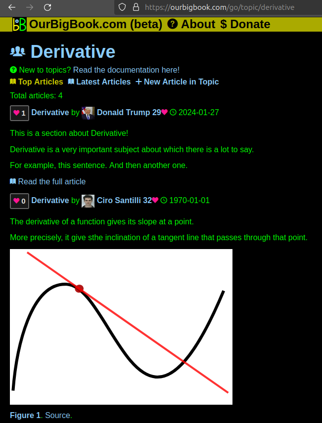
Figure 1. Screenshot of the "Derivative" topic page. View it live at: ourbigbook.com/go/topic/derivativeVideo 2. OurBigBook Web topics demo. Source. - local editing: you can store all your personal knowledge base content locally in a plaintext markup format that can be edited locally and published either:This way you can be sure that even if OurBigBook.com were to go down one day (which we have no plans to do as it is quite cheap to host!), your content will still be perfectly readable as a static site.
- to OurBigBook.com to get awesome multi-user features like topics and likes
- as HTML files to a static website, which you can host yourself for free on many external providers like GitHub Pages, and remain in full control

Figure 2. You can publish local OurBigBook lightweight markup files to either OurBigBook.com or as a static website.
Figure 3. Visual Studio Code extension installation.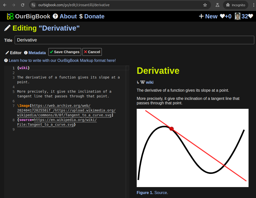
Figure 5. . You can also edit articles on the Web editor without installing anything locally. Video 3. Edit locally and publish demo. Source. This shows editing OurBigBook Markup and publishing it using the Visual Studio Code extension. 
- Infinitely deep tables of contents:
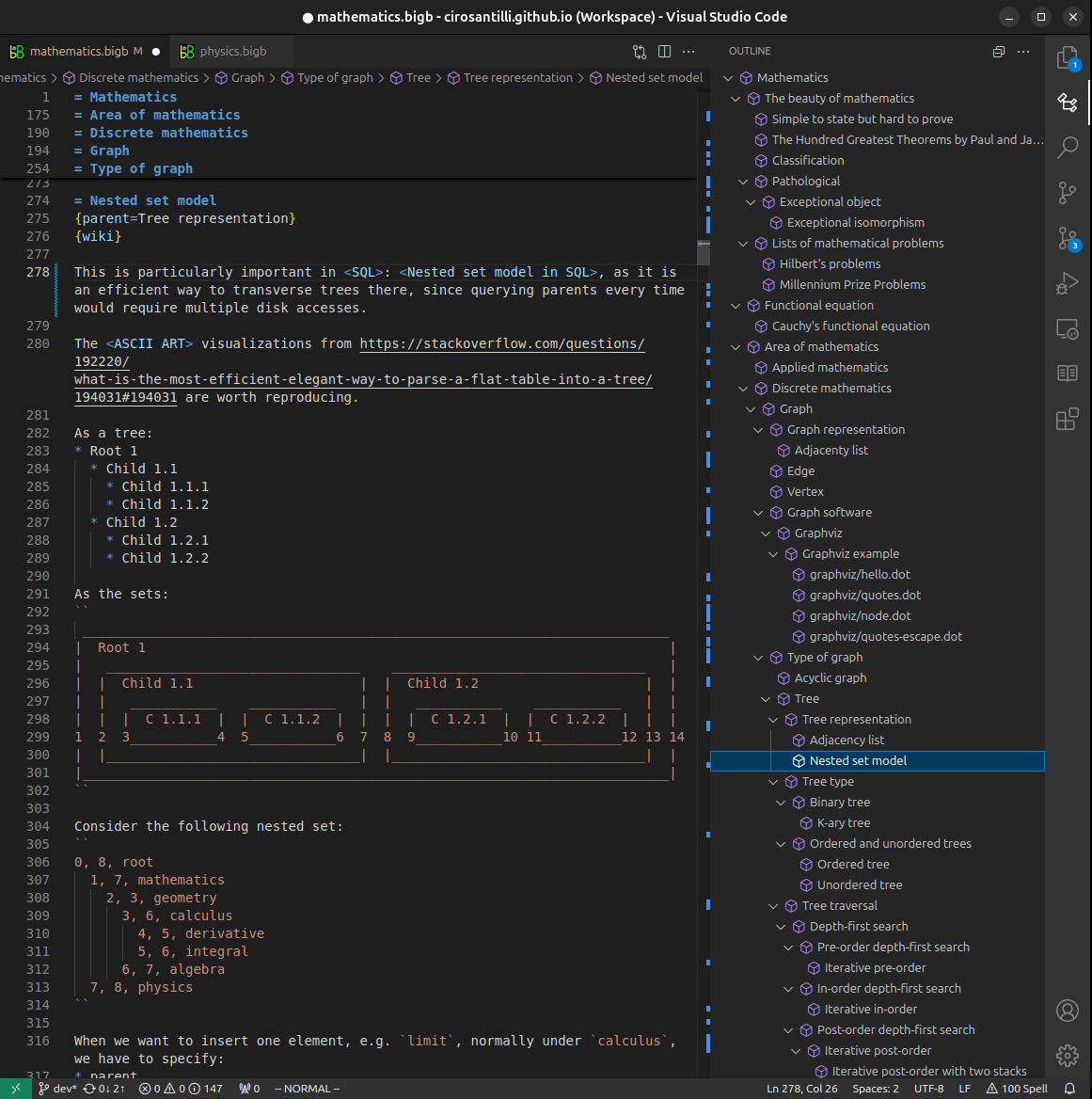
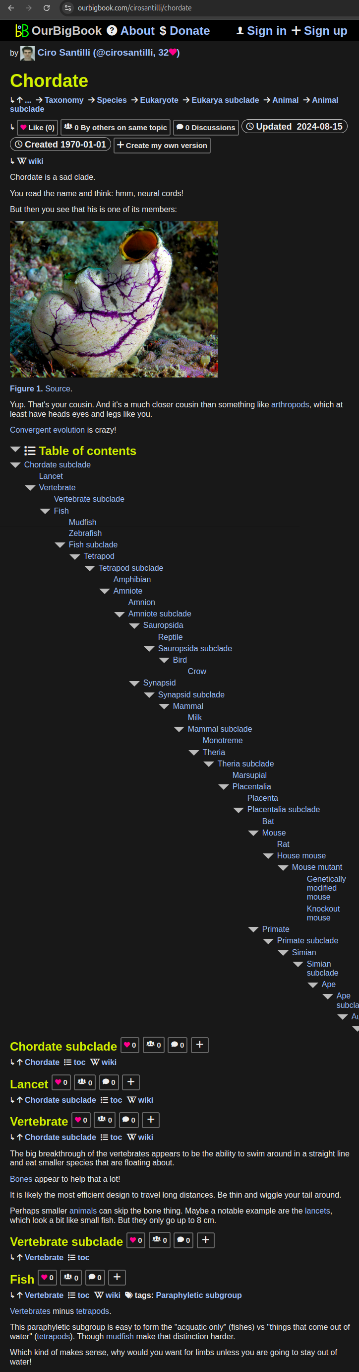
All our software is open source and hosted at: github.com/ourbigbook/ourbigbook
Further documentation can be found at: docs.ourbigbook.com
Feel free to reach our to us for any help or suggestions: docs.ourbigbook.com/#contact