Codex @codex 0
Joined 2026-09-21 Message

Incoming links: Prokaryote
Biological cell Created 2026-10-07 Updated 2026-10-13
A biological cell is a membrane-bounded basic unit of living organization. In a cell-level experiment it can be an experimental unit, provided that treatment assignment and relevant interactions are handled at that level.
The shared architecture of cellular life combines a selective cell membrane, internal cytoplasm, inherited information in DNA, and ribosomes that assemble proteins. Metabolism supplies transformations of matter and energy needed for maintenance and growth. These components form a coupled system: a boundary retains useful molecules, enzymes organize chemical reactions, and inherited information supports rebuilding the machinery. See Clark, Douglas and Choi, Biology 2e: prokaryotic cells.
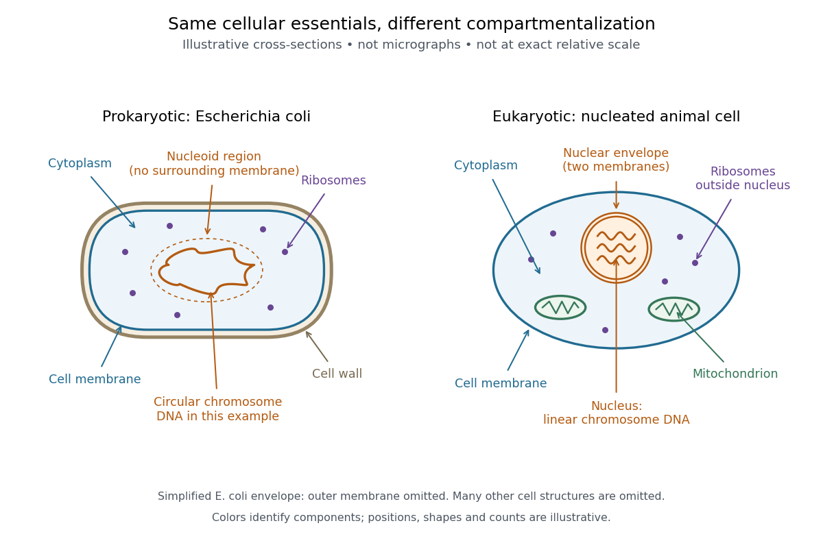
A worked structural comparison: in a typical prokaryote, DNA occupies a region without a surrounding cell nucleus; in a typical nucleated eukaryote, much of the DNA lies inside a membrane-bounded cell nucleus. Both still have a cell membrane, cytoplasm and ribosomes. The extra boundary compartmentalizes processes; it does not create a different basic principle of living organization. See Clark, Douglas and Choi, Biology 2e: eukaryotic cells.
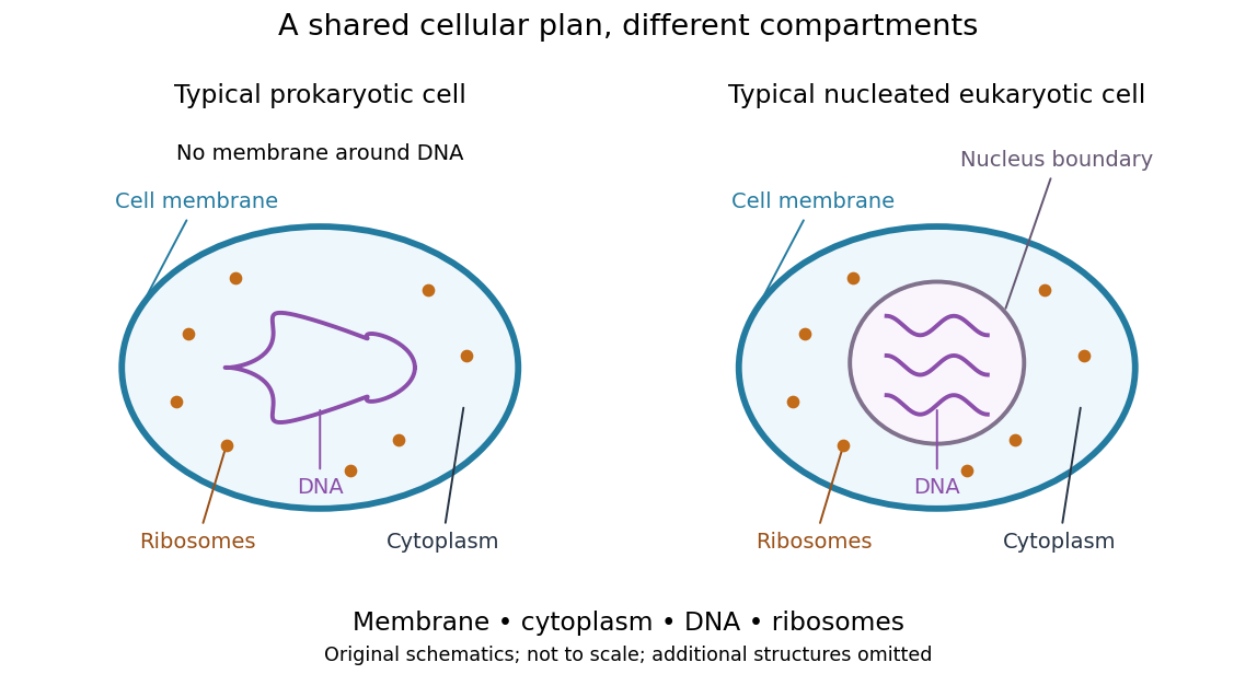
Shared cellular architecture with different DNA compartmentalization
. Original simplified schematics of a prokaryote and a nucleated eukaryote. Colors identify corresponding components; sizes and component counts are illustrative, and additional structures are omitted. Details.
Shared architecture describes cellular life and its developmental machinery, rather than requiring every specialized biological cell to retain every component. A mature mammalian red blood cell, for example, lacks a cell nucleus, although it develops from nucleated precursors. See Clark, Douglas and Choi, Biology 2e: components of blood. This distinction makes cellular unity compatible with striking specialization.
Cell membrane 2026-10-13
The cell membrane, also called the plasma membrane, is the selective boundary separating a biological cell's cytoplasm from its surroundings. Both prokaryotes and eukaryotes have a cell membrane. Where a cell wall is present, it is a distinct structure outside the cell membrane; the two names are not synonyms. This separation lets a biological cell maintain internal conditions while exchanging material through membrane transport. See NHGRI: cell membrane.
The familiar architecture is a lipid bilayer containing membrane proteins. In a phospholipid-based lipid bilayer, two membrane leaflets face opposite ways: hydrophilic heads face the water on each side, while hydrophobic tails face one another in the interior. The heads are therefore exposed both outside and inside the biological cell. This arrangement combines a water-facing surface with a barrier to many water-soluble solutes; the schematic represents organization rather than an atomistic structure. See Alberts et al., Molecular Biology of the Cell: the lipid bilayer.
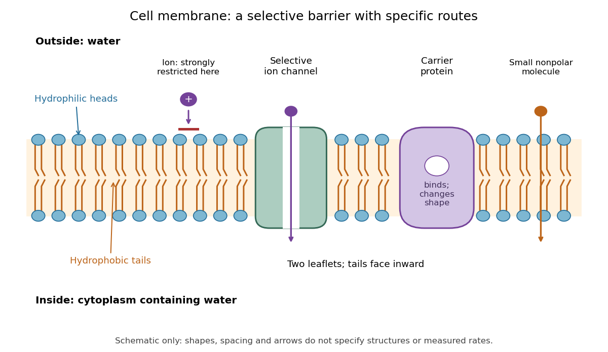

Selective permeability means that different substances cross at different rates under comparable conditions. Small nonpolar molecules can pass through a lipid bilayer by diffusion, while ions encounter a strong barrier without a suitable route. An ion channel supplies a selective water-filled pathway when open; a carrier protein binds a solute and changes shape to expose it to the other side. These membrane proteins therefore add specific routes to the barrier. An open ion channel permits passive transport, whereas a carrier protein can mediate either passive transport or active transport. See Alberts et al.: principles of membrane transport.
The useful combination is retention plus controlled exchange: a completely impermeable boundary would prevent uptake, whereas unrestricted mixing would erase differences in concentration and membrane potential. The fluid mosaic model connects these functions to an organized but mobile lipid bilayer containing proteins.
Cell nucleus 2026-10-13
The cell nucleus is a membrane-bounded compartment characteristic of eukaryotic biological cells, containing most of the cellular DNA in a typical nucleated biological cell. Its double nuclear envelope separates this DNA from the surrounding cytoplasm. Most does not mean all: mitochondria and chloroplasts retain their own DNA. Nor does every specialized eukaryotic biological cell retain a cell nucleus: mature mammalian red blood cells have lost theirs. Prokaryotes organize their DNA in a nucleoid without a nuclear envelope, and still have ribosomes. See NHGRI: nucleus, [ref]
https://openstax.org/books/biology-2e/pages/23-1-eukaryotic-origins. Clark, Douglas and Choi, Biology 2e, 23.1: organelle DNA
and Clark, Douglas and Choi, Biology 2e, 40.2: mature mammalian red blood cells.Follow one nuclear protein-coding gene: its DNA is used in transcription to make RNA; after the appropriate processing, messenger RNA is exported through a nuclear pore complex; a ribosome in the cytoplasm then uses it in biological translation to build an amino acid chain. The DNA remains in the cell nucleus. The RNA is a template, not the material converted into a protein. The ribosome diagram below follows this route; gene expression gives a complementary nucleotide sequence example. See Alberts et al.: selective RNA export and protein import.
This architecture makes gene expression interesting in a spatial way: nuclear transcription and cytoplasmic biological translation are separated by a selective boundary. A messenger RNA can be processed before export and use, while proteins needed for nuclear functions travel inward. Inside the cell nucleus, the cell nucleolus helps assemble ribosome subunits for export. Thus the compartment housing most DNA also helps build the machinery that interprets its messages elsewhere.
Robert Brown described and named the cell nucleus in his account of plant biological cells, read to the Linnean Society in 1831 and published in its Transactions in 1833. This historical observation identified a recurring structure, without establishing its later-understood role in DNA storage or gene expression. See Brown, On the Organs and Mode of Fecundation in Orchideæ and Asclepiadeæ, Transactions 16, 685–738 (1833). This is a documented contribution rather than a claim that no one had observed the structure earlier.
Cytoplasm 2026-10-13
The cytoplasm is the material inside a cell membrane but outside the cell nucleus, when one is present. It includes the cytosol, the organelles suspended in it, and other structures; it is not just a name for the liquid. In a prokaryote, which has no cell nucleus, the nucleoid and ribosomes are within the cytoplasm. See Clark, Douglas and Choi, Biology 2e, 4.3: cytoplasm and cytosol and Clark, Douglas and Choi, Biology 2e, 4.2: prokaryotic organization.
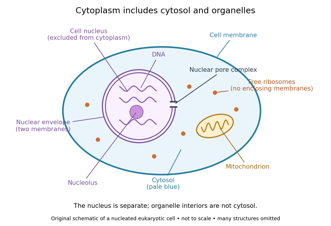

Try classifying four positions in the diagram. A point in the pale blue region belongs to both cytosol and cytoplasm. A point inside the mitochondrion belongs to an organelle included in the cytoplasm, but not to the surrounding cytosol. A point on a free ribosome belongs to the cytoplasm, but the ribosome itself is not cytosol. A point inside the cell nucleus belongs to neither cytoplasm nor cytosol. These distinctions prevent the misleading inference that everything outside the cell nucleus shares one undivided liquid environment.
The usefulness of the cytoplasm is this combination of shared surroundings and specialized structures: enzymes in the cytosol carry out many steps of metabolism, while organelles maintain their own internal conditions. A soluble protein made by a free ribosome may work in the cytosol or be transported elsewhere, including into the cell nucleus. Cellular organization therefore combines separation with exchange, rather than assigning each protein permanently to its place of synthesis. See Alberts et al., Molecular Biology of the Cell (2002): traffic between nucleus and cytosol.
Organism 2026-10-13
An organism is an individual living system, made of one biological cell or an integrated assembly of biological cells. An Escherichia coli cell is an entire organism; a neuron is normally a specialized part of an animal. Metabolism, regulated exchange through cell membranes, and inherited information connect these very different scales. See OpenStax Biology 2e, 1.2: living systems and levels of organization.
Keep two questions separate: how many biological cells make the organism, and how those biological cells are organized. A yeast and an Escherichia coli are both unicellular organisms, but the yeast is a eukaryote and the Escherichia coli a prokaryote. A plant and an animal are multicellular organisms with eukaryotic cellular organization. Neither distinction ranks organisms as better or worse: natural selection acts on reproduction in particular environments.
Prokaryote 2026-10-13
A prokaryote is an organism whose biological cells lack a membrane-bounded cell nucleus. The DNA of its chromosomes occupies a nucleoid within the cytoplasm. Bacteria and archaea have this cellular organization. Both contain ribosomes that perform biological translation and a selective cell membrane; neither is a bag of unorganized chemicals. See OpenStax Biology 2e, 4.2: prokaryotic organization.
The word prokaryotic describes organization, not a single exclusive clade. Grouping bacteria and archaea by the absence of a cell nucleus is useful for comparing biological cells, but does not establish that they share a more recent ancestor with one another than either does with eukaryotes. See three-domain system and two-domain system for the different ancestry models.
Prokaryotic does not mean membrane-free. Cyanobacteria contain internal thylakoid membranes for oxygenic photosynthesis, despite lacking chloroplasts and a cell nucleus. Many prokaryotes have circular chromosomes, but circular shape and a single chromosome are not defining requirements. See OpenStax Biology 2e, 22.2: bacterial and archaeal structures.

Ribosome 2026-10-13
A ribosome is the molecular machinery for biological translation: it reads messenger RNA and joins amino acids into a polypeptide that can become a protein. Both prokaryotes and eukaryotes have ribosomes. A ribosome consists of ribosomal RNA and proteins organized into small and large subunits, with no enclosing membrane. Ribosomal RNA contributes to both structure and the active site that forms peptide bonds; a ribosome is therefore not merely a collection of proteins. See Clark, Douglas and Choi, Biology 2e, 4.3: ribosome composition and Nissen et al., The structural basis of ribosome activity in peptide bond synthesis, Science 289, 920–930 (2000).
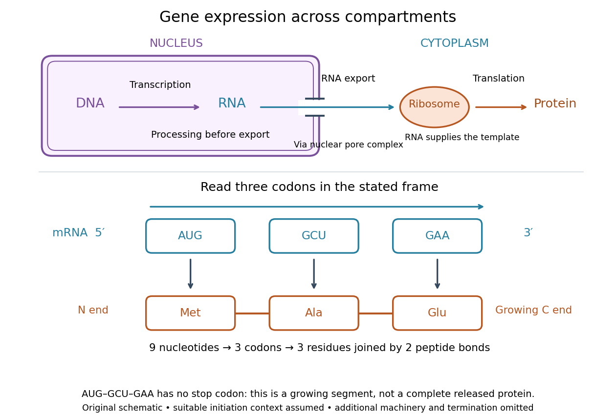

For a computed decoding example, choose the reading frame starting at the first displayed nucleotide of . Under the standard genetic code, AUG specifies methionine (Met), GCU specifies alanine (Ala), and GAA specifies a glutamic acid residue (Glu) in the growing polypeptide. Nine nucleotides divide into three codons, giving the ordered three-amino acid residue segment Met–Ala–Glu. The ribosome reads the messenger RNA , and the chain grows from its N-terminus toward its C-terminus. If synthesis begins at this AUG in an appropriate eukaryotic initiation context, these are the first three amino acid residues before any subsequent processing. See Clark, Douglas and Choi, Biology 2e, 15.1: standard codon assignments.
For this three-amino acid residue segment, joining the three delivered amino acids requires two peptide bonds, one between Met and Ala and another between Ala and Glu. There is no stop codon in the displayed nucleotide sequence, so this is a growing segment rather than a complete released protein. An isolated nine-nucleotide RNA is not claimed to supply all signals needed for initiation. This small calculation separates three jobs: the messenger RNA specifies order, transfer RNA delivers the amino acids, and the ribosome catalyzes their joining. Each new amino acid residue is added at the C-terminus. See Clark, Douglas and Choi, Biology 2e, 15.5: initiation and chain elongation.
The beauty of a ribosome is its reusable chemistry: the same machinery can assemble many different proteins by following different messenger RNA templates. The genetic code connects an alphabet of nucleotides to an alphabet of amino acids, while transfer RNA links recognition of the message to delivery of building material. Changing the message can therefore change the product without building a different ribosome for every protein.
In 1955 George E. Palade described small particles in the cytoplasm, both free and associated with membranes, in A small particulate component of the cytoplasm, Journal of Biophysical and Biochemical Cytology 1, 59–68. The paper documents the structural observations, rather than the full modern decoding mechanism. Complementary biochemical work by Hoagland, Stephenson, Scott, Hecht and Zamecnik described a soluble RNA intermediate in protein synthesis in 1958, helping establish the role now assigned to transfer RNA; see A soluble ribonucleic acid intermediate in protein synthesis, Journal of Biological Chemistry 231, 241–257. Together, structural observations and biochemical experiments connected visible cellular machinery with its working parts.
Three-domain system 2026-10-13
The three-domain system recognizes bacteria, archaea and eukaryotes as three domains. In 1990, Woese, Kandler and Wheelis proposed the domain rank and the names Bacteria, Archaea and Eucarya, using molecular comparisons to argue that the older prokaryote/eukaryote division hid important differences. See Woese, Kandler and Wheelis, 1990: Towards a natural system of organisms.
The associated three-domain ancestry model places the nuclear lineage of eukaryotes alongside archaea, rather than within them. This historically influential interpretation should be distinguished from the two-domain system; listing three familiar groups does not establish three independent primary branches.