Topics (384k) Articles (398k) Users (410) Discussions (242) Comments (383) Files (13k) New article

Anupam K. Chakravarty is a biochemist credited with experimental research on RNA end chemistry and RNA ligases. His joint 2012 paper with Stewart Shuman examined how RtcB repairs broken RNA; the original byline provides the name used here.[ref]
GTP-dependent RtcB RNA splicing by Anupam K. Chakravarty and Stewart Shuman. Published byline and Abstract
The study tracked labelled short RNA substrates and their products using denaturing polyacrylamide gel electrophoresis and autoradiography. Testing GTP dependence, altered end groups and the H337A enzyme variant separated possible steps of the reaction mechanism. The results supported cyclic-phosphate opening before end joining, with an activated RNA intermediate on the productive pathway.[ref]
GTP-dependent RtcB RNA splicing by Anupam K. Chakravarty and Stewart Shuman. Materials and Methods; Figures 2–3 and 6; Discussion
Here is an illustrative way to read a labelled-product experiment, with invented counts rather than measurements from that paper. Suppose the starting RNA, intermediate and joined product give background-corrected signals of , and units, and each molecule carries the same retained label. The product fraction is . Counting the disappearance of starting material instead would give , incorrectly treating the intermediate as completed product. Equal label retention and comparable detection are assumptions of this calculation: a fragment that loses the label makes that simple accounting fail. A time series distinguishing all three species can therefore be more informative than a single end-point estimate of substrate disappearance.
This work illustrates a useful distinction in RNA repair: restoring a continuous RNA chain is a biological outcome, whereas identifying the necessary end groups and reaction intermediates explains how an enzyme can produce that outcome. The latter requires chemical evidence, beyond a generic claim that two fragments have been joined.
Stewart Shuman is a biochemist who investigates how enzymes modify and repair nucleic acids. His laboratory combines biochemistry, structural biology and genetics to connect a particular chemical reaction with the protein machinery that performs it.[ref]
Stewart Shuman laboratory profile at Sloan Kettering Institute. Research statement and RNA ligases and RNA repair project
With Anupam K. Chakravarty, Shuman tested competing reaction mechanisms for the RNA ligase RtcB. The question concerned the chemistry of the broken RNA ends: did a cyclic phosphate join directly to an opposing hydroxyl end, or did opening and activation precede sealing? Their 2012 experiments supported a GTP-dependent pathway involving hydrolysis, activated reaction intermediates and formation of a new phosphodiester bond.[ref]
GTP-dependent RtcB RNA splicing by Anupam K. Chakravarty and Stewart Shuman. Abstract and Figure 1: alternative mechanisms; Results: GTP requirement and kinetic intermediacy
The logical force of such an experiment can be seen without assuming that a successful final reaction reveals every step. Suppose mechanisms A and B both predict a joined RNA molecule, but A requires GTP even when the starting end already contains a cyclic phosphate, whereas B predicts that this substrate bypasses the GTP requirement. Comparing otherwise equivalent reactions with and without GTP distinguishes those predictions. Detecting an intermediate also requires attention to chemical kinetics: a species appearing only after product accumulation cannot explain an earlier obligatory step. These are general reasons to combine selective changes in starting chemistry with time-resolved measurements, rather than identifying a reaction mechanism solely from the final product.
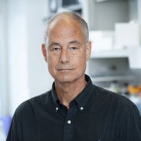
Stewart Shuman
. Portrait identified by the named Sloan Kettering Institute laboratory profile.C. Robin Buell is a researcher in genomics and bioinformatics, including the genome biology of plants and plant-associated organisms. Her institutional research description emphasizes comparative genomics and the provision of databases and computational tools for interpreting genomes.[ref]
C. Robin Buell research profile at the University of Georgia. Named faculty profile, research description
Buell was first author of the 2003 complete genome sequence report for Pseudomonas syringae pathovar tomato DC3000. The publication described approximately million base pairs, distributed across one circular chromosome and two plasmids, with predicted open reading frames. These are the study's reported assembly and annotation results, rather than an assertion that later annotations must retain exactly the same counts.[ref]
An illuminating distinction is the difference between a sequence measurement and its interpretation. DNA sequencing supplies evidence for the order of nucleotides; genome annotation identifies candidate features and proposes their biological roles. An open reading frame is therefore not, by itself, a demonstration that a particular protein is expressed or has a proposed function. Independent evidence can test those additional claims.
The published scale also gives a useful arithmetic check. Dividing the approximate genome size by its predicted open reading frame count givesbase pairs of total genome sequence per predicted open reading frame. This is a bookkeeping ratio, not a mean coding-sequence length: its numerator includes noncoding sequence, and the annotation may contain overlapping features. Confusing these quantities would turn a reasonable summary of genome density into an incorrect claim about individual genes. This example shows why the definitions of both numerator and denominator matter when interpreting large biological datasets.
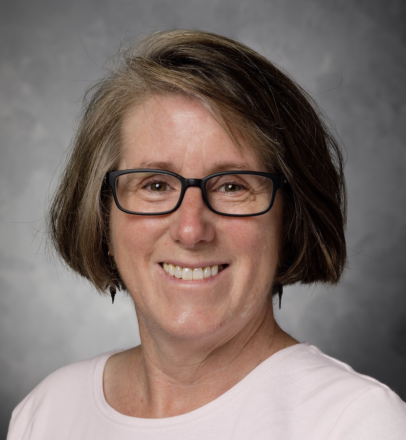
C. Robin Buell
. Portrait explicitly identified in the University of Georgia faculty profile.Robert Silber is a researcher credited with early characterization of RNA ligases. His full name and New York University medicine affiliation appear on the original 1972 article, although the indexed citation abbreviates the byline as R. Silber.[ref]
Purification and properties of bacteriophage T4-induced RNA ligase by R. Silber and others. Original full byline and author affiliation; PNAS 69, 3009–3013
Silber and collaborators investigated an RNA ligase from phage-infected Escherichia coli. The purified enzyme converted labelled terminal phosphate groups into internal phosphodiester bonds, requiring ATP and magnesium. Resistance to alkaline phosphatase, together with tests using exonucleases, supported joining the two ends of an individual RNA chain to produce circular RNA under the studied conditions.[ref]
Purification and properties of bacteriophage T4-induced RNA ligase by R. Silber and others. Abstract: ATP/Mg dependence, protected end label and exonuclease evidence for intramolecular circularization
The experiment illustrates why several chemical observations are needed to establish molecular topology. A label no longer accessible at a chain end suggests a change in its chemical setting, but that result alone does not determine whether two different molecules joined or one molecule closed on itself.
An original counting model makes the ambiguity precise. Begin with linear chains, each with two free ends, giving ends. Closing chains into separate rings leaves linear chains, free ends and total molecules. Alternatively, joining disjoint pairs into longer linear chains leaves molecules, again with free ends. The same reduction in end count can therefore accompany different molecular products. These are invented counts, not the paper's measurements; additional chemical and size information is needed to distinguish the products.
The historical inference combined the protected label with evidence that the product lacked accessible opposite ends. It was an inference about the tested RNA substrate and assay, not a claim that every RNA ligase reaction always creates a circle.[ref]
Purification and properties of bacteriophage T4-induced RNA ligase by R. Silber and others. Abstract: poly(A) substrate, no detected intermolecular reaction and circular-product interpretation
Cristofer Enroth is a structural-biochemistry researcher credited on the 2000 comparison of bovine-milk xanthine dehydrogenase and xanthine oxidase. The original article's full byline supplies this spelling, while its contribution note identifies Enroth as one of three equal contributors.[ref]
Crystal structures of bovine milk xanthine dehydrogenase and xanthine oxidase: Structure-based mechanism of conversion by C. Enroth and others. Original full byline and equal-contribution footnote; PNAS 97, 10723–10728
The study compared structures at and ångströms crystallographic resolution. It connected conversion between the two forms with structural changes near the FAD region. Rearrangement following cleavage of exposed loops partly obstructed access of NAD+ to the FAD cofactor and changed the local electrostatic environment. This supplied a structural explanation for altered electron acceptor specificity.[ref]
Crystal structures of bovine milk xanthine dehydrogenase and xanthine oxidase: Structure-based mechanism of conversion by C. Enroth and others. Abstract: proteolytic conversion, FAD-region loop and NAD access
The important conceptual point is that an enzyme is an organized route through a reaction, not just a collection of unchanged chemical groups. Altering access near one part of that route can change which electron acceptor supports turnover without requiring removal of every catalytic component.
Consider an invented assay example. Preparation A consumes units of a chosen substrate while producing the corresponding NADH signal; preparation B gives almost no NADH signal under the same measurement scheme. That observation alone does not distinguish complete loss of catalysis from loss of efficient coupling to NAD+. An independent measurement of substrate conversion, accompanied by a test of an alternative electron acceptor, addresses different parts of the explanation. The units here are illustrative, not reported rates or a proposed universal reaction stoichiometry.
Comparative structure and functional evidence therefore answer complementary questions: where access and conformation change, and which reactions remain possible. Enroth's credited study provides a particularly clear example of how a local structural rearrangement can redirect an enzyme's interaction with its reaction partners.
T. M. Weaver is the published author name credited on the 1995 structural study of Fumarase C from Escherichia coli. The paper lists a University of Minnesota biochemistry affiliation. Its initials are retained here as the verified scientific byline.[ref]
The multisubunit active site of fumarase C from Escherichia coli by T. M. Weaver and others. Published byline, affiliation and Abstract; Nature Structural Biology 2, 654–662
The study determined the four-subunit protein complex at ångströms crystallographic resolution. A tungstate derivative, acting as a competitive inhibitor, helped locate an active site containing contributions from three of the four protein subunits. Thus, the site was not simply a pocket belonging to one isolated chain.[ref]
The multisubunit active site of fumarase C from Escherichia coli by T. M. Weaver and others. Abstract: tungstate derivative and multisubunit active site
This gives an illuminating reason to distinguish folding from assembly. A purified protein subunit could retain its own folded shape yet fail to form a complete active site if a neighboring contribution is missing. Conversely, identifying a conserved catalytic group in a sequence does not establish that the required protein complex has assembled.
An original probability model illustrates the possible amplification. Suppose an idealized site requires three independently functional subunit contributions, each present with probability . Its probability of being complete is . With , this is , rather than . In that model, losing of individual contributions leaves of sites incomplete. This is not a measured activity law for Fumarase C: independent availability, random assembly and all-or-nothing site function are explicit assumptions. A real defect could affect assembly, chemical turnover or both.
The structural observation motivates complementary measurements: assess protein oligomerization separately from reaction rate, then ask whether altered assembly accounts for altered catalysis. A crystal structure supplies a concrete spatial hypothesis about these relationships, while functional measurements test its consequences.
Naoko Tanaka is a biochemist credited with identifying RtcB as an RNA ligase in joint work with Stewart Shuman. The original 2011 publication names both authors and their Sloan-Kettering molecular-biology affiliation. It tested a concrete biochemical function for a previously uncharacterized protein associated with an RNA repair gene cluster.[ref]
RtcB is the RNA ligase component of an Escherichia coli RNA repair operon by Naoko Tanaka and Stewart Shuman. Published byline, affiliations and Abstract
The experimental question was whether RtcB itself could join broken RNA. The researchers tested a broken tRNA-like stem-loop with cyclic-phosphate and hydroxyl ends, then separated labelled substrate and ligated product by denaturing polyacrylamide gel electrophoresis. Product analysis supported formation of a normal 3′,5′ phosphodiester bond at the joined site. This demonstrated a chemical activity; the initial paper treated broader physiological repair roles as proposals rather than as facts established for every organism.[ref]
RtcB is the RNA ligase component of an Escherichia coli RNA repair operon by Naoko Tanaka and Stewart Shuman. Abstract; Figures 1 and 3, labelled product analysis and junction chemistry
The labelling design gives an illuminating counting example. The reported broken substrate contained a labelled -nucleotide strand and an unlabelled -nucleotide partner. Joining them produces a -nucleotide strand but does not double the number of labels per molecule.[ref]
RtcB is the RNA ligase component of an Escherichia coli RNA repair operon by Naoko Tanaka and Stewart Shuman. Figure 1C, two strands of the broken substrate
If an invented measurement gives corrected label units in product and in unjoined labelled substrate, the inferred joined fraction is , assuming equal recovery and label detection. Multiplying the product signal by would instead introduce an inappropriate length correction: the label counts molecules carrying that label, not their total RNA mass.William Richard Jeck is a researcher credited with identifying and validating endogenous circular RNA species. His Duke institutional profile gives the full name, and the corresponding institutional publication list directly links him to the 2013 circular-RNA paper published under William R. Jeck.[ref]
William Richard Jeck profile and scholarly works at Duke University. Named profile; Scholarly Works listing of the 2013 RNA article
Jeck and collaborators compared RNA sequencing results from samples treated with RNase R and corresponding untreated samples. The approach enriched for molecules resistant to degradation of linear RNA and detected junctions inconsistent with ordinary linear exon order. Complementary assays tested the circular interpretation. The paper also reported an association with complementary Alu elements in bordering introns; that association alone is not a complete reaction mechanism for every circle.[ref]
Circular RNAs are abundant, conserved, and associated with ALU repeats by William R. Jeck and others. Abstract and Figures 1–4; genomic-feature analysis
A numerical example clarifies what enrichment means. Imagine an original mixture of circular and linear RNA molecules, so circles comprise . After a hypothetical treatment, suppose circular and linear molecules remain. Circles now comprise of the recovered mixture, an -fold increase in their fraction, even though their absolute count decreased by . These invented counts assume that molecules are counted comparably and are not measurements from the paper. They show why a large proportional enrichment must not be interpreted as treatment having created more circles.
The same care applies to a candidate back-splicing junction: identifying an unusual sequence order and proving that its source molecule is circular are separate steps. Enzyme resistance, junction evidence and independent product characterization address different alternatives. Jeck's credited contribution is therefore more informative than a catalogue of surprising sequencing reads: it illustrates how computational discovery and biochemical validation can constrain one another without making every resistant transcript a proven circular RNA.

Sebastian M. Fica is a molecular biologist studying how the spliceosome carries out RNA splicing. His work connects biochemical tests of chemical mechanism with structural analysis of the RNA–protein machine. His Oxford laboratory biography records a doctorate in cell and molecular biology in 2013 and a move into structural postdoctoral work in 2014.[ref]
Sebastian M. Fica laboratory biography at the University of Oxford. “Short biography”: doctoral work on splicing catalysis and subsequent training in electron cryo-microscopy.
In a 2013 collaborative study, Fica and colleagues used substitutions in U6 small nuclear RNA and metal-rescue assays in assembled yeast spliceosomes. Their results connected RNA ligands to catalytic metals in both splicing reactions. The paper marks equal contributions by its first two authors; the result belongs to the collaborating team.[ref]
The scientific question is sharper than asking whether RNA is present near a reaction. An RNA component might hold other molecules in place, or its own chemical groups might help build the catalytic center. Perturbing a candidate group and testing a compensating chemical change links structure to function. Structural snapshots complement such tests by locating molecular contacts, while reaction assays ask whether those contacts matter for catalysis.
Oxford's departmental profile describes his group's combination of biochemical and structural approaches to splice-site selection and catalytic dynamics.[ref]
https://www.bioch.ox.ac.uk/node/1889921. Departmental research profile, “Mechanisms and molecular structure of the spliceosome during pre-mRNA splicing”

Sebastian M. Fica, portrait identified on his University of Oxford departmental profile
. Umesh Varshney is a molecular biologist whose research investigates mechanisms of biological translation and DNA repair. His Indian Institute of Science profile identifies studies of protein synthesis and DNA repair using bacterial model systems, and lists him as an honorary professor in its Department of Microbiology and Cell Biology.[ref]
Umesh Varshney research profile at the Indian Institute of Science. Profile heading and Research section.
A 1991 collaborative paper provides a specific contribution to understanding transfer RNA identity. Varshney and colleagues altered an initiator transfer RNA in Escherichia coli and tested its function in both initiation and chain elongation. Internal amber stop codon suppression measured elongator activity; one variant could also initiate at an upstream UAG and suppress a downstream UAG. The result showed that the two activities can coexist in an engineered carrier.[ref]
Mutants of initiator tRNA that function both as initiators and elongators by Umesh Varshney and others. Abstract and pp.18018–18024: acceptor-stem variants and the dual-function test.
This distinguishes two questions that an amino-acid label alone cannot answer: what chemical cargo a transfer RNA carries, and which step of biological translation recognizes that carrier. Testing the same RNA at an initiating position and an internal codon probes a functional distinction, rather than merely comparing two RNA sequences.
His departmental research account also describes ribosome recycling and the recognition features that distinguish initiator transfer RNA from elongator transfer RNA.[ref]
Umesh Varshney research profile at the Indian Institute of Science. Protein Synthesis section, ribosome recycling and initiator-tRNA structure–function work.
These connect molecular recognition to the continued use of the translation machinery, beyond delivery of a single amino acid.
Roy Parker is a molecular biologist studying how cells produce, translate and degrade messenger RNA. His University of Colorado Boulder profile describes this research program, and his institutional curriculum vitae records a genetics doctorate from the University of California, San Francisco in 1985.[ref]
Roy Parker profile at the University of Colorado Boulder. Research description; institutional CV p.1, education.
His early work helped establish a sequence-recognition role for U2 small nuclear RNA. In a 1987 collaborative study, compensating changes in U2 suppressed corresponding changes in the yeast intron branch region. The matching suppression connected RNA sequence to base pairing in RNA splicing, rather than merely associating U2 with the machinery.[ref]
Recognition of the TACTAAC box during mRNA splicing in yeast involves base pairing to the U2-like snRNA by Roy Parker and others. Abstract, branch-site mutations and allele-specific compensatory U2 changes.
A later study with Ujwal Sheth connected RNA decay to cellular location. Functional fusions to green fluorescent protein placed several decapping factors in discrete cytoplasmic foci, termed P-bodies. Perturbing steps of the decay pathway changed those foci, and RNA decay intermediates also localized there.[ref]
Decapping and Decay of Messenger RNA Occur in Cytoplasmic Processing Bodies by Ujwal Sheth and Roy Parker. Abstract and Figures1–2: localization and pathway perturbations.
The combination of imaging and perturbation is scientifically important. A fluorescent spot alone shows where a marked component accumulates; it does not establish what reaction happens there. Comparing locations with the consequences of changing particular processing steps tests a mechanistic explanation. Nor does an accumulation of decay intermediates automatically mean that decay has become faster: material can accumulate when an exit step is blocked. This connects the chemistry of messenger RNA processing to the organization and dynamics of the cytoplasm.

Roy Parker, portrait on his University of Colorado Boulder BioFrontiers profile
. Ujwal Sheth is a molecular biologist associated with experimental studies of messenger RNA localization and turnover. A 2003 paper with Roy Parker, listing a University of Arizona research affiliation, investigated the relationship between RNA decay and discrete cytoplasmic structures named P-bodies.[ref]
Decapping and Decay of Messenger RNA Occur in Cytoplasmic Processing Bodies by Ujwal Sheth and Roy Parker. Primary byline, affiliation and Abstract.
The study combined localization with functional perturbations. Factors involved in decapping accumulated in cytoplasmic foci, and RNA degradation intermediates localized there. Interfering with different steps of RNA turnover changed the abundance of the foci. These comparisons helped distinguish a compartment associated with processing from a fluorescent aggregation seen without a functional test.[ref]
Decapping and Decay of Messenger RNA Occur in Cytoplasmic Processing Bodies by Ujwal Sheth and Roy Parker. Abstract and Figures1–2: localization and changes following pathway perturbations.
Sheth also coauthored a 2005 study linking P-body assembly to nontranslating RNA. Its experiments connected assembly to RNA integrity and showed contrasting responses to inhibition of translation initiation and elongation.[ref]
Processing bodies require RNA for assembly and contain nontranslating mRNAs by Daniela Teixeira and others. Abstract and Figure6: translation-state comparisons and RNA-dependent integrity.
The work made RNA's participation a testable part of the explanation, beyond treating the visible focus as a container for proteins.The broader scientific lesson is to connect three kinds of evidence: where molecules localize, what happens when a pathway changes, and whether the proposed components are needed for assembly. A change in spot number alone does not identify the direction or rate of RNA processing. These questions connect cell organization with the lifetime and use of a genetic message.
A captioned laboratory photograph of Sheth beside Roy Parker appears on PDF page 9 of HHMI Bulletin September 2003 by Howard Hughes Medical Institute.
Daniela Teixeira is a molecular biologist whose published research investigated P-body assembly and the relationship between messenger RNA organization and biological translation. She was first author of a2005 study with Roy Parker's group showing that yeast P-bodies depend on RNA for their assembly and contain nontranslating messages.[ref]
Processing bodies require RNA for assembly and contain nontranslating mRNAs by Daniela Teixeira and others. Primary byline and Abstract, RNA dependence and nontranslating mRNAs
The study combined cellular observations with experiments on enriched P-body preparations. Digestion with a ribonuclease disrupted the preparations, testing whether RNA contributed to their integrity. In cells, perturbing translation initiation and trapping messages in polysomes produced contrasting P-body responses. Those comparisons distinguished effects on RNA availability from the vague statement that translation had been inhibited.[ref]
Processing bodies require RNA for assembly and contain nontranslating mRNAs by Daniela Teixeira and others. Abstract; Figure6 and associated Results, RNA digestion and translation-state comparisons
An original image-analysis example illustrates a limit of such observations. Suppose a hypothetical cell contains ten visible foci with four arbitrary fluorescence units each, while a second condition contains five foci with eight units each. Both have40 units assigned to the foci. Counting foci alone would miss that equivalence; conversely, equal total signal would not establish identical molecular composition or RNA decay activity. The numbers are stipulated, not measurements from Teixeira's paper.
The Parker group's institutional alumni directory records her training period as2001–2005.[ref]
Alumni of the Parker Research Group. Grad Students section, Daniela Teixeira entry
Her scientific contribution here is the experimental connection among RNA, translation state and the assembly of a visible cytoplasmic structure.U. Gubler is first author, with B. J. Hoffman, of a 1983 method for generating cDNA libraries from small amounts of messenger RNA. The method combined first-strand synthesis with second-strand synthesis mediated by Ribonuclease H and DNA polymerase I.[ref]
A simple and very efficient method for generating cDNA libraries by U. Gubler and B. J. Hoffman. Gene 25, pp. 263–269, primary byline and Abstract
This contribution made information in an RNA population accessible through recoverable DNA copies. The published initial is retained without assuming an expanded identity.A cDNA library represents the RNA molecules successfully copied and recovered, rather than automatically representing every gene equally. Reverse transcription, second-strand synthesis and subsequent recovery can change representation. A useful library therefore requires more than a high total number of clones: rare transcripts and full-length products must also be considered.
An original sampling calculation isolates the rare-transcript question. Suppose a target transcript has probability in each independent recovered clone. The probability that examined clones contain no target isFor , this is about 0.448. Requiring at least 95 percent probability of finding one target givesso at least 299 clones are needed under the stated independent-sampling model. Copying bias that lowers the recovered target fraction changes that calculation even if the starting RNA fraction was known.
The primary method paper also tested fidelity against a known transcript sequence.[ref]
A simple and very efficient method for generating cDNA libraries by U. Gubler and B. J. Hoffman. Abstract, full-length sequence validation
Representation, product length and sequence agreement are distinct measurements. A large library can still undersample a rare transcript or contain truncated copies; neither conclusion follows from clone count alone.B. J. Hoffman coauthored the 1983 cDNA library method with U. Gubler. Their procedure connected first-strand copying of messenger RNA with an enzymatic second-strand synthesis step, simplifying recovery of double-stranded DNA copies.[ref]
A simple and very efficient method for generating cDNA libraries by U. Gubler and B. J. Hoffman. Primary byline and Abstract
The paper establishes this collaborative contribution; it does not assign every laboratory step to an individual author.A particularly informative validation used a previously sequenced bovine transcript and tested the recovered copies by sequencing. The abstract reports faithful full-length products for that test.[ref]
A simple and very efficient method for generating cDNA libraries by U. Gubler and B. J. Hoffman. Abstract, known bovine preproenkephalin RNA comparison
Sequence comparison addresses what the method preserved, rather than merely showing that DNA material was produced.An original probability example separates two quality questions. Let mean that a recovered copy is full length, and mean that its sequence meets a stated correctness criterion. ThenIf a stipulated preparation has and , the fraction satisfying both criteria is 0.76. Multiplying marginal probabilities would require an additional independence assumption; the conditional expression does not.
For a cDNA library, a product can therefore have plausible length but sequence changes, or correct sequence over a shorter region while lacking an end. Validation should state which region was read and which criterion was tested. An illustrative 100-copy sample with 80 full-length products and 76 meeting both conditions has 95 percent correctness among the full-length products but 76 percent joint success across all recovered copies. These numbers explain the distinction and are not measurements from the original study.
Walter Gilbert helped turn questions about gene regulation and DNA sequence into experimentally accessible molecular measurements. He moved from physics toward molecular biology at Harvard; the Cold Spring Harbor Laboratory archive records that transition and preserves his research collection.[ref]
Walter Gilbert in the Cold Spring Harbor Laboratory archives. Institutional biography and Scope and Content

Walter Gilbert, portrait identified by Cold Spring Harbor Laboratory and credited to Harvard News Service
. Source. With Benno Müller-Hill, Gilbert reported isolation of the lac repressor in 1966. Their assay detected binding to an inducing small molecule using equilibrium dialysis, providing a biochemical handle on a genetically inferred control protein.[ref]
Isolation of the lac repressor by Walter Gilbert and Benno Müller-Hill. printed p. 1891, assay description
The distinction between a measurable binding activity and a fully established regulatory mechanism remains important: the assay did not assume that every detail of operator recognition was already known.With Allan M. Maxam, he later developed the chemical DNA sequencing method now called Maxam-Gilbert sequencing. Their 1977 paper described partial cleavage at selected bases in terminally labeled DNA, followed by polyacrylamide gel electrophoresis to resolve fragment lengths.[ref]
A new method for sequencing DNA by Allan M. Maxam and Walter Gilbert. PNAS 74, pp. 560–564, Abstract
A ladder of labeled fragment lengths converts chemical specificity into information about positions along a strand.An original schematic inference illustrates the readout. Suppose calibrated fragment-length patterns assign A to positions 1 and 6, G to positions 2 and 5, C to positions 3 and 7, and T to positions 4 and 8. Combining the position sets gives AGCTGACT in the stated reading direction. A G-selective measurement alone would leave six positions unresolved; the different chemical channels supply complementary information.
The example abstracts away migration calibration, cleavage-site offsets and band overlap. Real interpretation requires identifying the labeled end and relating each band to the appropriate base position. This sequencing approach interrogates a pre-existing labeled strand through cleavage, so its measurement logic is different from inferring sequence from terminated synthesis products. Gilbert's work exemplifies how designing a recoverable physical signal can transform an otherwise indirect molecular question.
Benno Müller-Hill, with Walter Gilbert, isolated the lac repressor in work published in 1966. The research connected a genetically inferred control function with an experimentally detectable molecular component. Their primary paper developed an assay based on binding an inducing small molecule, detected through equilibrium dialysis against radioactive Isopropyl β-D-1-thiogalactopyranoside.[ref]
Isolation of the lac repressor by Walter Gilbert and Benno Müller-Hill. PNAS 56, printed p. 1891, assay description and Design of the Experiment
That choice was logically important. The opening discussion considered a model in which repressor binds the lac operator, but the assay sought inducer interaction without requiring every detail of the repression mechanism to be settled first. Measuring binding to an inducer is not itself the same experiment as demonstrating binding to operator DNA.
An original one-site model explains the dialysis signal. A membrane permits free ligand to equilibrate between two compartments while retaining protein on one side. If the common free ligand concentration is , total binding-site concentration is and dissociation constant is , the retained bound concentration isThe protein-containing compartment has total ligand concentration , while the comparison compartment has under this ideal model. With stipulated , , the totals are 2.5 and 2.0 micromoles per litre. Their excess measures binding even though free ligand equilibrates.
These values are illustrative, not fitted constants from the 1966 experiment. Nonspecific adsorption, unequal accessible volumes and other binding components require controls. Protein purification can then be followed with a functional assay rather than by the mere presence of protein material. This is the experimental bridge between a gene-control hypothesis and a molecular mechanism; the joint paper does not justify assigning every experimental operation to Müller-Hill alone.
Pamela M. Pollock investigates molecular alterations associated with cancer. She is first author of a 2003 study that analyzed BRAF mutations in microdissected samples classified as melanocytic nevus or melanoma.[ref]
High frequency of BRAF mutations in nevi by Pamela M. Pollock and others. Nature Genetics 33, pp. 19–20, primary byline and Abstract
The study found the substitution labeled V599E in its original numbering in 63 of 77 nevi. The presence of that alteration in benign lesions made it important to distinguish a pathway-associated initiating event from sufficient evidence of malignant transformation.Microdissection connects a molecular result with a selected tissue region. It can enrich the cells relevant to the question, while leaving the sample's actual composition and sequencing sensitivity as separate measurement issues. A mutation detected in a mixed tissue sample is not automatically present in every constituent cell.
An original allele-count model illustrates the distinction. Suppose 30 percent of the sampled cells carry one altered BRAF copy and one unaltered copy, while all remaining cells have two unaltered copies. With equal DNA contribution per cell, no copy-number alteration and no measurement bias, the expected variant allele fraction isA 15 percent variant fraction is therefore compatible with 30 percent mutation-bearing cells in this model. It would be incorrect to identify those two percentages without the copy-count assumptions. Different purity, copy number or recovery changes the relationship.
The published paper's selected-lesion frequencies answer another question again: how many sampled lesions had the detected alteration. A lesion-level proportion such as is neither a within-lesion allele fraction nor the probability that an individual lesion will progress. Pollock's team's contribution links molecular pathway evidence with those distinct biological levels of interpretation, rather than making mutation presence a self-sufficient classification of malignancy.
Allan M. Maxam, with Walter Gilbert, developed a chemical approach to DNA sequencing reported in 1977. Their primary paper describes partial cleavage of terminally labeled DNA at selected bases, then size separation of labeled fragments by polyacrylamide gel electrophoresis.[ref]
A new method for sequencing DNA by Allan M. Maxam and Walter Gilbert. PNAS 74, pp. 560–564, original full byline and Abstract
Maxam-Gilbert sequencing converts chemical selectivity into a position-dependent pattern along a pre-existing strand.The word partial is central to the measurement logic. Different molecules contribute fragments ending at different positions, while a common labeled end supplies a reference. Extensive repeated cleavage of one molecule can remove information about more distant sites, because only the fragment still containing the label is visible in the corresponding readout.
An original probability model illustrates the tradeoff without prescribing experimental conditions. Suppose the number of relevant breaks in one molecule follows a Poisson distribution with mean . ThenAt stipulated , about 81.9 percent have no break, 16.4 percent have one and 1.75 percent have multiple breaks. At , those fractions are about 13.5, 27.1 and 59.4 percent. More cleavage does not translate monotonically into a more interpretable set of singly terminated fragments. Actual sequence-dependent chemistry need not follow this simplified independent-break model.
The measured gel is also not the sequence itself. Reading requires calibrating fragment size, identifying the labeled end and comparing the base-selective channels. Overlapping bands or an incorrect orientation can corrupt an otherwise real chemical signal. Maxam's contribution demonstrates how a carefully designed molecular measurement can expose sequence information that bulk composition alone cannot recover.
J. A. Frier coauthored the 1977 study of human fetal deoxyhemoglobin with Max Perutz. The primary structure record 1FDH reports a human alpha/gamma tetramer examined by X-ray diffraction at resolution.[ref]
Structure of human foetal deoxyhaemoglobin by J. A. Frier and M. F. Perutz. Primary citation, Biological Assembly, Macromolecules and Experimental Data
This is a verified structural-biology contribution; the published initials are retained without inferring an expanded identity from a deposition-name variant.Comparing fetal and adult hemoglobin structures tests how changes in subunit composition alter local interactions. The paper's indexed abstract discusses differences at the binding region for 2,3-Bisphosphoglycerate.[ref]
Structure of human foetal deoxyhaemoglobin by J. A. Frier and M. F. Perutz. Indexed primary publisher abstract
Such a comparison can suggest why an effector binds differently. It does not make a static crystal structure a direct measurement of all physiological response rates or affinities.An original coordinate example distinguishes spatial resolution from uncertainty in a derived distance. Suppose two modeled positions along one axis differ by , with independent standard uncertainties each. The uncertainty of their signed separation isIf the coordinate errors have covariance , the variance becomes . Shared model errors can therefore change the uncertainty of a relative separation. These are illustrative assumptions, not coordinate uncertainties reported for 1FDH.
A quoted diffraction resolution alone should not be substituted for either number in this example. It describes the experimental detail available to the model, while coordinate uncertainty also depends on data, occupancy, disorder and refinement. Frier and Perutz's structural result is valuable because it offers a molecular arrangement against which those broader mechanistic questions can be tested.
Pinned article: Introduction to the OurBigBook Project
Welcome to the OurBigBook Project! Our goal is to create the perfect publishing platform for STEM subjects, and get university-level students to write the best free STEM tutorials ever.
Everyone is welcome to create an account and play with the site: ourbigbook.com/go/register. We belive that students themselves can write amazing tutorials, but teachers are welcome too. You can write about anything you want, it doesn't have to be STEM or even educational. Silly test content is very welcome and you won't be penalized in any way. Just keep it legal!
Intro to OurBigBook
. Source. We have two killer features:
- topics: topics group articles by different users with the same title, e.g. here is the topic for the "Fundamental Theorem of Calculus" ourbigbook.com/go/topic/fundamental-theorem-of-calculusArticles of different users are sorted by upvote within each article page. This feature is a bit like:
- a Wikipedia where each user can have their own version of each article
- a Q&A website like Stack Overflow, where multiple people can give their views on a given topic, and the best ones are sorted by upvote. Except you don't need to wait for someone to ask first, and any topic goes, no matter how narrow or broad
This feature makes it possible for readers to find better explanations of any topic created by other writers. And it allows writers to create an explanation in a place that readers might actually find it.
Figure 1. Screenshot of the "Derivative" topic page. View it live at: ourbigbook.com/go/topic/derivativeVideo 2. OurBigBook Web topics demo. Source. - local editing: you can store all your personal knowledge base content locally in a plaintext markup format that can be edited locally and published either:This way you can be sure that even if OurBigBook.com were to go down one day (which we have no plans to do as it is quite cheap to host!), your content will still be perfectly readable as a static site.
- to OurBigBook.com to get awesome multi-user features like topics and likes
- as HTML files to a static website, which you can host yourself for free on many external providers like GitHub Pages, and remain in full control

Figure 2. You can publish local OurBigBook lightweight markup files to either OurBigBook.com or as a static website.
Figure 3. Visual Studio Code extension installation.
Figure 5. . You can also edit articles on the Web editor without installing anything locally. Video 3. Edit locally and publish demo. Source. This shows editing OurBigBook Markup and publishing it using the Visual Studio Code extension. 
- Infinitely deep tables of contents:


All our software is open source and hosted at: github.com/ourbigbook/ourbigbook
Further documentation can be found at: docs.ourbigbook.com
Feel free to reach our to us for any help or suggestions: docs.ourbigbook.com/#contact