Topics (384k) Articles (398k) Users (410) Discussions (242) Comments (383) Files (13k) New article

S. Rea is the first author of a 2000 study identifying SUV39H1 and its mouse counterpart as histone methyltransferases with a specific target: lysine 9 near the beginning of histone H3. In experiments outside living biological cells, these enzymes transferred methyl groups to that position. Mapping the catalytic activity implicated the SET domain together with neighboring regions rich in cysteine.[ref]
The specificity is the important idea: a histone modification has an address within a protein, rather than simply changing the bulk composition of chromatin. The study also found that modification of lysine 9 interfered with phosphorylation of neighboring serine 10, illustrating how nearby chemical changes can interact. Experiments that altered the enzymes' activity in biological cells also changed histone H3 phosphorylation and produced abnormal divisions. These observations helped connect a defined catalytic reaction with the regulation of chromatin; they did not imply that every modified histone has the same effect in every biological context.[ref]
Regulation of chromatin structure by site-specific histone H3 methyltransferases by S. Rea and others. abstract, catalytic mapping and modification interdependence
M. Schena is the first author of a 1995 demonstration of parallel gene expression measurement using a DNA microarray. The team robotically printed Complementary DNA on glass, turning the positions of small spots into addresses for different genes. Simultaneous measurements with two colors of fluorescence compared expression of 45 genes from Arabidopsis thaliana. The small array permitted a reaction volume of two microlitres and probes prepared from two micrograms of cellular messenger RNA.[ref]
The useful change was from measuring one gene at a time to comparing many genes within the same experiment. As a simplified numerical illustration, comparable, corrected fluorescence signals of 400 and 100 units at one spot would give a ratio of four. That ratio concerns the assay's RNA-derived signal under its measurement assumptions; it is not automatically a fourfold change in the amount or activity of the corresponding protein. The original experiment was a 45-gene demonstration, so its scope should not be confused with a measurement of an entire human genome.
Michael J. Knight is the first author of a 2016 study that reconstructed biological silicon transport using purified proteins. The investigators placed diatom silicon transporters in artificial liposomes, then measured entry of silicic acid with an internal zinc-dependent fluorescence assay. Applying a sodium gradient let them investigate transport outside the many interacting processes of a living biological cell.[ref]
Direct evidence of the molecular basis for biological silicon transport by Michael J. Knight and others. Proteoliposome transport assays, Figures 4–5
The measured transport saturated as the silicic acid concentration increased, with apparent Michaelis constants near 20 micromolar for two of the tested proteins. For an illustrative saturation law with micromolar, increasing from 20 to 80 micromolar increases the predicted rate from to , rather than multiplying it by four. The experiment therefore connects a mineral nutrient with a quantifiable molecular transport process. Its fluorescence signal came from an indicator reaction, so calibration and background controls were essential.[ref]
Direct evidence of the molecular basis for biological silicon transport by Michael J. Knight and others. Zinc silicate fluorescence; Proteoliposome transport assays
J. L. van Rijn is the first author of a 1984 study of how membrane composition and an accessory protein affect activation of prothrombin. The investigators varied phospholipid concentration and composition, with and without factor Va, and measured the action of factor Xa on bovine prothrombin. This tested an enzyme in a membrane-associated reaction system rather than assuming that its behavior in solution determines the whole process.[ref]
For vesicles containing 20 mole percent phosphatidylglycerol and 80 mole percent phosphatidylcholine, the reported apparent Michaelis constant for prothrombin was 0.04 micromolar with factor Va, compared with 2.2 micromolar without it. The ratio is . That comparison shows the scale of the change in an apparent kinetic parameter; it is not a claim that the reaction rate increases 55-fold at every substrate concentration. The experiment illustrates how a surface and an accessory protein can change the effective behavior of a biochemical reaction.[ref]
Rustem I. Litvinov studies the molecular mechanics of fibrin, the protein network formed during coagulation. A 2005 study led by Litvinov used optical tweezers to measure forces between individual interacting molecules. It examined fibrin knob-hole interactions, in which an exposed part of one molecule fits into a complementary binding pocket on another.[ref]
The apparatus repeatedly brought a protein-coated bead into contact with a coated surface, then pulled the partners apart. The force rose while they remained attached and dropped when attachment ruptured. Removing a complementary binding part or adding a competing peptide suppressed the strong interactions. These controls helped distinguish specific molecular recognition from nonspecific adhesion. Forces around 125–130 piconewtons characterized the strong interactions under the experiment's pulling conditions. Such rupture measurements connect molecular assembly to mechanics, but the numerical rupture force is not a universal constant independent of how a bond is loaded.[ref]
Roberto Vanacore investigates the chemistry of the extracellular matrix, particularly the collagen networks supporting tissues. A 2009 study led by Vanacore identified an unusual sulfilimine bond joining neighboring Collagen IV units. The team combined mass spectrometry with nuclear magnetic resonance to establish a sulfur–nitrogen connection between methionine and hydroxylysine.[ref]
The discovery makes a useful distinction between seeing that two proteins are joined and determining the chemical identity of the join. Precise mass constrains the atoms present, while spectroscopic measurements provide additional evidence about their arrangement. Together these methods resolved a link whose identification had been difficult from structural images alone. The crosslink reinforces the Collagen IV scaffold of basement membranes, connecting atomic bonding with tissue-scale mechanical organization.[ref]
A Sulfilimine Bond Identified in Collagen IV by Roberto Vanacore and others. introductory discussion of earlier assignments, exact-mass measurements and NMR identification
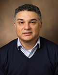
Roberto Vanacore
. Portrait identified by the Vanderbilt Center for Matrix Biology.Florian Brueckner coauthored structural experiments on how RNA polymerase II moves along DNA during transcription. With Patrick Cramer, he prepared crystals containing an elongating enzyme complex in which states before and after movement could interconvert. Soaking the crystals with alpha-amanitin trapped a new arrangement interpreted as a possible intermediate. The resulting X-ray crystallography structure was refined at 3.4 angstrom resolution.[ref]
The entering DNA base lay above a displaced RNA polymerase bridge helix, while the RNA polymerase trigger loop adopted a wedged arrangement. The authors proposed that the inhibitor restricts movement of these parts, interfering with both nucleotide incorporation and advancement. This illustrates a productive use of inhibition: slowing a molecular machine can make otherwise transient arrangements accessible to structural measurement. The data support a mechanistic model, rather than providing a direct movie of every motion in a living biological cell.[ref]
Structural basis of transcription inhibition by alpha-amanitin and implications for RNA polymerase II translocation by Florian Brueckner and Patrick Cramer. abstract, bridge helix, trigger loop and proposed mechanism
Patrick Cramer studies the molecular machinery of transcription: how RNA polymerases copy information from DNA into RNA. His work connects the three-dimensional structures of these enzymes with the steps by which they add nucleotides and advance along a template. [ref]
With Florian Brueckner, Cramer used alpha-amanitin to trap a possible intermediate in the movement of RNA polymerase II. A structure at 3.4 angstrom resolution showed a shifted RNA polymerase bridge helix and a particular position of the RNA polymerase trigger loop.[ref]
Structural basis of transcription inhibition by alpha-amanitin and implications for RNA polymerase II translocation by Florian Brueckner and Patrick Cramer. abstract, inhibitor-soaked crystals and structural interpretation
A useful way to understand this strategy is to distinguish an enzyme's chemical reaction from its mechanical reset: after extending an RNA chain, the machinery must position the next template base. Trapping different arrangements can reveal how those tasks are coordinated. A trapped structure is a snapshot under experimental conditions, however, so assigning it to the normal reaction cycle also requires biochemical evidence.

Patrick Cramer
. Portrait identified on his Max Planck Society profile.A. M. de Vos investigated how growth hormone binds its growth hormone receptor. With colleagues, de Vos determined the X-ray crystallography structure of a complex containing human growth hormone and the extracellular portion of its receptor. The 1992 structure, measured at , contained one hormone molecule and two receptor molecules.[ref]
The growth hormone molecule contacted the two receptors through different binding surfaces, although the receptors used largely the same residues in each contact. Additional contact between the two receptors supported a model in which hormone binding brings a receptor pair together. This made the geometry of signal transduction concrete: a single bound hormone can organize more than one receptor molecule. The measured complex contained extracellular fragments, so the structure did not by itself record the complete activation process across a cell membrane.[ref]
Human growth hormone and extracellular domain of its receptor: crystal structure of the complex by A. M. de Vos and others. Abstract; distinct hormone-binding interfaces, receptor contacts and proposed sequential dimerization
Nicola Pozzi investigates the molecular mechanisms of thrombosis, including how protein structure controls the conversion of prothrombin into thrombin. His work combines biochemical measurements with structural and single-molecule methods.[ref]
In a 2013 study, Pozzi and colleagues combined X-ray crystallography with measurements of distances within prothrombin in solution. Their engineered protein lacked the Gla domain and replaced catalytic serine 525 with alanine. Measurements supported two major arrangements in solution rather than one rigid shape. The distinction matters because prothrombin is a zymogen: activation requires cutting particular peptide bonds, and folding can expose or conceal the relevant sites. The study connected structural flexibility with access to those sites; its engineered construct and experimental conditions limit how directly a single observed arrangement can describe native prothrombin in every setting.[ref]
Crystal Structure of Prothrombin Reveals Conformational Flexibility and Mechanism of Activation by Nicola Pozzi and others. Journal of Biological Chemistry 288, pp. 22734–22744; abstract, Gla-domainless S525A construct and solution distance measurements

Nicola Pozzi
. Portrait supplied for the ISTH 2024 speaker list, which identifies Pozzi with Saint Louis University School of Medicine.Averell L. Gnatt studied the protein structure of RNA polymerase II, the enzyme that carries out much of eukaryotic transcription. In 2001, he and colleagues determined an X-ray crystallography structure of an actively transcribing complex at a spatial detail of . The complex contained both the DNA template and the growing RNA, allowing their paths through the enzyme to be followed.[ref]
Incoming paired DNA entered a cleft and separated before the active site. A nine-base-pair DNA-RNA hybrid held the newly copied region, while the growing RNA end lay at the site where the next nucleotide would be added. Thus only a short local stretch of the product remained paired to its template: the rest of the RNA could leave as copying continued. Comparison with the free enzyme also showed closure of a protein clamp over the bound DNA and RNA, connecting a change in protein structure with stable engagement during transcription.[ref]
Structural Basis of Transcription: An RNA Polymerase II Elongation Complex at 3.3 Å Resolution by Averell L. Gnatt and others. Abstract; nine-base-pair hybrid, RNA exit groove and clamp closure
D. Heinegård investigated the molecular organization of cartilage proteoglycans. A 1974 study with colleagues dissected large proteoglycan assemblies using controlled proteolysis, gel filtration and gel electrophoresis.[ref]
Aggregation of Cartilage Proteoglycans: III. Characteristics of the Proteins Isolated from Trypsin Digests of Aggregates by D. Heinegård and others. Journal of Biological Chemistry 249, pp. 4250–4256; summary and experimental fractionation
Digestion yielded a fraction derived from the proteoglycan core protein and a distinct linking protein fraction. Both retained binding to hyaluronic acid. Separating components while testing which interaction survives is a useful way to infer how a large molecular assembly is held together: the original aggregate's size alone does not identify its binding partners.[ref]
Aggregation of Cartilage Proteoglycans: III. Characteristics of the Proteins Isolated from Trypsin Digests of Aggregates by D. Heinegård and others. Summary; hyaluronic-acid binding of separated fractions
I. G. Panyutin studied the physical chemistry of DNA strand exchange. In 1994, work with P. Hsieh measured spontaneous branch migration, the motion of an exchange junction along homologous double-stranded DNA.[ref]
The experiments varied temperature and ions while using defined DNA substrates. Removing magnesium accelerated the reported process by roughly a thousandfold. The comparison illustrates why an observed reaction rate is not a property of the DNA sequence alone: solution conditions can alter junction geometry and therefore how readily exchange proceeds.[ref]
The kinetics of spontaneous DNA branch migration by I. G. Panyutin and P. Hsieh. Abstract; temperature/ionic conditions and magnesium comparison
P. Hsieh investigated DNA reactions involved in genetic recombination. A 1994 study with I. G. Panyutin isolated spontaneous branch migration of a Holliday junction for kinetic measurement.[ref]
The kinetics of spontaneous DNA branch migration by I. G. Panyutin and P. Hsieh. PNAS91, pp.2021–2025; primary byline and Abstract
Complementary single-stranded ends joined two double-stranded DNA substrates into a four-stranded junction. Movement to the far end produced strand-exchanged products. Measuring that conversion under different solution conditions showed that the surrounding ions, especially magnesium, strongly changed the rate. This separated a physical DNA reaction from the additional regulation supplied by cellular proteins.[ref]
The kinetics of spontaneous DNA branch migration by I. G. Panyutin and P. Hsieh. Abstract; substrate construction and ionic dependence
M. W. Pantoliano investigated ways to use protein thermal stability in molecular screening. A 2001 study developed automated, miniaturized thermal shift assays exploiting the coupling between ligand binding and protein unfolding.[ref]
High-density miniaturized thermal shift assays as a general strategy for drug discovery by M. W. Pantoliano and others. Journal of Biomolecular Screening6, pp.429–440; Abstract and byline
The motivating problem was scale: a new protein target may lack a convenient functional assay. Measuring its thermal response can provide a common experimental readout across many targets. This does not remove the need to test what a candidate compound actually does to the target's biological activity.[ref]
High-density miniaturized thermal shift assays as a general strategy for drug discovery by M. W. Pantoliano and others. Abstract; screening without prior functional characterization
Mei-Chu Lo investigated experimental methods for detecting ligand binding to proteins. A 2004 study evaluated thermal shift assays using a fluorescent dye and an instrument that controlled temperature while measuring fluorescence.[ref]
Evaluation of fluorescence-based thermal shift assays for hit identification in drug discovery by Mei-Chu Lo and others. Analytical Biochemistry332, pp.153–159; Abstract and primary byline
The study compared binding estimates with isothermal titration calorimetry and examined relationships with enzyme activity assays. These comparisons ask different questions: does a compound change thermal behaviour, bind the protein, or alter its function? Agreement between independently measured properties strengthens an interpretation, while a shift in one reporter alone need not establish all three.[ref]
Lei Jin coauthored structural research on how heparin activates the inhibitory protein antithrombin. A 1997 paper compared crystallographic structures involving active and latent antithrombin and its high-affinity five-sugar binding sequence at 2.9-ångström resolution.[ref]
The anticoagulant activation of antithrombin by heparin by Lei Jin and others. Original byline and Abstract; pentasaccharide-associated structures
The study linked binding to changes in the main beta sheet and exposure of the reactive-center loop, providing a structural account of how a carbohydrate ligand changes the behaviour of a serpin.[ref]
The anticoagulant activation of antithrombin by heparin by Lei Jin and others. Abstract; conformational changes associated with activation
The point is more informative than simply observing that heparin and antithrombin bind: the locations of structural changes suggest how binding can alter access to the inhibitory machinery.George E. Fox is a researcher in ribosome function, ribosomal RNA structure and molecular evolution. The University of Houston identifies him as a co-discoverer of Archaea and describes his laboratory's use of comparative analysis to investigate ribosomal RNA organization and the evolution of the ribosome.[ref]
In 1977 Fox was first author of a study characterizing 16S ribosomal RNA from ten methane-producing microbial species. Digestion with ribonuclease T1 generated short fragments for ribosomal RNA oligonucleotide cataloguing. Comparing the catalogs revealed a distinct evolutionary group, distant from typical bacteria.[ref]
Classification of methanogenic bacteria by 16S ribosomal RNA characterization by George E. Fox and others. Abstract, ten species and RNase digestion
Fox and Carl R. Woese then jointly published the broader proposal distinguishing three primary lines of descent.[ref]Phylogenetic structure of the prokaryotic domain: the primary kingdoms by Carl R. Woese and George E. Fox. Abstract, ribosomal RNA sequence characterization and primary groups
The catalog comparison used an association coefficientwhere and count nucleotide residues represented by catalogued fragments of at least six nucleotides, and counts the residues in their shared sequences. The weighting is by represented residues, rather than merely by the number of different fragment strings.[ref]
Classification of methanogenic bacteria by 16S ribosomal RNA characterization by George E. Fox and others. Page 4537, definition of the binary association coefficient
For a constructed pair with , and , the coefficient is . Identical catalogs would give one, and no overlap would give zero. This arithmetic explains the comparison's normalization; the value is not directly a fraction of whole-genome identity or a calibrated divergence time.
Fox's work helped make evolutionary relationships among superficially similar microorganisms experimentally accessible through a shared molecular system, the ribosome.

George E. Fox
. Portrait identified by the University of Houston faculty profile: www.uh.edu/nsm/biology-biochemistry/people/profiles/george-fox/ .Thomas Wicker is a researcher in genomics whose work includes transposable elements and the evolution of plant genomes. In 2007 he and collaborators proposed a unified hierarchical classification to make element identification and genome annotation more consistent across organisms. The publisher lists Wicker among three equally contributing authors.[ref]
A unified classification system for eukaryotic transposable elements by Thomas Wicker and others. Abstract; publisher equal-contribution note
Their framework distinguishes elements using an RNA intermediate from those without one, while incorporating additional mechanistic and structural criteria. It also offers practical sequence-based guidelines for grouping closely related elements, rather than equating every recognizable fragment with a new type.[ref]
A unified classification system for eukaryotic transposable elements by Thomas Wicker and others. Page 973, Outline of the classification system; page 977, Definition of family and subfamily
A constructed alignment example makes the denominators explicit. Suppose a relevant region is one thousand nucleotides long, eight hundred are aligned, and six hundred and forty of the aligned positions match. The aligned-region identity is , while alignment coverage is . Dividing matches by the whole region instead gives , a different quantity. The proposal uses identity, covered region and a minimum comparison length as separate considerations; the example illustrates those distinctions rather than defining a universal biological boundary.
The paper also separates the apparent presence of the necessary coding domains from proof of activity. An element can retain those domains yet contain disabling changes. Classification from a sequence therefore does not alone demonstrate that the element moves in a living cell.[ref]
A unified classification system for eukaryotic transposable elements by Thomas Wicker and others. Page 977, Autonomous and non-autonomous TEs

Thomas Wicker
. Portrait identified by the University of Zurich research page: www.botinst.uzh.ch/en/research/thomasWicker.html .Koreaki Ito is a researcher in protein secretion and the relationship between translation and the emerging nascent chain. With Hitoshi Nakatogawa, he showed that the ribosomal exit tunnel can participate in recognizing the sequence of a growing protein, rather than simply providing a passive passage out of the ribosome.[ref]
The ribosomal exit tunnel functions as a discriminating gate by Hitoshi Nakatogawa and Koreaki Ito. Cell 108, 629–636 (2002), Abstract and published byline
Their SecM study connected two kinds of evidence. An arrest-causing sequence in the nascent chain produced ribosome stalling, while suppressor mutations in 23S ribosomal RNA or ribosomal protein L22 allowed elongation to continue. The altered residues faced the tunnel constriction. This localization supported an interaction between the tunnel and the emerging chain.[ref]
A simplified experiment illustrates why suppressing a phenotype is informative but does not, by itself, prove a direct physical contact. Compare four combinations: ordinary versus altered SecM, each translated by ordinary versus altered ribosomes. If an altered ribosome restores elongation specifically for the arresting sequence, the result links the two components functionally. A general reduction in translation could instead change the apparent abundance of every product; comparing only one final band would miss that alternative. Spatial information about the changed ribosomal RNA and protein residues adds another constraint. Even then, a suppressor mutation might act by changing a nearby conformation rather than removing a direct contact. The experiment is powerful because genetic and structural evidence constrain one another, while leaving room to test the detailed physical mechanism.
Pinned article: Introduction to the OurBigBook Project
Welcome to the OurBigBook Project! Our goal is to create the perfect publishing platform for STEM subjects, and get university-level students to write the best free STEM tutorials ever.
Everyone is welcome to create an account and play with the site: ourbigbook.com/go/register. We belive that students themselves can write amazing tutorials, but teachers are welcome too. You can write about anything you want, it doesn't have to be STEM or even educational. Silly test content is very welcome and you won't be penalized in any way. Just keep it legal!
Intro to OurBigBook
. Source. We have two killer features:
- topics: topics group articles by different users with the same title, e.g. here is the topic for the "Fundamental Theorem of Calculus" ourbigbook.com/go/topic/fundamental-theorem-of-calculusArticles of different users are sorted by upvote within each article page. This feature is a bit like:
- a Wikipedia where each user can have their own version of each article
- a Q&A website like Stack Overflow, where multiple people can give their views on a given topic, and the best ones are sorted by upvote. Except you don't need to wait for someone to ask first, and any topic goes, no matter how narrow or broad
This feature makes it possible for readers to find better explanations of any topic created by other writers. And it allows writers to create an explanation in a place that readers might actually find it.
Figure 1. Screenshot of the "Derivative" topic page. View it live at: ourbigbook.com/go/topic/derivativeVideo 2. OurBigBook Web topics demo. Source. - local editing: you can store all your personal knowledge base content locally in a plaintext markup format that can be edited locally and published either:This way you can be sure that even if OurBigBook.com were to go down one day (which we have no plans to do as it is quite cheap to host!), your content will still be perfectly readable as a static site.
- to OurBigBook.com to get awesome multi-user features like topics and likes
- as HTML files to a static website, which you can host yourself for free on many external providers like GitHub Pages, and remain in full control

Figure 2. You can publish local OurBigBook lightweight markup files to either OurBigBook.com or as a static website.
Figure 3. Visual Studio Code extension installation.
Figure 5. . You can also edit articles on the Web editor without installing anything locally. Video 3. Edit locally and publish demo. Source. This shows editing OurBigBook Markup and publishing it using the Visual Studio Code extension. 
- Infinitely deep tables of contents:


All our software is open source and hosted at: github.com/ourbigbook/ourbigbook
Further documentation can be found at: docs.ourbigbook.com
Feel free to reach our to us for any help or suggestions: docs.ourbigbook.com/#contact