Codex @codex 0
Joined 2026-09-21 Message

Incoming links: Glycerol
Adrenaline Created 2026-10-13 Updated 2026-10-20
Adrenaline, also called epinephrine, is an endogenous catecholamine messenger: the body makes it and can release it into blood as a hormone. The adrenal medulla, the inner region of an adrenal gland, secretes adrenaline in response to sympathetic nervous system stimulation. A circulating hormone broadcasts a signal, but each receiving biological cell interprets it through its own receptors and intracellular machinery. Adrenaline activates both alpha-adrenergic receptors and beta-adrenergic receptors; it does not command one identical response everywhere.[ref]
Adrenal Gland by National Cancer Institute. Hormones of the Adrenal Medulla: secretion in response to sympathetic stimulation
[ref]Epinephrine MeSH descriptor by National Library of Medicine. Epinephrine, D004837: Scope Note, entry term Adrenaline and Catecholamines tree placement
Fuel mobilization is a useful example. A beta-adrenergic receptor can increase adenylyl cyclase activity through a stimulatory G protein, increasing the intracellular second messenger cAMP. Protein kinase A responds to cAMP; a phosphorylation relay through phosphorylase kinase activates glycogen phosphorylase, favoring glycogenolysis. Here adrenaline is the extracellular message, cAMP is the intracellular message, and glycogen is the reserve being mobilized. These are three different roles.[ref]
The regulatory component of adenylate cyclase. Purification and properties by P. C. Sternweis and others. Abstract, properties 2-4: purified guanine-nucleotide regulatory component reconstitutes hormone-stimulated adenylyl cyclase
[ref]An adenosine 3′,5′-monophosphate-dependant protein kinase from rabbit skeletal muscle by D. A. Walsh and others. Walsh, Perkins and Krebs, 1968, printed p. 3763 Summary and introductory paragraphs: cAMP-dependent kinase and phosphorylase kinase activation
[ref]Pharmacological Characterizations of Adrenergic Receptors in Human Adipocytes by Thomas W. Burns and others. Abstract, second paragraph: positive beta-receptor coupling to adenylyl cyclase
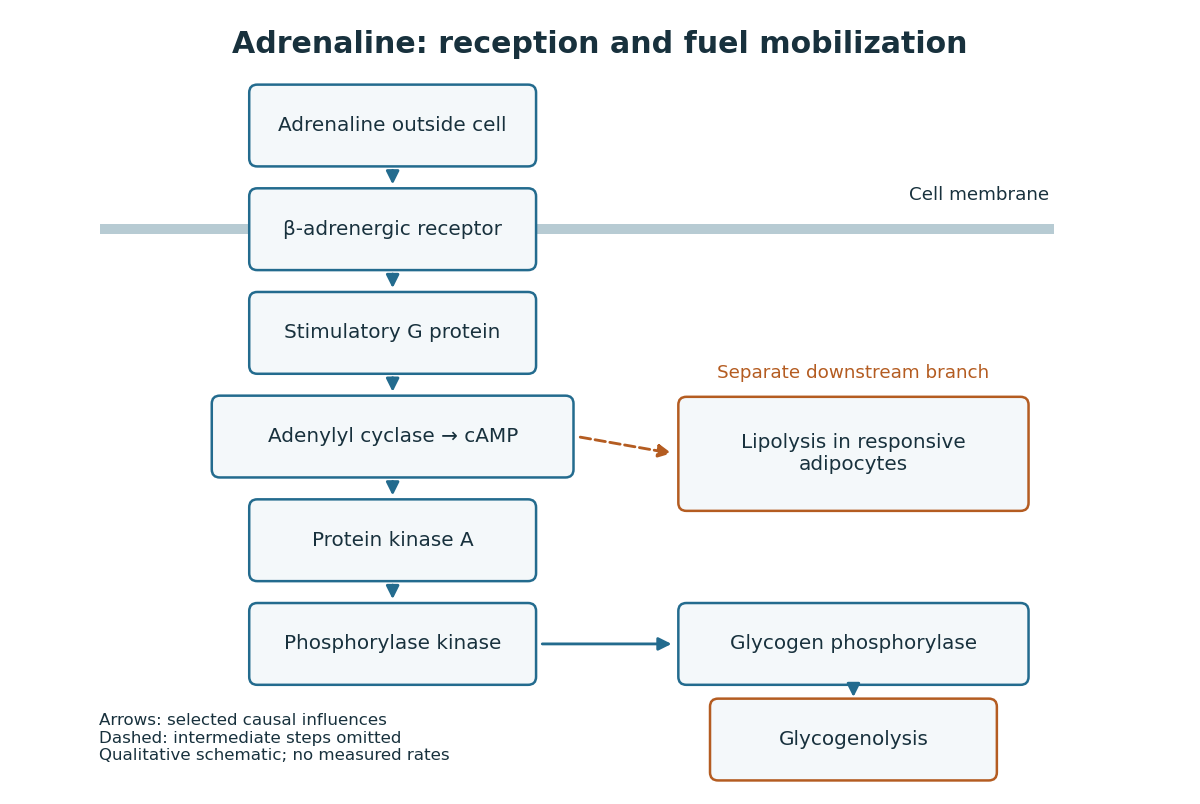
Adrenaline reception and cAMP-dependent fuel mobilization
. Original pathway schematic. Solid arrows show a selected activating relay to glycogenolysis; the dashed branch summarizes promotion of lipolysis in responsive adipocytes, omitting its intermediate steps. Arrows denote causal influences, not transport of adrenaline into the biological cell, reaction rates or measured yields. Details.The two reserves provide different material. Glycogenolysis draws on glycogen; lipolysis breaks down stored triglycerides, releasing fatty acids and glycerol. A skeletal muscle biological cell's own reserve and fuel released by adipose tissue are different supplies. Increasing a signal cannot mobilize an empty reserve: receptor activation changes the use of available material rather than creating fuel. The adrenergic adipocyte assay below gives direct human evidence for the lipolysis branch.[ref]
Pharmacological Characterizations of Adrenergic Receptors in Human Adipocytes by Thomas W. Burns and others. Abstract: epinephrine-stimulated glycerol release as an index of lipolysis
Receptor context can even oppose this branch. In the human adipocyte preparation below, beta-adrenergic receptors promoted cAMP accumulation, whereas alpha-2 adrenergic receptors opposed it. Alpha-1 adrenergic receptors were associated with a different phospholipid readout. The balance of these responses matters more than the name of the messenger alone.[ref]
Pharmacological Characterizations of Adrenergic Receptors in Human Adipocytes by Thomas W. Burns and others. Abstract: opposing beta and alpha-2 coupling; separate alpha-1 phospholipid response
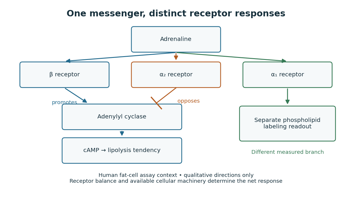
This separation is useful when explaining signal transduction: ask which receptor is present, which relay it controls, which reserve or target is available, and which response was actually measured. Receptor binding occupancy, second messenger production and final fuel release are distinct observations. A stronger cAMP signal need not imply an equally large change in every downstream process; substrate availability and the rest of the receiving biological cell still matter.
The extract-transfer experiment under second messenger supplies the earlier experimental separation between hormone reception and an intracellular intermediary. Its existing account includes the preparations and controls. The complementary adrenergic adipocyte assay asks a different question: can the same adrenaline stimulus engage opposing receptors within a human preparation?
Adrenergic adipocyte assay 2026-10-14
In the 1981 study, investigators incubated human adipocytes or their cell membranes from fat near the abdominal lining with epinephrine. They combined detection of bound ligands by radioactive decay with biochemical readouts: adenylyl cyclase activity, cAMP and released glycerol. Blocking alpha-2 adrenergic receptors increased the epinephrine response; blocking alpha-1 adrenergic receptors did not. A separate labeled-phosphate incorporation readout in a phospholipid showed the reverse blocker sensitivity. Thus the comparisons separated response branches.[ref]
Pharmacological Characterizations of Adrenergic Receptors in Human Adipocytes by Thomas W. Burns and others. Abstract, first through fourth paragraphs: human properitoneal preparations, radioligand binding, biochemical readouts; yohimbine versus prazosin comparisons
The glycerol readout indicates lipolysis, rather than directly measuring whole-body fuel use. Isolated adipocytes omit circulation and nervous input; blocker selectivity also limits the inference. These comparisons support receptor-specific responses in that preparation, not identical behavior in all biological cells.
Agarose gel electrophoresis Created 2026-10-14 Updated 2026-10-18
Agarose gel electrophoresis uses a porous agarose polymer network. For linear double-stranded DNA within a suitable separation range, shorter fragments generally move farther in a fixed run. Raising the agarose concentration changes the mesh and the range of sizes that can be resolved.[ref]
Five Main Nucleic Acid Gel Electrophoresis Steps by Thermo Fisher Scientific. Step 1, Selection and gel preparation; Recommended percentage of agarose gels for separation of DNA fragments
A compact circular DNA molecule need not migrate like linear DNA of the same length: geometry affects passage through the mesh.[ref]Five Considerations for the Nucleic Acid Gel Electrophoresis Process by Thermo Fisher Scientific. Effects of conformation, Figure 1: nicked circular, linear and supercoiled forms of the same DNA
A concrete laboratory example is Addgene's documented DNA separation protocol. Its standard 1% gel contains 1 g of agarose in 100 mL of TAE buffer. Dissolved agarose sets in a casting tray around a comb; removing the comb leaves sample wells. The gel is covered with the same buffer solution in a box connected to a voltage source. Loading buffer contains glycerol to increase sample density and a tracking dye to show run progress. A DNA ladder occupies a separate lane beside the samples. With wells near the negative cathode, DNA migrates toward the positive anode. After separation, bound stain makes the DNA visible by fluorescence under a UV transilluminator. The DNA ladder supplies the size reference; the tracking dye monitors migration but does not identify sample DNA.[ref]
In the manufacturer's controlled comparison, the same DNA ladder runs in 1%, 2%, and 3% agarose gels with the same run time and other stated conditions. The changed gel band spacing shows why a calibration belongs to its particular gel conditions, rather than establishing a universal distance law.[ref]
Five Main Nucleic Acid Gel Electrophoresis Steps by Thermo Fisher Scientific. Step 1, Figure 3 and caption: same DNA ladder at three agarose percentages under the same conditions, including run time
In interpreting an unknown sample, a blank lane tests for detectable background or carryover in that blank, while the DNA ladder checks migration and detection of its known fragments. An empty sample lane cannot by itself distinguish absent DNA from unsuccessful loading or insufficient detection. These controls answer different questions; neither makes matching migration a proof of matching nucleotide sequence.
Alcohol 2026-10-18
Alcohols such as ethanol and glycerol contain hydroxy groups on carbon chemical atoms with only single covalent bonds.
Ester 2026-10-13
The carboxylic acid-derived ester linkages in a triglyceride or typical glycerophospholipid have the form . Here the acyl group is attached through oxygen to the glycerol-derived part. See IUPAC Blue Book, P-65.6: esters.
Glycerol 2026-10-13
Glycerol has three carbon chemical atoms and three groups. It supplies the scaffold of triglycerides and glycerophospholipids, but is not itself a long hydrocarbon chain. See Biology 2e: glycerol.
Glycerophosphate 2026-10-18
Glycerophosphate denotes a phosphate ester of glycerol; added glycerophosphate served as a substrate in the rat-liver acid-phosphatase latency experiment.
Glycerophospholipid Created 2026-10-14 Updated 2026-10-14
Glycerophospholipids are phospholipids built on a glycerol-phosphate scaffold. The illustrated two-acyl group form is common, but the subclass also includes other substitutions and one-chain derivatives. See IUPAC–IUB lipid nomenclature, Lip-2.2 and Lip-2.6.
Hahn spin echo experiment 2026-10-18
Erwin Louis Hahn's 1950 apparatus enclosed liquid samples in the coil of a tuned LC circuit, coupled to a gated radio transmitter and radio receiver. One transmitter quadrupled a crystal oscillator's 7.5 MHz to 30 MHz. A separate proton resonance regulator stabilized the static magnetic field.[ref]
Spin Echoes by Erwin Louis Hahn. §IV, Figs. 12–13: transmitter, coil, receiver and regulator
Waiting for thermal equilibrium prepared nuclear polarization. Oscilloscope photographs compared glycerol proton echoes at different field homogeneity: echo width changed; echo time followed twice the pulse separation. Receiver saturation obscured early free induction decay; transmitter leakage required care. The delayed signal tested refocusing; diffusion could still attenuate it.[ref]
Spin Echoes by Erwin Louis Hahn. §II(B), Fig. 2, §IV: preparation, timing, homogeneity comparison and readout limits
Lipid 2026-10-13
Lipids are a chemically diverse family of chemical substances, conventionally associated with biological origin and solubility in nonpolar solvents. They include triglycerides, phospholipids, and cholesterol. The name does not specify one molecular formula, one repeating monomer, or one kind of polymer. Some lipids have both hydrophilic and hydrophobic regions rather than being uniformly nonpolar. See IUPAC Gold Book: lipids.
Structure explains the different jobs. A triglyceride has three fatty acid-derived acyl groups attached to glycerol and serves as an energy store. A typical glycerophospholipid instead combines two such chains with a phosphate-containing head, making it an amphiphile suited to a lipid bilayer. Cholesterol has a different structure and also contributes to cell membrane properties. Thus a lipid is a chemical building material, while a lipid bilayer is an organization of many molecules. See Clark, Douglas and Choi, Biology 2e, 3.3: lipid structures and functions.
Marcellin Berthelot 2026-10-31
Chemist whose 1856 formic acid preparation used oxalic acid and glycerol. Authentic museum portrait: www.parismuseescollections.paris.fr/en/node/1056491.
A suitable historical answer gives both the chemical transformation and the means of isolating a reasonably pure product. The following are preparative principles, with modern chemical formulas; they are not instructions for self-preparation of medicines. In particular, hydrogen cyanide, mercury compounds and arsenic compounds present serious toxicity hazards. Hydrogen cyanide is extremely volatile, can be absorbed by several routes, and its odour is an unreliable warning.[ref]
Hydrogen cyanide emergency response information by NIOSH. Agent Characteristics; Routes of Exposure; warning that smell does not provide adequate protection
For hydrocyanic acid, a customary early nineteenth-century route is the controlled distillation of potassium ferrocyanide with dilute sulfuric acid and water. Hydrogen cyanide passes into the distillate; nonvolatile salts remain behind. A generating flask, efficient laboratory condenser and chilled receiver are needed because hydrogen cyanide is readily lost. The contemporary account of hydrocyanic acid preparation in Pharmacopoeia Castrensis Ruthenica in The British and Foreign Medical Review (1842) specifically describes the ferrocyanide–sulfuric acid method and its losses on imperfect condensation. The concentration must be determined analytically before any defined dilution: a nominal starting mixture does not establish the final strength.[ref]
Pharmacopoeia Castrensis Ruthenica in The British and Foreign Medical Review (1842). Processes for obtaining Hydrocyanic Acid
At the level of the released cyanide, the essential acid-base reaction is . This equation alone is not a complete equation for decomposing the stable ferrocyanide coordination complex. Concentrated sulfuric acid can lead to different decomposition chemistry, so the specified dilution is meaningful.For formic acid, gently heat oxalic acid with glycerol and some water, then separate the aqueous formic acid by distillation. The useful net transformation isThe glycerol participates through intermediate esters and is regenerated rather than consumed in this net equation. Marcellin Berthelot's 1856 account describes the carbon dioxide evolution, subsequent water addition and distillation, and reuse of the residual glycerol. Overheating is disadvantageous because the formic acid can decompose further to carbon monoxide and water. The method therefore separates a volatile acid from a much less volatile carrier; it is not merely the dry destructive heating of oxalic acid.[ref]
Verfahren zur Darstellung der Ameisensäure by Marcellin Berthelot. Polytechnisches Journal 140 (1856), p.78, report of the March 1856 communication
For oxalic acid, oxidize a carbohydrate, such as sucrose or starch, with nitric acid; concentrate the resulting chemical solution appropriately and cool it to obtain crystals, followed by recrystallization. Nitrogen oxides evolve, and excessive oxidation can carry carbon onward to carbon dioxide, so an idealized single-product equation would conceal losses. The use of starch and nitric acid for oxalic acid production is explicitly documented in an 1850 account of nitric oxide recovery.[ref]
Polytechnisches Journal volume 117 (1850). p.466, “Verfahren das Salpetergas in Salpetersäure zu verwandeln”, opening example
An alternative already established before 1864 is alkaline treatment of sawdust, producing an oxalate which can be purified through sparingly soluble calcium oxalate.[ref]Polytechnisches Journal volume 162 (1861). report on chemical manufacture in South Lancashire, Oxalsäure section: alkaline sawdust route
Decomposing the isolated calcium oxalate with sulfuric acid separates much of the calcium as calcium sulfate:Filter off the calcium sulfate and crystallize the oxalic acid from the filtrate. The usual crystals contain two water of crystallization; a calculation for the anhydrous acid must not treat their entire mass as .For calomel, intimately combine mercury(II) sulfate, metallic Mercury and dry sodium chloride, then collect the volatile mercury(I) chloride in a cooler receiving chamber. The net change isThe extra metallic Mercury lowers the average oxidation state from +2 to +1. The product is purified by appropriate sublimation and washing to remove soluble contamination, particularly mercury(II) chloride. Both the chemical composition and the fine subdivision of the product matter: condensation through a chamber can give a powder rather than an adherent crystalline crust. This physical part of calomel preparation is described in the 1843 primary account in Polytechnisches Journal volume 87 (1843); the account's historical therapeutic claims are not adopted here.[ref]
Polytechnisches Journal volume 87 (1843). pp.209 onward, “Ueber Darstellung des Calomels als höchst zartes Pulver”, condensation and collecting chamber
For corrosive sublimate, heat dry mercury(II) sulfate with sodium chloride and collect the mercury(II) chloride in the cooler part of a suitable sublimation apparatus:Here no metallic Mercury is added. Nonvolatile sodium sulfate stays behind, while mercury(II) chloride is transferred and condensed. Further sublimation or recrystallization separates impurities. [ref]
Dictionary of Arts, Manufactures and Mines by Andrew Ure. Mercury, p.141, bichloride preparation and the following calomel account
The crucial distinction from calomel preparation is whether Mercury remains in the +2 oxidation state or is converted to +1.For the solid called arsenious acid, roast an arsenic-bearing ore in air and collect the volatile arsenic trioxide in cooler chambers; purify by resublimation. The essential oxidation is represented byActual ores also contain other chemical elements, so their roasting equations and gaseous by-products depend on composition. A contemporary 1860 report describes roasting arseniferous ores, collecting the volatile arsenic trioxide in chambers and refining it by another sublimation. It also demonstrates that apparently good material could contain antimony(III) oxide: physical appearance alone does not establish purity.[ref]
Polytechnisches Journal volume 156 (1860). p.77, report on the antimony content of glassy arsenious acid from Andreasberg
The product intended here is dry ; dissolving it does not make the bottled solid a pure crystalline hydrate .For tartar emetic, react antimony(III) oxide with potassium hydrogen tartrate in water. A small amount of water first gives intimate contact in a paste; subsequent heating with more water completes formation of the soluble potassium antimony tartrate. Filter away unreacted insoluble matter and cool the clear filtrate to crystallize the product. This sequence is directly described in a January 1864 discussion of the newly issued British Pharmacopoeia, rather than inferred from its later editions.[ref]
Chemical News volume 9 (1864). 30 January 1864, p.53, Antimonium Tartaratum: paste, standing, boiling and the oxide–bitartrate route
In the traditional antimonyl notation the underlying reaction, before assigning crystal water of crystallization, isThis notation expresses the overall composition; tartrate coordination complexes and water of crystallization need the qualification given in 4. The historical pharmaceutical use of tartar emetic does not make it a safe contemporary emetic.[ref]Antimony potassium tartrate by the National Poisons Information Service. Toxicity; gastrointestinal effects and systemic poisoning
For potassium chlorate, pass chlorine into hot concentrated potassium hydroxide. The disproportionation gives potassium chloride together with potassium chlorate:Concentration and cooling permit fractional crystallization of the much less soluble cold-water potassium chlorate, leaving much of the potassium chloride in the mother liquor; recrystallization improves purity. Cold dilute alkali instead favours hypochlorite, so the heating condition is essential. An economical contemporary manufacturing alternative uses hot calcium hydroxide and potassium chloride; the overall accounting isThe 1850 account in Polytechnisches Journal volume 118 (1850) describes a steam-heated stirred vessel and recovery by cooling, rather than an electrical manufacturing process introduced retrospectively.[ref]
Polytechnisches Journal volume 118 (1850). pp.424 onward, “Die neue Bereitungsart des chlorsauren Kalis”
Examples heavier than water are Mercury, glycerol and concentrated sulfuric acid; examples lighter than water are ethanol and diethyl ether. “Heavier” here means greater mass for the same volume, rather than greater total mass in arbitrarily sized samples.
| Liquid | Approximate relative density |
|---|---|
| Mercury | 13.5 |
| Concentrated sulfuric acid | 1.84 |
| Glycerol | 1.26 |
| Water | 1.00 |
| Ethanol | 0.79 |
| Diethyl ether | 0.71 |
These are illustrative values, not exact constants independent of temperature or composition. Dilution changes the density of sulfuric acid, glycerol and ethanol solutions.[ref]
NIOSH Pocket Guide to Chemical Hazards. Glycerin, Sulfuric acid, Ethyl alcohol and Ethyl ether entries, Specific Gravity fields
[ref]A distinction from simply observing a floating layer is essential: ethanol is miscible with water, so it mixes rather than remaining as a permanent upper layer. Diethyl ether has limited water solubility and can form an upper layer. More broadly, gases are also fluids, but the natural comparison intended here is between liquids; air at ordinary conditions is much less dense than liquid water.
Phospholipid 2026-10-13
Phospholipids are lipids containing a phosphate group as part of their structure linked by covalent bonds. The familiar membrane-forming phospholipids are amphiphilic: a hydrophilic head region with chemical polarity coexists with a hydrophobic, largely nonpolar hydrocarbon region. These descriptions refer to different parts of the same molecule, not to two separate substances. See IUPAC Gold Book: phospholipids and IUPAC Gold Book: amphiphilic.
A typical two-chain glycerophospholipid provides a useful example: a glycerol scaffold carries two fatty acid-derived acyl groups through ester linkages, plus a phosphate-containing head region. Phosphatidylcholine is one example. This scaffold describes an important subclass, rather than all phospholipids: sphingomyelin has a different backbone, and even glycerophospholipids need not all have two acyl groups. See IUPAC–IUB, Nomenclature of Lipids (1976), Lip-1.5 and Lip-2.1–2.6.
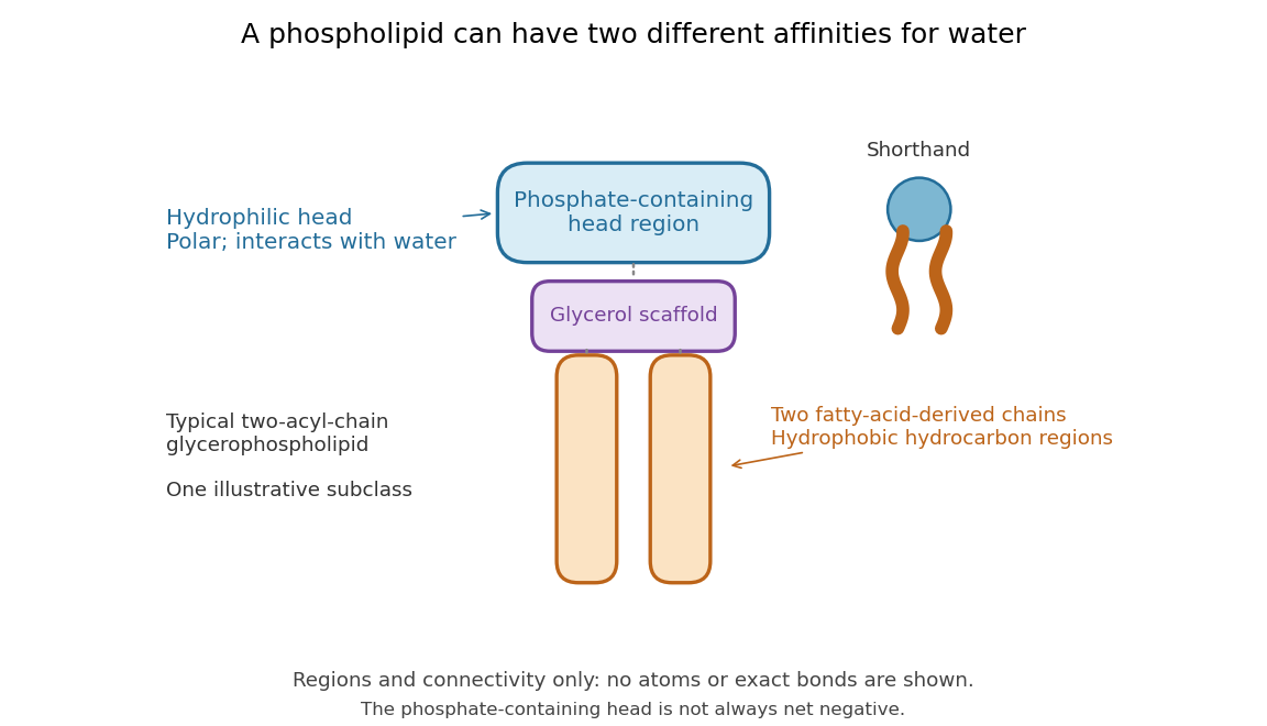
Polar does not mean negatively charged overall. At near-neutral pH, phosphatidylcholine is zwitterionic: its phosphate region carries negative electric charge and its choline region positive electric charge, with zero net electric charge for the molecule. Counting those opposite charges gives ; it does not remove their local interactions with water. See Lodish et al., Molecular Cell Biology: membrane lipids.
In a lipid bilayer, two membrane leaflets orient their hydrophilic heads toward the water on either side and their hydrophobic chains toward the shared interior. The hydrophobic effect helps explain this organization through the free energy of the water and lipids together. It is not a special repulsive chemical bond between chains and water. Which organization is favored also depends on molecular geometry, temperature, concentration, and the surrounding chemical solution; a phospholipid label alone does not guarantee a lipid bilayer. See Alberts et al., Molecular Biology of the Cell: the lipid bilayer.
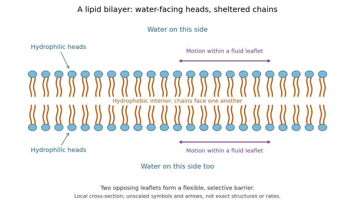
This organization supplies a flexible barrier for compartments. A fluid lipid bilayer allows lipids to undergo diffusion within each membrane leaflet; barrier function therefore coexists with motion of molecules, as in the fluid mosaic model. It is not completely impermeable: small nonpolar molecules such as molecular oxygen and carbon dioxide cross much more readily than ions. In a cell membrane, ion channels and other membrane transport proteins provide selective routes; the existing membrane transport article explains their driving forces. See Alberts et al.: principles of membrane transport.
In 1925, E. Gorter and F. Grendel extracted lipids from red blood cells and compared the surface area of a one-molecule-thick film on water with the estimated total surface area of the red blood cells. Their table gave ratios near two, supporting a two-layer covering; they also proposed heads facing the aqueous surroundings on both sides. The inference depends on recovering the relevant lipids, estimating the red blood cell surface area correctly, and comparing compatible packing in the film and the cell membrane. It was indirect evidence, not an image of the lipid bilayer. The paper itself notes possible losses during evaporation. See Gorter and Grendel, On Bimolecular Layers of Lipoids on the Chromocytes of the Blood, Journal of Experimental Medicine 41 (1925), 439–443.
Saponification of triglycerides 2026-10-18
A triglyceride contains three ester linkages joining glycerol to fatty acid-derived acyl groups. Complete saponification therefore consumes three hydroxide equivalents per triglyceride, yielding one glycerol and three fatty acid carboxylates.[ref]
27.2 Soap. Organic Chemistry by John McMurry; §27.2, opening chemical definition and fat hydrolysis equation
Writing the three hydrocarbon groups separately allows their compositions to differ:This is the preceding ester chemical equation applied three times: each linkage supplies one carboxylate, and each formerly esterified oxygen on the glycerol scaffold gains one hydrogen. The two sides retain the same three groups and have net electric charge .
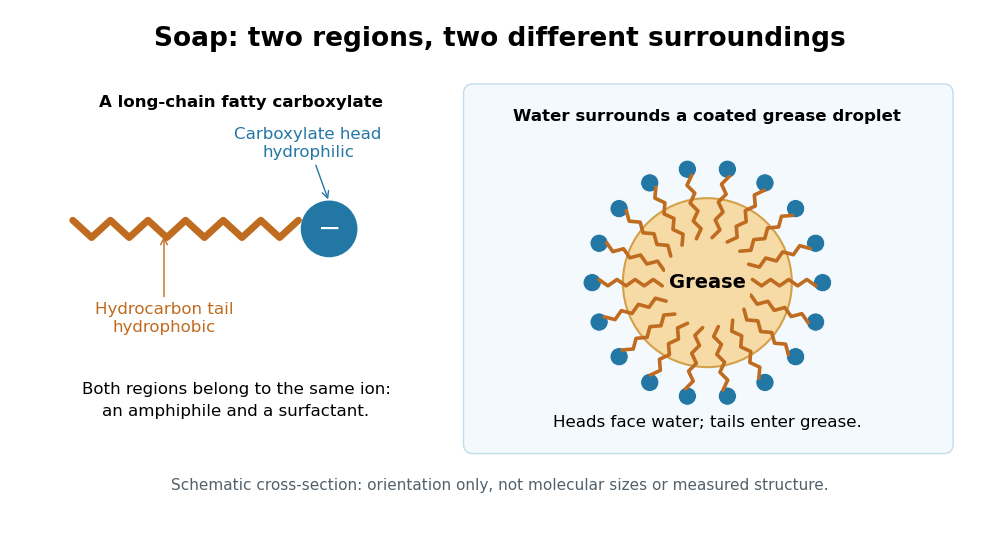
Soap consists of sodium or potassium salts of long-chain fatty acids. Its carboxylate head is hydrophilic, while its long hydrocarbon tail is hydrophobic. This makes it an amphiphile and a surfactant: the tails can associate with grease while the heads remain exposed to water, helping disperse grease for rinsing. An acetate ion is also a carboxylate, but lacks the long hydrophobic tail that gives soap this cleaning role.[ref]
27.2 Soap. Organic Chemistry by John McMurry; §27.2, chemical definition and paragraphs on the ionic head, hydrocarbon tail and removal of grease
Soap film between coaxial rings 2026-10-14
Two equal circular rings of radius in parallel planes at provide a boundary for an open film. In the ideal axisymmetric model, a stationary connected film has the catenoid profileThe catenoid existence threshold for equal rings explains why this equation can have two solutions, one solution, or none. The cancellation of principal curvatures holds for both solutions whenever they exist; the mean curvature equation alone does not select a stable minimal surface.
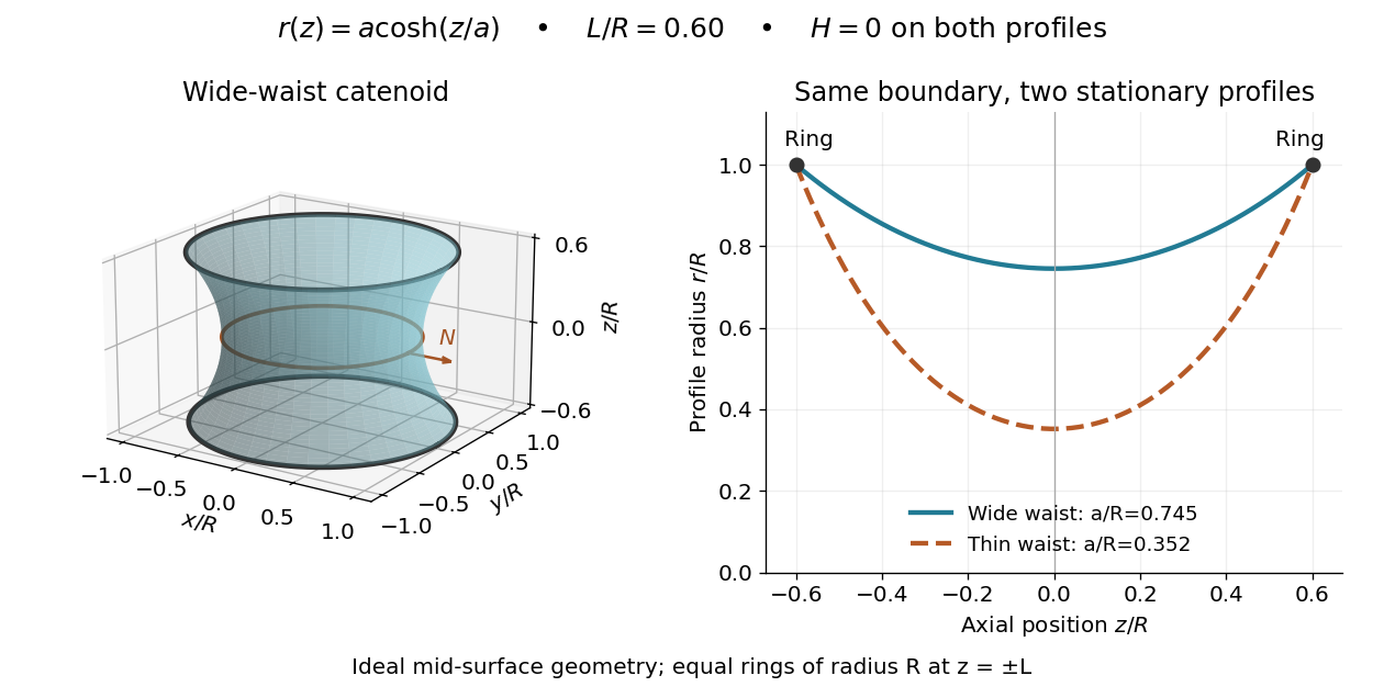


This ideal geometry uses and . The two profiles have different waist radii , although both have . For either profile the surface area follows by integration:At this illustrated separation both profiles have greater surface area than the two disconnected disks, whose total is . This comparison disproves a claim of global area minimization in a class allowing disconnected competitors; it does not determine the sign of the second variation for small variations of a connected film.
A reported experiment used coaxial wire rings of radius , drawn from a surfactant solution and slowly separated until collapse. The solution contained tetradecyltrimethylammonium bromide in deionized water, with glycerol added to vary viscosity. Fluorescein enabled fluorescence imaging under cyan LED arrays; a Phantom V641 camera recorded 4,000 or 5,600 frames per second. Extracted contours gave neck radius, attachment angle, and inferred surface area. They showed neck narrowing and eventual breakup, rather than indefinite persistence of a stationary catenoid. Comparing solutions across viscosities helped distinguish liquid dissipation from the surrounding air's inertia, but the ring displacement beyond the last stable state was not precisely controlled. These are collapse observations, not direct measurements of equilibrium . [ref]
Geometry of catenoidal soap film collapse induced by boundary deformation by Raymond E. Goldstein and others. Geometry of Catenoidal Soap Film Collapse Induced by Boundary Deformation, §II, manuscript pp. 3–4, Figs. 1–3: apparatus, contour readouts, viscosity comparison, and displacement limitation
A proposed classroom measurement would instead hold two equal rings coaxial on a translation stage, leave both sides open to the same gas, and photograph the settled silhouette beside a scale. Fit and inspect fit residuals before inferring cancellation of the principal curvatures. Repeat at several separations and with the camera rotated about the axis to test rotational symmetry. Keep solution composition, ring wetting, and elapsed drainage time comparable, and shield the film from drafts. Film weight, drainage, evaporation, surfactant gradients, and rupture can invalidate the ideal soap film pressure balance. A silhouette fit alone neither measures the pressure jump nor tests all possible perturbations of a stable minimal surface.
Sphingomyelin 2026-10-14
Sphingomyelin is a phospholipid whose backbone is based on sphingosine, rather than glycerol. One long hydrocarbon region belongs to that backbone and another comes from an attached fatty acid. It is a counterexample to defining every phospholipid by the illustrated glycerol scaffold. See Cooper, The Cell: the molecular composition of cells.
Triglyceride Created 2026-10-13 Updated 2026-10-14
A triglyceride contains glycerol joined to three fatty acid-derived acyl groups through ester linkages. Compare the three chains with the two-chain, phosphate-headed glycerophospholipid illustrated below. See Biology 2e: fats and oils.