A chemical substance is matter identified by a characteristic chemical composition and set of chemical properties. It can be an elemental substance, such as molecular nitrogen, or a chemical compound, such as water. A mixture instead contains components whose proportions can vary without requiring a different compound identity.
Composition alone may not specify the physical form. Diamond and graphite are elemental carbon with different bonding networks, and a single compound can have several crystalline phases. Describing a sample therefore calls for both chemical identity and, when relevant, its phase, structure, and purity.
The initial table of content was limited to the first level because it is larger than 1000 articles. There are 1378 articles in total. Expand a branch to load its children.
Dipyrromethene, also called dipyrrin, is a conjugated nitrogen-containing framework built from two pyrrole-derived rings joined through a methine-containing connection. The conventional dipyrrin name refers to the 2,2′-linked system. IUPAC retains dipyrromethane as a distinct name and introduces dipyrrin for the corresponding unsaturated framework.[ref]
Nomenclature of Tetrapyrroles by IUPAC-IUB Joint Commission on Biochemical Nomenclature. TP-6 and TP-7, retained and introduced trivial names
The unsubstituted neutral parent has formula ; its attachment pattern and protonation state matter when discussing derivatives.[ref] A dipyrrin-derived ligand and its boron complex are chemically different species, even when they share a recognizable skeleton.
An original atom-accounting example starts with neutral . Removing one proton gives a ligand composition . Combining that formal ligand with a boron-difluoride center gives the neutral simplest BODIPY composition . This is a charge and composition check, not a claim that free isolated fragments are the actual synthesis intermediates. Substituted dyes have additional atoms and their own molecular formulas.
The extended conjugated system makes the framework useful for optical dyes, while coordination and substitution alter the electronic structure. BODIPY dyes are therefore a family of complexes rather than another name for free dipyrromethene. The primary ChEBI dye-class definition identifies that coordinated boron-containing family.[ref]
BODIPY dyes in ChEBI. CHEBI:51123, Definition
A proposed analytical comparison would record absorption spectra before and after a defined coordination or protonation change, with concentration and solvent held known. A changed band can support a change in electronic structure, but its amplitude alone cannot distinguish conversion, concentration loss and altered molar absorption coefficient. A Beer-Lambert law calibration needs the identity of the absorbing species.
Dipyrrin protonation and optical response is consequently a separate question from merely recognizing the skeleton. Specify the free compound, ligand or complex, its substituents and the measured optical observable before transferring a property between members of the family.
Dipyrrin protonation and optical response concerns how an acid-base change alters which dipyrrin species absorb or emit light. A change in absorbance can reflect redistribution among protonation states without any loss of the total dye inventory. The free dipyrromethene, its deprotonated ligand and a coordinated BODIPY complex must be identified separately.[ref]
Dipyrrin in PubChem. CID 3083424, neutral parent identity
A primary example involving naphthalimide-fused dipyrrins reports that protonation changes conformation and electronic coupling, with large absorption shifts in selected derivatives.[ref]
Naphthalimide-Fused Dipyrrins: Tunable Halochromic Switches and Photothermal NIR-II Dyes. PMID 35174648, primary indexed Abstract
It demonstrates a real chemical route to an optical change; it does not assign the same shift or acid dissociation constant to every dipyrrin.An original two-species model writes . In a medium where a Henderson-Hasselbalch equation description is appropriate, the neutral fraction isFor fixed total concentration and path length , the Beer-Lambert law givesBoth species contribute. The solvent, activity convention and possibility of additional species belong to the model.
For stipulated , micromolar, cm, and , absorbances at pH 5, 6 and 7 are approximately 0.236, 0.400 and 0.564. No dye is consumed in this example. These chosen values are not measured constants of the unsubstituted parent or of the cited fused dyes.
Dipyrrin titration mass balance requires tracking the same total analytical amount while accounting for added volume and all significant species. At a wavelength where the two coefficients coincide, the ideal two-species absorbance is independent of their proportions: an isosbestic point. Such a crossing is consistent with this model but does not uniquely prove that only two chemical species exist.
A proposed experiment collects full spectra during a reversible acidity change, with matched solvent and dilution controls. Check recovery on returning conditions, changes in concentration and possible additional equilibria before interpreting one band's decrease as degradation. A calibration of one protonation state is not automatically valid after the state population changes.
Dipyrrin titration mass balance keeps track of the amount of dye while acid addition changes its protonation states and the solution volume. An absorbance decrease at one wavelength can result from redistribution or dilution, rather than destruction of dipyrromethene units. In a primary study of fused derivatives, acid titration produced several distinct spectral responses and, for some derivatives, more than one protonation step.[ref]
Naphthalimide-Fused Dipyrrins: Tunable Halochromic Switches and Photothermal NIR-II Dyes. Figures 3–4 and captions; acid-induced absorption spectra of the 1a–1e and 2a–2e series
For an original two-species model containing neutral and protonated , conservation givesThe relevant acid dissociation constant supplies a second relation. In an ideal dilute aqueous model,This chosen model does not assign an aqueous constant to the cited dyes in an organic solvent. If pH is not independently fixed, acid inventory and electroneutrality must also be solved; adding a known volume of acid does not prescribe the final free hydrogen ion concentration by itself.
The Beer-Lambert law predictsFor a stipulated 0.10 micromole of dye initially in 10 mL, adding 2.5 mL gives micromolar. At a measured condition with , cm, and , . Omitting dilution predicts 0.40 despite exactly the same dye amount and species fraction.
Aliquot-aware dye inventory in titration adds another correction. If aliquots of volume are removed from well-mixed solutions having total dye concentrations , the remaining amount is , unless other gains or losses occur. Adsorption, precipitation or chemical transformation require additional inventories. For species containing two dye units, their concentration contributes twice to the conserved dye-unit sum.
A useful titration records additions, withdrawals and full spectra, then checks whether returning the acid-base conditions recovers the predicted inventory-corrected response. Conservation of dye units, charge balance and the spectral measurement equation impose different constraints; agreement with only one does not establish the others.
An aliquot-aware dye inventory accounts for dye removed in samples as well as dilution by titrant. In dipyrrin titration mass balance, an absorbance change can result from changing protonation, changing solution volume or loss of material from the vessel. Those effects require separate terms even when the underlying acid-base transformation is reversible.
Let a well-mixed vessel contain volume , dye amount and total dye concentration . Suppose an aliquot is removed first, then dye-free titrant volume is added, with no other gain or loss. The next inventory issoThe order is part of the protocol. Adding titrant before withdrawing the same numerical volume would remove dye from a different concentration.
For repeated sampling followed by dye-free replacement to restore a fixed volume , let . Then . Three withdrawals leave of the original dye, so becomes . Subtracting three times the first aliquot's amount would incorrectly give , because later aliquots contain less dye.
More generally, the removed amount is , with each concentration evaluated at the actual sampling time. Under an additive Beer-Lambert law model,where the species fractions sum to one on the chosen dye-unit basis. Correcting alone does not correct . Returning to the initial protonation conditions need not restore the initial absorbance after sampling has removed material.
Primary acid titrations of fused dipyrrins show derivative-dependent spectral changes and multiple protonation stages.[ref]
Naphthalimide-Fused Dipyrrins: Tunable Halochromic Switches and Photothermal NIR-II Dyes. Figures 3–4 and captions, acid-induced spectral changes
The recurrence here is an explicit inventory model for interpreting such measurements, not a claim about aliquots in that paper. Adsorption, precipitation or chemical destruction would add separate loss terms; disagreement with the sampling-only prediction is a reason to test those possibilities.BODIPY denotes the boron-dipyrromethene family of compounds, widely used as optical dyes. A common dye skeleton contains a boron-difluoride centre coordinated within a dipyrromethene-derived framework. ChEBI defines BODIPY dyes structurally and lists many substituted members, rather than assigning one chemical formula or optical spectrum to the whole family.[ref]
BODIPY dyes in ChEBI. CHEBI:51123, definition and listed class members; broader class CHEBI:51108
The conjugated system, substituents and environment affect light absorption and fluorescence. Sharing a scaffold does not imply equal molar absorption coefficients, fluorescence quantum yields or chemical reactivity. A dye can also serve as a reaction participant, rather than an inert tracer.
A primary method by Evan A. Haidasz, Antonius T. M. Van Kessel and Derek A. Pratt followed co-oxidation of a hydrocarbon substrate with the signal carriers PBD-BODIPY or STY-BODIPY by visible-light spectrophotometry. The assay was designed to characterize radical-trapping antioxidants and distinguish reaction kinetics from trapping capacity.[ref]
A Continuous Visible Light Spectrophotometric Approach To Accurately Determine the Reactivity of Radical-Trapping Antioxidants by Evan A. Haidasz and others. Abstract, signal carriers and kinetic/stoichiometric interpretation
Its optical readout should not be described automatically as a fluorescence measurement simply because the compounds are dye derivatives.An original photon-counting model illustrates another distinction. For incident photon rate and absorbance , the fraction absorbed is . With fluorescence quantum yield and detection efficiency , an idealized detected emission rate isAt small , this is approximately , which can be proportional to concentration under a suitable Beer-Lambert law. Increasing from0.10 to0.20 instead increases absorbed fraction from approximately0.206 to0.369, a factor1.79 rather than2. This example assumes unchanged quantum yield, geometry and negligible reabsorption; it does not assign those conditions to every BODIPY experiment.
A quantitative application therefore specifies the derivative, solvent, wavelength, chemical state and observable. Absorption loss, emission change and reaction-product formation are related measurements only through a validated response model.
STY-BODIPY is the styrene-conjugated BODIPY signal carrier used in a visible-light assay of radical-trapping antioxidants. It co-autoxidizes with a hydrocarbon substrate, allowing reaction progress to be followed optically. The primary method distinguishes antioxidant reactivity from the number of radicals trapped.[ref]
A Continuous Visible Light Spectrophotometric Approach To Accurately Determine the Reactivity of Radical-Trapping Antioxidants by Evan A. Haidasz and others. Abstract, STY-BODIPY signal carrier and kinetic/stoichiometric aims
The manufacturer's record for the named material gives formula and formula weight approximately . Its method description follows loss of absorbance at 571 nm during co-autoxidation.[ref] This identifies a particular derivative and measurement channel; it does not assign one spectrum to every styrene-substituted BODIPY dye.
An original Beer-Lambert law example assumes a 1 cm path and calibrated molar absorption coefficient . Absorbance falling from 1.00 to 0.80 corresponds to signal-carrier concentration falling from 20 to 16 micromolar. The inferred 4 micromolar loss depends on that assumed coefficient and the identity of the absorbing species. Those numbers are illustrative, not a reported STY-BODIPY calibration.
A simple kinetic model writes , where represents a constant peroxyl-radical concentration. Then . An antioxidant that changes the radical population can change the optical decay, but the resulting trace must be interpreted through the co-oxidation model. Dye consumption alone does not count antioxidant molecules without further kinetic and stoichiometric information.
Dark-loss control for STY-BODIPY autoxidation tests one potential confounder. Proposed controls compare the signal carrier without radical initiation, under matched illumination and in the presence of test material that might itself absorb at the observation wavelength. These distinguish chemical oxidation from unrelated optical change or added absorbance.
Although the product is described as fluorogenic, the original assay discussed here uses visible-light spectrophotometry. Absorbance loss and fluorescence change are different observables. Specify the derivative, solvent, optical channel, blank correction and response model when using the named dye to compare antioxidant activity.
A dark-loss control for STY-BODIPY autoxidation distinguishes initiation-associated chemical loss from changes caused by illumination, handling or the measurement itself. The control must preserve the relevant experimental conditions while accounting for the photons needed to read the sample. “Dark” handling followed by an optical measurement does not mean zero photon exposure.
The primary antioxidant method uses STY-BODIPY as a co-autoxidizing signal carrier monitored by visible-light spectrophotometry.[ref]
A Continuous Visible Light Spectrophotometric Approach To Accurately Determine the Reactivity of Radical-Trapping Antioxidants by Evan A. Haidasz and others. Abstract
The manufacturer describes monitoring absorbance loss at 571 nm.[ref] That signal does not label its own cause.An original diagnostic design crosses initiation absent/present with low-exposure handling/continuous illumination. Let be the matched fractional absorbance loss, where marks initiation and illumination. The interaction contrast isIt is zero under a simple additive loss model. A nonzero result shows that subtracting an independently measured light-only loss may not isolate the initiated process.
For stipulated losses , , and , the additive prediction for the last condition is 0.16, while the observed-model value is 0.20. The interaction is 0.04. These are constructed observations, not published STY-BODIPY data. They illustrate why photobleaching and chemical oxidation need not combine as independent corrections.
Illumination-initiation interaction in dye-loss controls can arise in a measurement model without being uniquely diagnosed by this contrast. Temperature, oxygen availability and exposure timing also need matching. Compare spectral changes and final readout conditions, rather than assuming every difference is photobleaching.
A proposed control series includes carrier without initiation, initiation without test antioxidant and test material without carrier where its own absorbance could contribute. Record readout duration and photon exposure for each condition. A dark endpoint measured once and an illuminated sample monitored repeatedly have different acquisition histories. Interpret their difference using that history before converting absorbance loss into antioxidant kinetics or trapping capacity.
Illumination-initiation interaction in dye-loss controls tests whether changing illumination has the same response in the absence and presence of chemical initiation. A crossed factorial design separates the four conditions. For mean fractional losses , with indicating initiation and illumination, the contrastmeasures statistical interaction on the fractional-loss scale. In the STY-BODIPY assay context, the optical observable is carrier loss during co-autoxidation, not a direct label of its chemical cause.[ref]
A Continuous Visible Light Spectrophotometric Approach To Accurately Determine the Reactivity of Radical-Trapping Antioxidants by Evan A. Haidasz and others. Abstract: visible-light spectrophotometric co-autoxidation and kinetic/stoichiometric interpretation
The crucial qualification is response-scale dependence of dye-loss interactions. Consider an original first-order survival model with fully additive rate constants,ThenFor positive , and , this is negative even though the rates have no interaction term. With stipulated , in reciprocal time units and , the losses are , , and , giving . Saturation of fractional loss produces the contrast without a new chemical coupling.
On the transformed scale , the same noiseless model gives and zero interaction contrast. This transformation is appropriate only when the measured signal genuinely follows the stated survival model; product absorption or changing backgrounds can invalidate it. A transformed zero is therefore a conditional model check, not proof of a reaction mechanism.
For independent replicate measurements, the variance of the estimated loss contrast is . Four replicates per condition with common standard deviation 0.01 give a contrast standard uncertainty of 0.01. The modeled contrast above is smaller than that scale. Shared references instead introduce covariances that must be retained.[ref]
Match temperature, oxygen access, timing and readout exposure across conditions. Specify the response scale before interpreting a nonzero contrast as evidence about photobleaching or initiated oxidation.
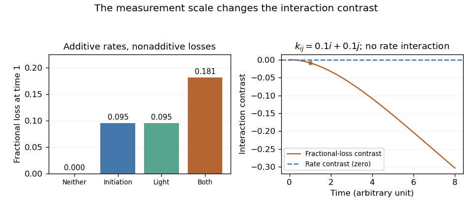
An original additive-rate model gives a negative interaction contrast on the fractional-loss scale. Transforming the same ideal observations back to rates gives zero rate interaction.
Details. A dye-loss interaction is a departure from additivity on a specified response scale. Changing from fractional loss to survival, logarithmic survival or a fitted rate can change that departure. The contrast must therefore be defined before it is interpreted as evidence about coupled chemical mechanisms.
For a crossed initiation/illumination experiment, write fractional losses at a common observation time and defineAn original example takes losses . The contrast is . Yet the surviving fractions obey , with . They are exactly multiplicative on the survival scale.
For first-order survival , transform to . The rate-scale interaction isThe example gives . More generally, additive rates yieldwhich is negative for positive . The fractional-loss ceiling creates the contrast without an additional rate interaction.
The transformation also changes noise. First-order uncertainty propagation gives , so near-complete loss can strongly amplify uncertainty. In addition, the mean of a nonlinear transformation need not equal the transformation of the mean; Jensen's inequality makes that distinction explicit. Averaging transformed replicates and transforming an average need not produce the same result.
The STY-BODIPY readout is a coupled oxidation signal. Its primary method distinguishes radical-trapping kinetics from stoichiometry.[ref]
A Continuous Visible Light Spectrophotometric Approach To Accurately Determine the Reactivity of Radical-Trapping Antioxidants by Evan A. Haidasz and others. Abstract, co-autoxidation method and kinetic/stoichiometric interpretation
A rate transformation requires a justified survival model after product absorption and background have been addressed. Report both the response definition and the uncertainty model; a transformed zero is conditional evidence about that model, rather than a unique identification of the chemistry.PBD-BODIPY is the 1-phenylbutadiene-conjugated BODIPY signal carrier in a spectrophotometric method for radical-trapping antioxidants. The conjugated appendage distinguishes it from the styrene-conjugated carrier STY-BODIPY. In the primary method, each can co-autoxidize with a hydrocarbon substrate so that the changing optical signal follows reaction progress.[ref]
The shared scaffold does not make the two carriers interchangeable calibrants. Additional conjugation can alter the electronic structure, while substitution and solvent affect the spectrum and chemical response. Chromophore-specific calibration of antioxidant signal carriers is therefore needed before converting an absorbance slope into a chemical rate.
An original comparison assumes a PBD-carrier calibration and path length 0.5 cm. Absorbance 0.40 then represents 10 micromolar carrier. Substituting a coefficient of 50000 from another hypothetical dye would instead infer 16 micromolar, a 60% overestimate. These stipulated coefficients illustrate a calibration error; neither is asserted as a measured value for these compounds.
The same issue affects a rate. Under a Beer-Lambert law with unchanged absorbing species and coefficient,With the first illustrative coefficient and path, an absorbance slope gives carrier-loss rate . A raw optical slope is not yet a molecular consumption rate, and a carrier-consumption rate is not automatically an antioxidant-trapping rate.
A proposed comparison records separate starting spectra, matched reaction conditions and appropriate carrier and test-material blanks. Differences in lag duration and decay slope can constrain different parts of a kinetic model. A longer lag can reflect trapping capacity, while a changed slope also depends on the competition between propagation and trapping; neither readout should be assigned a unique molecular explanation without testing the response model.
The primary study explicitly seeks both kinetic and stoichiometric information. Preserve that distinction when comparing PBD-BODIPY with STY-BODIPY, rather than describing every signal decrease as an interchangeable measure of antioxidant concentration.
Chromophore-specific calibration of antioxidant signal carriers determines how the optical response of a particular dye, its products and its environment maps to chemical amount. Sharing the BODIPY scaffold does not establish equal spectra or extinction coefficients. The original assay distinguishes the PBD- and STY-conjugated carriers and seeks kinetic as well as stoichiometric information.[ref]
An original two-species absorption model iswhere is the carrier, an absorbing product and the background. If one product molecule forms for each consumed carrier, , soThe optical slope depends on the difference in coefficients, not the carrier coefficient alone.
For stipulated cm, and , conversion of 10 micromolar carrier changes absorbance by 0.40. Treating the product as transparent would infer only micromolar loss. The chemical conversion has been underestimated by one third. No actual carrier coefficient is assigned by this example.
Product-absorbance correction in antioxidant signal carriers can be tested with multiple wavelengths. With known backgrounds, two independent spectral equations can recover two species if their coefficient matrix is invertible. For the illustrative coefficient matrixpath 1 cm and absorbances , solving gives and micromolar. Nearly proportional columns would make that separation unstable even if the matrix were formally invertible.
The experimental calibration should therefore specify derivative identity, solvent, wavelength, path, background and relevant chemical states. Acidity changes can alter species populations, and a reaction product may remain optically active. A good fit to a single trace does not independently establish those assumptions. Once the carrier-consumption rate is inferred, a further reaction model is still needed to interpret antioxidant reactivity or stoichiometry.
Product-absorbance correction in antioxidant signal carriers separates the optical response of unconsumed carrier from that of absorbing reaction products. It is needed before an absorbance slope is converted into carrier consumption. The primary co-autoxidation method distinguishes signal-carrier chemistry and uses its response for both kinetic and trapping-stoichiometry information.[ref]
For an original two-species Beer-Lambert law model with fixed volume and corrected background,If product yield is per consumed carrier molecule, , soBoth molar absorption coefficients and the assumed yield enter. When , conversion is optically invisible at that wavelength even if it proceeds to completion. An unchanged trace is then consistent with extensive chemistry.
Product-null spectral contrasts can eliminate one known interfering component. Suppose two background-corrected wavelengths have illustrative coefficient columns and in . The linear combinationcancels the product because . At cm, absorbances yield and micromolar. This separation does not require a product-yield assumption, but it does require valid coefficients, only the specified absorbing components and known backgrounds.
If the two absorbances have independent standard uncertainties of 0.001, the contrast has uncertainty , giving approximately 0.0203 micromolar uncertainty in . Correlated errors change that result through the covariance term: .[ref]
A product spectrum proportional to the carrier spectrum cannot be removed while retaining carrier sensitivity. Additional products, changing protonation or a drifting background also require a larger measurement model. Test the correction with independently prepared carrier/product mixtures and full spectra before attributing the corrected trace to a particular antioxidant reaction rate. The numerical coefficients here are constructed examples, not reported carrier constants.
A product-null spectral contrast is a weighted combination of spectral measurements that cancels a modeled product contribution while retaining sensitivity to the carrier of interest. It is useful when carrier consumption and product formation both change the absorbance recorded in an inhibited autoxidation assay.
After background correction and with path length included in the response coefficients, write a measurement vector asHere and are known carrier and product spectral response columns. A contrast removes the product and has unit carrier response whenIf the columns are proportional, these constraints are incompatible. More measurements do not help if they add no independent spectral information.
An original three-channel example uses scaled coefficient columns and . The weights satisfy both constraints. Stipulated concentrations give and the contrast returns . The coefficient and concentration units have been absorbed into the scaling; these are not measured dye coefficients.
For independent equal-variance channel noise , the example has variance . These are the minimum-variance weights under the stated noise model: project perpendicular to , then normalize that residual to unit carrier response. The Cauchy-Schwarz inequality shows that any other vector with the same constraints has at least as large a norm. A two-channel difference using just the first two measurements also isolates the carrier, but weights give the larger variance .
For general covariance matrix , the variance is , so correlated or unequal noise changes the optimal weighting.[ref]
Evaluation of measurement data — Guide to the expression of uncertainty in measurement by JCGM. Section 5.2, covariance propagation
The coupled chemical readout remains the one described by the primary signal-carrier method.[ref]A Continuous Visible Light Spectrophotometric Approach To Accurately Determine the Reactivity of Radical-Trapping Antioxidants by Evan A. Haidasz and others. Abstract
Additional products or changing protonation require additional response columns; a cancellation calibrated for one product does not remove every unknown spectral contribution.Quercetin is a flavonol with five hydroxy groups, at positions3,5,7, and , and neutral molecular formula . The primes distinguish positions on the attached phenyl group from positions on the fused ring system.[ref]
Five hydroxyl oxygens account for only five of the seven oxygen atoms: the flavone framework also contains a ring oxygen and a carbonyl group. Compared with kaempferol, the additional hydroxyl substitution adds one oxygen without changing the total hydrogen count, because a carbon-bound hydrogen is replaced by OH. Counting elemental composition is a useful structural check, but does not locate the groups without connectivity information.
An original solution calculation uses the average molar mass . Dissolving of the specified pure neutral compound to a final volume givesIf only 90 percent of the weighed mass is that compound and the balance contributes none, the actual concentration is . Salt form, retained solvent or incomplete dissolution require their own corrections; the molecular formula alone does not establish preparation purity.
Pradeep K. Sengupta and Michael Kasha included quercetin in their early study of unusual fluorescence from flavonols, connecting its optical behaviour with excited-state proton transfer.[ref]
Excited state proton-transfer spectroscopy of 3-hydroxyflavone and quercetin by Pradeep K. Sengupta and Michael Kasha. 1979, printed pp.382–384, molecular comparison and spectroscopy
Chemical identity, absorption and emission conditions matter when interpreting a signal. Classification as an antioxidant does not, by itself, establish protection of living cells or a clinical effect.An antioxidant is a substance acting to oppose oxidation or inhibit reactions driven by oxygen or peroxides in a specified system. ChEBI classifies antioxidant as a chemical role: the label describes what a substance does under relevant conditions, rather than one shared molecular structure.[ref]
Antioxidant in ChEBI. CHEBI:22586, definition and chemical-role classification
A radical-trapping antioxidant provides one possible mechanism. In a simplified reaction scheme, a donor intercepts a radical :This step is protective in the model only if the resulting species and subsequent reactions propagate less unwanted oxidation than the intercepted route. Removing one radical while producing another does not, by itself, prove radical chain termination. Other protective mechanisms need not follow this scheme.
An original antioxidant reaction competition model makes the importance of concentration explicit. Suppose a radical has just two consumption routes: interception by and attack on substrate . Their reaction rates areAt fixed concentrations, the fraction of radical-consumption events assigned to interception isWith and , this fraction is . Decreasing antioxidant concentration tenfold gives . The compound has not changed identity; its competition with the substrate has changed. Depletion, additional reactions and transfer between phases require a richer chemical kinetics model.
Experimental interpretation must specify the substrate, medium, time interval and measured outcome. Reducing an indicator is a chemical observation and need not establish protection of cells. A primary study found that kaempferol and resveratrol could produce formazan from MTT without cells.[ref]
Interference of plant extracts, phytoestrogens and antioxidants with the MTT tetrazolium assay by Regina Bruggisser and others. Abstract, cell-free tests
Such a response motivates a tetrazolium cell-free control, rather than interpreting a stronger color as increased cell viability.An antioxidant assay therefore measures a defined reaction or consequence under its own conditions. Extrapolation to a different reaction environment requires evidence about concentration, reaction partners and the outcome of interest.
Antioxidant reaction competition is competition between reactions that consume the same reactive intermediate, such as interception of a radical by an antioxidant and attack by that radical on a susceptible substrate. A large reaction rate constant alone does not establish which route dominates: the local concentrations of the reaction partners also enter the reaction rates.
For an original two-route model, let a radical react with donor or substrate . Hold both partner concentrations constant, with second-order rate constants . The corresponding pseudo-first-order reaction rates per radical areThe probability of surviving without either event for time is . Integrating the instantaneous interception probability giveswhere is the mean waiting time for the first event. This is a competing risks calculation for the stipulated constant-rate reaction model.
Take chosen , , and . Both effective rates are , so interception has probability and the mean waiting time is . Raising only the donor concentration tenfold gives and .
This fraction describes the first reaction, not necessarily final protection. If substrate attack regenerates another reactive radical, radical chain propagation can continue. In a simplified chain where each event either propagates with probability or terminates by trapping with probability , the expected number of propagation events satisfiesDepletion, additional termination and movement between phases alter this model.
An inhibited autoxidation assay measures a consequence of such chain chemistry under defined conditions. Primary work compared inhibited-oxidation measurements of lipid-soluble chain-breaking activity with independent chromatographic measurements of vitamin E in plasma and membrane preparations.[ref]
Is vitamin E the only lipid-soluble, chain-breaking antioxidant in human blood plasma and erythrocyte membranes? by G. W. Burton and others. Abstract, inhibited-autoxidation and HPLC comparison
That independent comparison is stronger evidence than assigning protection from the chemical label antioxidant alone.A radical-trapping antioxidant reacts with a chain-carrying radical in a way that reduces continued radical chain propagation. It is a mechanistic role in a particular reaction system, not a guarantee that every reaction product is harmless or that the substance improves health.
For a simplified oxidizing substrate , propagation can involve a peroxyl radical:The first step consumes substrate while regenerating a radical that can continue the chain. A hydrogen-donating trap offers a competing route,The newly formed must be less effective at continuing the unwanted chain or undergo radical chain termination for the trapping step to be protective overall. Producing a different radical does not alone establish termination.
Speed and capacity are separate properties. Antioxidant reaction competition depends on relative to competing routes. Capacity instead counts how many radical events a finite amount of trap can intercept before it is consumed or transformed.
An original capacity model assumes that each antioxidant molecule traps radicals, all newly initiated radicals are trapped during an inhibited interval, and initiation supplies radicals at a constant rate per unit volume. ThenWith chosen , and , the predicted inhibited interval is . The stipulated stoichiometry is not universal; incomplete trapping, regeneration or other reactive species change the relation.
A motivating primary experiment measured lipid-soluble chain-breaking activity by an inhibited autoxidation assay and compared it with independently measured vitamin E in human plasma and erythrocyte membrane preparations.[ref]
Is vitamin E the only lipid-soluble, chain-breaking antioxidant in human blood plasma and erythrocyte membranes? by G. W. Burton and others. 1983, Abstract, inhibited-autoxidation activity and HPLC composition
Activity under a defined chemical challenge and the amount of an identified substance are distinct observations. Their comparison can test an attribution in that system; the same conclusion cannot be read directly from unrelated colour-reduction or cell viability assays.Resveratrol is a hydroxylated stilbene with hydroxy groups at positions 3, 5 and . Its neutral molecular formula is . The ChEBI parent record specifies this connectivity while leaving the central double bond's geometry unspecified, and distinguishes cis-Resveratrol and trans-Resveratrol as separate members.[ref]
The stilbene backbone has two phenyl groups connected through . Replacing three ring hydrogen atoms with hydroxy groups adds three oxygen atoms without changing the total hydrogen count relative to . The labels 3 and 5 refer to one aromatic ring, whereas the prime in distinguishes a position on the other ring.
Cis-trans isomerism concerns arrangement around the central double bond, not the number of oxygen atoms. Like cis-Stilbene and trans-Stilbene, the two resveratrol geometries share a molecular formula. A formula measurement alone cannot distinguish them.
For an original amount calculation, using rounded molar mass givesBoth geometries give the same formula-based mass calculation. It establishes an amount-to-mass conversion for the specified neutral compound, not its isomeric purity or solution concentration after losses.
Resveratrol is also an instructive assay-interference example. Primary experiments observed cell-free MTT reduction in its presence.[ref]
Interference of plant extracts, phytoestrogens and antioxidants with the MTT tetrazolium assay by Regina Bruggisser and others. Abstract, resveratrol in the cell-free system
Formation of colored formazan can therefore reflect direct chemical reduction without any cells being present. A cell-free tetrazolium interaction contrast helps separate a reagent-dependent contribution from compound-only optical background; a separate validated observation is needed to infer cell viability.trans-Resveratrol is the resveratrol stereoisomer with the two aryl groups on opposite sides of the central double bond, corresponding to the configuration. It retains the three hydroxy groups and neutral molecular formula of resveratrol. ChEBI specifies the double-bond geometry explicitly, unlike the geometry-unspecified parent record.[ref]
Resveratrol in ChEBI. CHEBI:45713, definition, formula and stereochemical structure; CHEBI:27881, parent
The geometry affects the optical measurement. Brent C. Trela and Andrew L. Waterhouse reported an ethanol absorption maximum at and a molar absorption coefficient of for this isomer. They tested chemical stability under different light and pH conditions.[ref]
Resveratrol: Isomeric Molar Absorptivities and Stability by Brent C. Trela and Andrew L. Waterhouse. 1996, Abstract, trans-isomer maximum and stability observations
For an original Beer-Lambert law calculation under those stated spectral conditions, in a path givesThe calculation assumes a suitable homogeneous solution of the specified isomer, correctly subtracted background and the stated wavelength and solvent. A different solvent, wavelength or isomer mixture requires the corresponding optical model.
The trans and cis forms have the same molar mass, so weighing alone cannot determine their proportions. Illumination can also change a preparation after it has been weighed. Separating the species by chromatography and using appropriate wavelength-dependent response factors addresses a different question from merely measuring total formula-based mass. This chemical identity does not, by itself, establish a biological benefit.
cis-Resveratrol is the resveratrol stereoisomer with the two aryl groups on the same side of the central double bond, corresponding to the configuration. It has the same neutral molecular formula, , and formula-based molar mass as trans-Resveratrol, but a different spatial arrangement.[ref]
The isomers need different optical response factors. Brent C. Trela and Andrew L. Waterhouse measured an ethanol absorption maximum at with molar absorption coefficient for the cis form; their stability tests also depended on light exposure and pH.[ref]
Resveratrol: Isomeric Molar Absorptivities and Stability by Brent C. Trela and Andrew L. Waterhouse. 1996, Abstract, cis-isomer maximum and stability
At that maximum, an original solution in a path has ideal absorbanceThis is lower than the trans-isomer's corresponding example at its own maximum, but the two maxima occur at different wavelengths. Dividing their peak coefficients does not supply the cis/trans response ratio at one common detector wavelength.
For a mixture at a chosen wavelength , an additive Beer-Lambert law model isOne reading generally cannot identify both concentrations. Two wavelengths help only if their response rows are independent and the coefficients are appropriate to the solution. Multivariate calibration makes that requirement explicit; physical separation by chromatography supplies another route. Neither equal molecular formulas nor a single colour measurement establishes isomeric purity.
Kaempferol is a flavonol with four hydroxy groups at positions 3, 5, 7 and , and neutral molecular formula . ChEBI specifies this substitution pattern and records its structure and flavonol classification.[ref]
The four hydroxy groups do not account for all six oxygen atoms. The recorded structure also contains a ring oxygen and a carbonyl group. Counting the groups therefore provides a useful check on the molecular formula, while the numbered positions identify connectivity that an elemental formula alone cannot supply.
Using molar mass , an original amount has massThat calculation applies to the specified neutral compound. A different derivative, salt, impurity fraction or retained solvent can change the relation between sample mass and kaempferol amount.
A primary experimental discrepancy makes kaempferol particularly useful for understanding MTT assay interpretation. In treated breast-cancer-cell cultures, the authors' crystal violet cell-number readout decreased while MTT formazan production increased. They also observed direct reduction in a cell-free system and reduced the interference by washing treated cultures before adding reagent.[ref]
An original signal model illustrates the ambiguity. Let the net MTT signal be , where is cell number, the response per cell and a direct chemical contribution. A control with and gives . A treated sample with cellular contribution and direct contribution gives despite the smaller cellular contribution. These chosen numbers illustrate the mechanism; they are not measurements from the paper.
The experimental lesson is to compare an appropriate independent readout and a tetrazolium cell-free control. A stronger formazan optical response alone does not distinguish more cells, altered per-cell activity and direct reagent reduction.
Crystal violet is an organic chloride salt containing the singly charged crystal violet cation and one chloride counterion. Its formula unit is , conventionally combined as . ChEBI records the disconnected ionic structure and zero net charge for the complete salt.[ref]
The neutral formula unit should not be confused with an uncharged dye molecule. The organic cation carries charge and the chloride counterion carries . One millimole of ideal anhydrous salt therefore represents one millimole of organic cations and one millimole of chloride ions. With rounded molar mass , that formula-unit amount corresponds to about .
As a stain, crystal violet can provide a readout different from tetrazolium reduction. One primary study used crystal violet staining as its cell-number comparison and found that this readout fell while the MTT formazan signal rose in kaempferol-treated cultures.[ref]
Interference of plant extracts, phytoestrogens and antioxidants with the MTT tetrazolium assay by Regina Bruggisser and others. Abstract, crystal violet staining compared with MTT
The disagreement motivated controls on the chemical origin of the tetrazolium signal.A crystal violet biomass response still needs its own measurement model. For an original idealization, let each retained cell contribute average stain amount , with retained cells. After extraction into a matched fixed volume, a linear optical model givesHalving halves net absorbance only if and the optical factor remain constant. Doubling stain retention per cell while halving the retained cell count leaves this modeled signal unchanged. Changes in cell adhesion, material retained during washing, extraction or optical conditions can alter the interpretation.
Crystal violet staining and the MTT assay thus interrogate different aspects of a preparation. Agreement can support a model within validated conditions; the dye identity itself does not make a staining signal a universal count of living cells.
A crystal violet biomass response is a staining signal from material retained through a crystal violet assay's handling steps. In an adherent cell culture, it can track retained cellular material, but its interpretation as living cell number requires additional assumptions about adhesion, staining and recovery.
A primary protocol describes crystal violet binding to proteins and DNA in attached cells. Detachment during cell death can remove material before measurement, reducing the signal; the authors explicitly require additional methods to identify the cause of reduced staining.[ref]
Crystal Violet Assay for Determining Viability of Cultured Cells by Maria Feoktistova and others. 2016, Abstract, retained adherence, binding and interpretation limitation
This is an indirect readout, rather than a dye reaction that intrinsically counts every living cell.An original linear model writes the blank-corrected absorbance aswhere is the number of relevant cells before handling, the fraction retained, the average retained stain per retained cell, and the extraction and optical conversion factor. The last equality is an aggregate definition, not proof that different cells bind equal amounts of dye.
Suppose a matched control gives from 10000 cells. A treated preparation with the same initial number but half retained gives if the other factors stay fixed. A second preparation with 5000 cells, all retained and the same per-cell response, gives the same signal. Doubling retained stain per cell while halving retained number would instead preserve the original signal. These chosen examples demonstrate different explanations of one optical observation.
A dilution or cell number series can test a chosen linear response range. It does not establish invariant staining per cell after a treatment that changes cell size, protein content, attachment or material lost in washing. Independent counting or a validated cell viability readout addresses information missing from the stain signal alone.
The distinction is experimentally important when comparing dye methods. Primary work found lower crystal violet cell-number readouts but increased MTT formazan formation under kaempferol treatment, followed by cell-free tests of direct tetrazolium interference.[ref]
Interference of plant extracts, phytoestrogens and antioxidants with the MTT tetrazolium assay by Regina Bruggisser and others. Abstract, discordant readouts and cell-free experiment
Agreement or disagreement between assays must be interpreted through their different measurement mechanisms.The crystal violet cation is the positively charged organic component of the crystal violet salt. Its molecular formula is , with net charge . The complete chloride salt additionally contains a chloride counterion; the isolated organic cation does not contain a chlorine atom.[ref]
Crystal violet in ChEBI. CHEBI:41688, has-part relation; CHEBI:77181, cation formula, charge and structure
The cation contains three substituted aromatic rings and nitrogen-containing groups in a conjugated system. A drawn charged Lewis structure is a representation of that electronic system, rather than a claim that the molecular charge is a classical point permanently confined to the atom carrying the printed plus sign. Resonance distinguishes alternative electronic representations from different chemical substances.
An original mass check uses the ChEBI average masses for the cation and for the chloride formula unit. Their difference, , is the chloride contribution under that atomic-mass convention. For of ideal anhydrous salt, the sample contains of dye cations and of chloride ions; the total sample mass is , not .
The organic cation accounts for about of that ideal salt's mass. Confusing the two molar masses therefore changes an amount calculation even when the desired number of dye ions is specified correctly. This stoichiometric distinction does not itself predict dye binding, solution speciation or the crystal violet biomass response.
A formazan belongs to the family defined by the parent connectivity and derivatives formed by substitution at carbon or nitrogen. The chain contains four nitrogen atoms and one central carbon atom. A particular substituted formazan is a chemical compound; the family name does not identify one universal dye.[ref]
The linked nitrogen–carbon framework provides a conjugated system. Substitution and chemical conditions affect its optical behavior, so a measured formazan optical response should identify the derivative and its state. The strongly colored product obtained by reducing MTT is a specific example, not the definition of every member of the class.
In the manufacturer's MTT test principle, yellow tetrazolium salt is reduced to purple formazan crystals, then the product is dissolved for optical measurement.[ref] Product formation, crystal recovery and final dissolved-product absorbance are different stages; incomplete dissolution or optical scattering can change their relationship.
For an original dissolved-product model, the Beer-Lambert law giveswhere is product amount, solution volume, optical path length and the appropriate molar absorption coefficient. At fixed , doubling while keeping unchanged halves the ideal net absorbance. Doubling the amount and the volume together preserves concentration and therefore preserves that ideal signal.
The equation requires a suitable solution measurement regime and a specified product. A tetrazolium-derived color can arise from cellular or noncellular reduction; identifying a formazan product does not by itself identify its electron source. This is why tetrazolium-reduction interference matters when formazan is used as a readout of cell viability.
A formazan optical response is an observed optical signal associated with a specified formazan derivative and its physical/chemical state. It can depend on dissolved product concentration, wavelength, solvent, optical geometry and suspended material. The family name alone does not supply one universal molar absorption coefficient.
For a suitable dissolved-product measurement, the Beer-Lambert law givesHere is absorbance, the relevant background, the geometric light path, dissolved amount and solution volume, with compatible units. In a fixed-path cuvette, doubling volume at fixed amount halves the ideal product absorbance.
A vertical measurement through an ideal parallel-sided well behaves differently. If well area is , fill height is , soThis microplate fill-height optical cancellation assumes the same represented area and optical geometry. Dilution lowers concentration while increasing path length, so the volume factors cancel. A real well's meniscus, beam geometry and instrument processing can require a more detailed model; this is not a universal correction for every plate reader.
A controlled dilution check should therefore specify whether path length is fixed or changes with fill height. In an original fixed-path example, product contribution plus a constant optical background gives raw absorbance . Halving product concentration with that same background gives , not half the initial raw value. Subtracting the correct background recovers product contributions and .
The manufacturer's MTT principle separates formation of formazan crystals from their later dissolution and multiwell measurement.[ref] Visible particles can introduce light scattering, which is not automatically described by a dissolved-dye absorption law. An MTT formazan solubilization check addresses this physical stage.
A spectrum or a single absorbance value is an optical observation. Inferring cell viability additionally requires a validated biochemical response model, because tetrazolium-reduction interference can produce the same colored material without the intended cellular mechanism.
Microplate fill-height optical cancellation is the cancellation between dilution and increasing light-path length when a fixed dissolved amount is measured vertically through an ideal parallel-sided microplate well. It is a geometric limit of the Beer-Lambert law, not a general guarantee that changing liquid volume leaves a microplate reader signal unchanged.
Let be the dissolved amount of formazan, the solution volume, the constant internal area perpendicular to the beam and the liquid height. For a homogeneous solution and a beam traversing that whole height, the background-subtracted absorbance isHere is the molar absorption coefficient, with units compatible with the units of concentration and length. The formazan optical response depends on amount per illuminated well area in this idealization.
For example, increasing volume from to in a well of area increases the ideal path from to . At fixed dissolved amount, concentration halves and path length doubles, leaving their product unchanged. A fixed-path cuvette would instead show half the product absorbance. If concentration is held fixed while adding more solution to the well, amount increases and the cancellation argument does not apply.
A tapered microplate optical path model shows why well shape matters. Suppose horizontal area at height is , with positive area throughout the filled region. ThenFor an outward taper, increasing fill height now reduces the ideal product absorbance at fixed amount. The parallel-sided result is recovered at .
This model assumes absorption by dissolved product. A meniscus, an oblique or finite beam, suspended particles, evaporation and instrument-applied microplate path-length correction can change the measured response. The manufacturer's MTT procedure explicitly separates formation of product crystals, dissolution and multiwell measurement.[ref] A dilution experiment should therefore specify the optical geometry, product recovery and processing rather than treating volume independence as evidence of complete dissolution or unchanged cell viability.
A tapered microplate optical path model accounts for a well's changing horizontal area when relating liquid volume to the vertical light path. The ideal microplate fill-height optical cancellation is exact for parallel sides; a tapered well generally breaks that cancellation even for a fully dissolved, homogeneous dye.
Let the internal area at height be and the filled height be . For dissolved amount distributed uniformly in volume ,The last equality uses the Beer-Lambert law for an axial beam that traverses the whole liquid height. Thus the relevant area is the height-averaged area , not automatically the bottom area or the area at the meniscus.
For an original area-linear model , with positive area throughout the liquid,For outward taper , raising the fill height at fixed dissolved amount lowers the ideal absorbance. With stipulated and , heights and correspond to volumes and . The signal ratio is , despite unchanged product amount. These are geometric examples, not dimensions assigned to a commercial plate.
Area increasing linearly with height is not an exact conical frustum. If the radius instead follows , integration givesBoth models recover the constant-area limit when taper vanishes. They assume the same molar absorption coefficient and full product recovery at every volume. The manufacturer separates MTT product formation from dissolution and optical measurement.[ref] Suspended particles, a curved meniscus, an oblique beam or a reader's microplate path-length correction need additional treatment. A change in signal with dilution therefore does not, by itself, establish a change in formazan amount or cell viability.
A tetrazolium salt contains a tetrazolium cation, based on a five-membered heterocyclic compound ring with four nitrogen atoms and one carbon atom, together with a charge-balancing counterion. Substitution around the ring changes the compound's physical properties. The family is useful because reduction can produce a strongly coloured formazan product.
MTT is a familiar member: the bromide salt used in a colour-forming cell assay. The manufacturer's chemistry diagram and test principle describe yellow MTT being reduced to purple formazan crystals, followed by solubilization and optical measurement.[ref]
Cell Proliferation Kit I MTT instructions by Roche. version 20, December 2020, §3.1 and Fig. 4, printed p. 9
The crystals and dissolved dye are different physical stages of the readout; incomplete dissolution can change the measured signal.An original ideal optical model uses the Beer-Lambert law after complete product solubilization:where is absorbance, a background contribution, the optical path length and the product's molar concentration. With stipulated , and , the product contributes absorbance 0.10. These are chosen optical parameters, not an MTT calibration curve.
A fixed background matters when interpreting ratios. If one specimen produces net absorbance 0.20 and another 0.10, their product-signal ratio is one half. Adding an unremoved background 0.10 to each changes the raw ratio to . Background subtraction, path length and detector behaviour belong in the measurement model.
In an MTT assay, colour production is a biochemical response rather than a direct count of cells. The original 1983 study explicitly says that the signal depends on cellular activation as well as survival.[ref]
Rapid colorimetric assay for cellular growth and survival: application to proliferation and cytotoxicity assays by T. Mosmann. Abstract, dependence on activation and multiwell spectrophotometer readout
A reagent-only blank and appropriate cellular controls help test tetrazolium-reduction interference. A positive bulk readout alone does not locate the reducing activity within a particular organelle or establish that a cell population doubled.Different tetrazolium reagents can require different response and solubilization models. Specify the chemical reagent, product, assay conditions and optical observable rather than transferring a numerical calibration solely because both reagents belong to the same salt family.
Tetrazolium-reduction interference occurs when chemistry or optical effects outside the intended cellular-response model alter a tetrazolium assay signal. Direct noncellular reduction, a test compound's own absorbance, incomplete product dissolution and a change in cellular reducing activity can affect interpretation in different ways.
A primary study found that kaempferol treatment could decrease a cell-number readout while increasing MTT formazan production. The investigators also observed direct formazan formation in cell-free tests with several compounds.[ref]
Interference of plant extracts, phytoestrogens and antioxidants with the MTT tetrazolium assay by Regina Bruggisser and others. Abstract, crystal-violet/MTT comparison and cell-free tests
Thus a larger color signal need not mean a larger surviving cell population.A tetrazolium cell-free control asks whether the reagent, medium and test compound produce signal without cells. A separate control lacking tetrazolium helps assess pre-existing optical background. These answer different questions; a single blank cannot necessarily identify both mechanisms.
An original additive measurement model uses four matched observations, all containing the same test compound: with cells and reagent, with cells but no reagent, with reagent but no cells, and with neither. The contrastremoves simple additive cell-associated optical background and cell-free reagent signal under that model. Chosen readings give , rather than the raw . The cell-free reagent contribution is .
This subtraction is not a universal repair. A compound can alter intracellular reduction per cell, interact differently in the presence of cells, or change product recovery. Such interactions violate the simple additive separation. An independent cell-number or other appropriate biological readout tests a different part of the interpretation.
The original MTT paper states that its signal depends on cellular activation as well as survival.[ref]
Rapid colorimetric assay for cellular growth and survival: application to proliferation and cytotoxicity assays by T. Mosmann. Abstract, activation dependence and multiwell optical readout
Controls should therefore support the specific conclusion being drawn, rather than converting any corrected absorbance into a direct count of living cells.A tetrazolium cell-free control measures the reagent system without cells while preserving the relevant medium, test compound and processing conditions. It tests whether chemistry or optical background outside living cells can contribute to a tetrazolium readout. It does not measure cellular survival.
A primary study reported formazan formation in cell-free mixtures containing MTT and selected test compounds, alongside a disagreement between cell-number and MTT readouts.[ref]
Interference of plant extracts, phytoestrogens and antioxidants with the MTT tetrazolium assay by Regina Bruggisser and others. Abstract, direct cell-free reduction and cell-number comparison
This makes the absence of cells an experimental control on one proposed source of signal, rather than a purely mathematical blank.Controls with and without reagent answer different questions. Let and be absorbances with and without tetrazolium in the presence of the test compound; let and be the corresponding vehicle controls. An original cell-free tetrazolium interaction contrast isIt subtracts the compound's pre-existing optical contribution and the vehicle/reagent contribution under an additive model.
For chosen readings in that order, . Subtracting only the reagent-containing vehicle value would instead give and combine distinct contributions. A positive contrast identifies a reagent-dependent cell-free change, but product identity can require further spectral or chemical evidence.
The comparison should retain the intended compound concentration, medium, reagent amount, observation interval, temperature and product-handling conditions. A reference measured under a different chemical environment can fail to reproduce the interference present in the actual assay.
A negative cell-free control does not establish that a treatment leaves the per-cell reduction rate unchanged. Cells can transform compounds or alter the chemical environment in ways absent from the control. Likewise, a positive control does not quantify how much of the cell-containing signal has the same origin unless the model supports that transfer. Appropriate cellular controls and an independent readout remain needed for a cell viability interpretation.
A cell-free tetrazolium interaction contrast isolates the part of an optical change that depends jointly on adding the test compound and adding the tetrazolium salt, in a specified tetrazolium cell-free control. All four mixtures lack cells; the comparison tests chemical or optical interference rather than cellular survival.
Write for reagent present, for reagent absent, for test compound and for its matched vehicle. From the four absorbance readings defineThe final term restores a baseline that would otherwise be subtracted twice. In the additive modelthe contrast equals the interaction . The terms and describe the reagent-only and compound-only contributions relative to the vehicle baseline. This is an algebraic separation under stated conditions, not an assumption that the underlying chemical reactions are always additive.
An original example with readings in that order gives . Subtracting only gives and retains the compound-only increment . Adding the same constant optical offset to every reading leaves unchanged, whereas multiplying every reading by an unknown gain also multiplies .
For tetrazolium contrast uncertainty, let the four readings have covariance matrix in the order above and put . Linear uncertainty propagation givesFour independent readings with equal standard uncertainty give . A single shared additive offset contributes zero because the coefficients sum to zero; treating it as four independent offsets would overstate that contribution. Correlations require the appropriate input model.[ref]
Evaluation of measurement data — Guide to the expression of uncertainty in measurement by JCGM. Sections 5.1–5.2, uncorrelated and correlated input quantities
Direct cell-free MTT reduction by selected compounds motivated this kind of control.[ref]
Interference of plant extracts, phytoestrogens and antioxidants with the MTT tetrazolium assay by Regina Bruggisser and others. Abstract, cell-free formazan formation
A nonzero contrast establishes a reagent-dependent cell-free optical effect under the matched conditions. Identifying that effect as a particular formazan requires suitable chemical or spectral evidence; transferring its magnitude to a cell-containing sample additionally requires a validated response model.Tetrazolium contrast uncertainty quantifies uncertainty in the four-reading cell-free tetrazolium interaction contrast, including correlations between its inputs. In the order reagent/test, no-reagent/test, reagent/vehicle and no-reagent/vehicle, writeFor finite input variances and known covariance matrix , the variance is exactlyThe exactness here follows from linearity of the contrast. JCGM's general uncertainty propagation framework uses sensitivity coefficients and correlated input quantities; this contrast has coefficients independent of the readings.[ref]
An original example gives each reading an independent residual with standard uncertainty and all four readings the same additive offset with standard uncertainty . ThenSince , the common offset cancels, giving . Treating those four appearances of one shared offset as independent would instead give . The distinction concerns the physical source of variation, not the number of numbers in the subtraction.
A shared multiplicative gain does not cancel. If , with independent zero-mean gain error , common offset and independent residuals , thenFor chosen , and , the combined standard uncertainty is approximately . The gain contribution is a relative uncertainty, whereas the residual contribution is in absorbance units.
The input model should distinguish repeated readings of one preparation from independently prepared controls. A small propagated uncertainty does not test whether controls reproduce the sample's chemistry or whether a nonzero optical contrast identifies a particular formazan. Those are separate questions about the measurement model.
MTT is the tetrazolium salt 3-(4,5-dimethylthiazol-2-yl)-2,5-diphenyltetrazolium bromide used as a color-forming reagent. It contains a substituted tetrazolium cation and a bromide counterion. The manufacturer's documentation names the reagent and describes its reduction to a purple formazan product.[ref]
The reagent is not the same thing as an MTT assay. The assay combines cellular or chemical reduction, product handling, optical measurement and an interpretation model. In the manufacturer example, product crystals are solubilized before a multiwell optical readout. An MTT formazan solubilization check addresses product recovery and dissolution rather than proving how many cells were present.
An original response model makes the distinction quantitative. Let each of cells produce product at constant rate during observation time , with no other source. Product amount is and, after complete dissolution,At fixed optical conditions, cells with per-cell rate and cells with rate give the same modeled signal. A signal doubling could result from more cells, a higher rate per cell or both. This is a chosen proportional-response example, not a universal MTT calibration.
The primary 1983 method used tetrazolium reduction and a multiwell scanning spectrophotometer, while explicitly noting dependence on cellular activation.[ref]
Rapid colorimetric assay for cellular growth and survival: application to proliferation and cytotoxicity assays by T. Mosmann. Abstract
Later primary work showed that direct chemical reduction can generate formazan without cells.[ref]A useful interpretation therefore specifies reagent identity, cell conditions, observation interval, product dissolution, background subtraction and the optical observable. If the goal is cell viability or cell number, an appropriate independent comparison should test whether the signal model remains valid in the treatment conditions. The chemical color reaction alone does not supply that conclusion.
An MTT formazan solubilization check tests whether the colored product formed from MTT has been recovered and dissolved consistently for the intended optical readout. It addresses a measurement stage between biochemical product formation and interpretation of absorbance, rather than directly testing cell viability.
The manufacturer describes formation of purple formazan crystals, followed by dissolution and multiwell optical measurement.[ref] A sample with undissolved particles is not necessarily equivalent to a homogeneous solution containing the same total product amount.
For an original model, let be total formed product and let be the dissolved formazan recovery fraction. With a suitable fixed-path solution measurement,Two samples with equal but recovery fractions and give a signal ratio under these assumptions. Interpreting that ratio entirely as a difference in cell number would assign a handling effect to biology.
A controlled check can compare additional dissolution or mixing with a matched handling control while accounting for any change in volume, path length, temperature or solvent. In a chosen fixed-volume, fixed-path example, net absorbance rising from to after further handling means the initial signal was of the later one. It does not independently prove that the later measurement recovered every molecule.
A suitable known-product addition provides another test: the expected incremental signal is when the same solution model applies. A smaller observed increment can indicate loss, incomplete dissolution or a matrix-dependent optical response. The added product must be chemically appropriate and the solvent/geometry matched before calling the ratio a recovery estimate.
An apparent signal plateau is useful evidence of repeatability under that procedure, but can also occur because of an instrumental limit or another chemical change. The formazan optical response and tetrazolium-reduction interference should therefore be considered separately from product recovery. A reproducible dissolution step strengthens a readout model without making MTT color a direct cell count.
The dissolved formazan recovery fraction is the fraction of a specified amount of formed formazan that is represented as dissolved product in the solution being measured. Definefor a model in which the same chemical product is conserved apart from incomplete dissolution or loss. The denominator and the stage at which it is established must be stated. Product formation, product recovery and the optical response per dissolved amount are distinct parts of an MTT assay.
The manufacturer's procedure forms product crystals before solubilization and multiwell optical measurement.[ref] A lower signal can therefore arise even when the same amount of product was initially formed.
For a homogeneous solution obeying the Beer-Lambert law in a fixed optical path,If complete recovery would give net absorbance , a matched observation of gives under that model. This inference requires the reference amount, molar absorption coefficient, volume and path length to be valid for the sample. A different solvent response or erroneous background can otherwise mimic incomplete recovery.
Formazan recovery identifiability is the central limitation: the optical observation alone determines the product , not its two factors. For example, arbitrary amount units formed with and units formed with give the same dissolved amount. Repeating the same optical measurement more precisely does not separate these explanations.
A known-product addition can test the recovery of that addition at a chosen processing stage. For matched optical conditions, the expected increment is , and the observed-to-expected increment estimates the recovery of the added product. An addition after dissolution does not test losses that occurred before it. Nor does a plateau after further mixing prove complete recovery without an independent amount reference.
An MTT formazan solubilization check should consequently record where product was added or formed, which liquid was retained, dilution, solvent and optical geometry. Microplate fill-height optical cancellation can change the volume dependence of the readout, but it does not restore product that was discarded or establish cell viability.
Formazan recovery identifiability asks whether measurements distinguish the amount of formazan formed from the fraction recovered in the optical reading. Better repeatability does not necessarily separate those quantities.
Let be total product formed, the dissolved formazan recovery fraction, and a known optical conversion factor in a suitable fixed-path solution model. Then net absorbance isThe manufacturer's procedure separates formation of crystals, dissolution and measurement, motivating these distinct model quantities.[ref] Knowing and determines the dissolved amount , but not the two parameters separately.
For any admissible scale , replacing by leaves unchanged, provided . For observed , both and fit. In this conserved-product model the bound implies , with no finite upper bound supplied by that measurement alone. This is an identifiability limitation, rather than merely a large measurement uncertainty.
Changing volumes or optical paths can provide several known factors , but measurements still determine only the same product if and stay fixed. Such changes can test the optical law without resolving the underlying ambiguity.
An original matched-addition model illustrates what extra information could help. Suppose a known amount is added before the recovery step and experiences exactly the same unknown fraction as the native product. Converting both readings to dissolved amounts givesIf and adding increases the dissolved amount by , the model gives and . Adding already dissolved product after the recovery step would instead test later optical response and would not identify earlier losses.
The shared-recovery assumption must be justified: a dissolved spike can behave differently from product crystals formed inside a preparation. A second measurement helps only if its relation to the unknown quantities is independently established. Repeated measurement of the same product cannot supply that missing relation.
An ionic liquid is a liquid composed of ions, conventionally a low-melting salt. Usage often restricts the term to salts with melting or glass-transition temperatures below ; a room-temperature ionic liquid must additionally be liquid at the stated room temperature. IUPAC notes the conventional restriction rather than making a physical discontinuity.[ref]
A neat ionic liquid differs from a salt dissolved in water. The latter contains a molecular solvent as well as ions; a dry pure ionic liquid does not require that solvent to remain liquid. Its cation and anion need not be small monatomic species. Molecular shape, charge distribution and interactions affect the solid–liquid free-energy balance; the presence of electric charge alone does not determine melting temperature.
For an original number-density example, stipulate a pure 1:1 ionic liquid with molar mass and density . One litre contains , hence moles of formula units, or moles of each ionic species. The combined stoichiometric ion count is therefore moles per litre. That is a composition calculation, not an ideal-solution activity or a prediction of conductivity: ionic correlations and mobility matter.
Przemyslaw Kubisa's polymerization review gives a motivating use. An ionic medium can dissolve reactive components and affect polymerization kinetics or facilitate separation of polymer from a catalyst-containing phase.[ref]
Application of ionic liquids as solvents for polymerization processes by Przemyslaw Kubisa. Abstract, polymerization-solvent role and catalyst separation
“Ionic” does not by itself guarantee low viscosity, chemical inertness, environmental harmlessness or compatibility with every reaction. The selected ions, water content, temperature and intended measurement must be specified.Phenazine methosulfate is a salt containing a methylphenazinium cation and a methyl sulfate counterion. Its overall formula is . The redox-active phenazinium system makes the compound useful as an artificial electron carrier in coupled biochemical measurements.[ref]
Phenazine methosulfate in PubChem. CID9285, component compounds, structure and formula
The original methanol dehydrogenase work used phenazine methosulfate between the enzyme reaction and an optical readout involving DCPIP. A reducing equivalent transferred from substrate through the carrier can reduce the dye, so a change in absorbance reports the coupled system. The investigators used a spectrophotometer with 10-mm optical paths and calibrated the dye's absorption at 600 nm.[ref]
The microbial oxidation of methanol. 2. The methanol-oxidizing enzyme of Pseudomonas sp. M 27 by C. Anthony and L. J. Zatman. p.614, Materials and Methods, Spectrophotometry and dye standardization
An original measurement example separates concentration from mechanism. Suppose a dye's initial absorbance is 0.80 and its corrected final absorbance is 0.40. If the Beer-Lambert law applies with unchanged optical path length and coefficient, the absorbing dye concentration has halved. That observation alone does not determine the concentration of the enzyme or prove which substrate supplied the electrons.
The carrier and reporter are separate experimental components. Controls without enzyme or without substrate help distinguish the intended coupled reaction from background reduction. A carrier compatible with one enzyme system is not automatically the natural physiological electron acceptor of that enzyme.
Pyrroloquinoline quinone, or PQQ, is a redox-active cofactor with a fused-ring quinone structure and three carboxylic acid groups. Its neutral-acid formula is . Protonation and counterions must be specified when comparing a solution species or salt with that neutral formula.[ref]
Pyrroloquinoline quinone in PubChem. CID1024, formula and chemical structure
One established role is in the cytochrome-linked class of methanol dehydrogenase. There, PQQ is associated with calcium at the active site; oxidation of an alcohol supplies reducing equivalents that can pass onward to a cytochrome c acceptor. This is a different cofactor system from NAD-dependent methanol oxidation.[ref]
A schematic redox balance isThis represents a two-electron reduced state and suppresses the separate ionization states of the carboxylic groups. It is an electron-accounting model, not a complete enzyme mechanism.
The cofactor's regeneration explains why cofactor amount and substrate conversion need not match one-to-one. In a stipulated example, ten cofactor molecules each completing ten cycles could support 100 substrate turnovers, provided the rest of the system restores the active cofactor state each time. Measuring substrate conversion therefore cannot by itself count PQQ molecules. Its role as a bacterial enzyme cofactor also does not establish a human dietary requirement.
Indole-3-pyruvic acid is an indole-substituted oxo carboxylic acid, systematically 3-(1H-indol-3-yl)-2-oxopropanoic acid. Its neutral molecular formula is . Its conjugate base is indole-3-pyruvate, which retains the same carbon skeleton after loss of a proton.[ref]
Indole-3-pyruvic acid in PubChem. CID803, Names and Identifiers, Molecular Formula and structure; anion CID5460337.
The acid/base relationship can be represented asBoth sides have eleven carbon atoms and three oxygen atoms; the change concerns protonation and charge. This is different from the biochemical conversion of indole-3-pyruvate to indole-3-acetate, which releases carbon dioxide and changes the carbon inventory of the organic product.[ref]
Enzyme Nomenclature by the IUBMB. EC1.14.13.168, substrate/product Glossary and accepted net reaction.
An illuminating comparison is therefore between changing a solution's acid/base conditions and changing a metabolite's carbon skeleton. Those operations can alter names or measured signals for different reasons. Identifying the chemical structure and protonation state prevents the acid, its anion and the shorter-side-chain auxin product from being treated as one interchangeable chemical identity.
Indole-3-pyruvate is the indole-substituted pyruvate anion named systematically 3-(1H-indol-3-yl)-2-oxopropanoate. It is the conjugate base of indole-3-pyruvic acid. The three-carbon side chain distinguishes it from indole-3-acetate, whose corresponding side chain has two carbons.[ref]
Enzyme Nomenclature by the IUBMB. EC1.14.13.168, substrate and product Glossary.
A biological example is the plant route from tryptophan to the auxin indole-3-acetate. A transamination supplies indole-3-pyruvate; a YUCCA flavin monooxygenase then participates in its conversion to indole-3-acetate. The accepted net reaction uses NADPH and molecular oxygen and releases carbon dioxide, so the step is not simply a change in protonation.[ref]
An original carbon inventory makes the one-carbon loss clear. The indole framework contributes eight carbon atoms, giving eleven in indole-3-pyruvate and ten in indole-3-acetate. Converting 20 nmol of substrate according to the stated reaction distributes nmol of carbon atoms between nmol in the organic product and 20 nmol in carbon dioxide. The balance checks the net stoichiometry, without establishing the provenance of individual oxygen atoms or a reaction rate.
Homocysteine is a sulfur-containing, non-proteinogenic amino acid with the side chain . Its neutral-form molecular formula is . The terminal is a thiol group. The name without a stereochemical qualifier does not itself distinguish the possible configurations; L-homocysteine specifies the biologically important L form.[ref]
Homocysteine in ChEBI. CHEBI:17230, Definition, Structure, Formula and Ontology; CHEBI:17588 for the L form.
A concrete metabolic connection is the reaction catalyzed by adenosylhomocysteinase:Thus homocysteine is not another name for S-adenosylhomocysteine: hydrolysis separates the sulfur-linked adenosyl portion from the amino-acid portion. The accepted IUBMB entry is now EC 3.13.2.1.[ref]
An atom-inventory check makes the separation clear: the 14 carbons in S-adenosylhomocysteine partition into the four-carbon homocysteine portion and ten-carbon adenosine portion, . This checks one part of the stoichiometry; it does not establish the equilibrium position or net reaction direction in a living cell.
L-homocysteine is the L-configured form of homocysteine, a sulfur-containing amino acid. ChEBI specifies its systematic name as (2S)-2-amino-4-sulfanylbutanoic acid. The neutral representation has molecular formula , but the formula alone does not specify the stereochemistry.[ref]
Homocysteine in ChEBI. CHEBI:17588, Definition, IUPAC Name, Formula and structure; compare generic CHEBI:17230.
Its stereocenter has four different attachments: hydrogen, an amino group, a carboxyl group and the side chain . A mirror-image arrangement can retain the same atom inventory and connectivity while differing in configuration. This makes L-homocysteine a concrete example of why a molecular formula cannot distinguish enantiomers.
The stereochemical qualifier also matters in biochemical reaction notation. The accepted adenosylhomocysteinase reaction names L-homocysteine as one product of hydrolyzing S-adenosylhomocysteine. That explicit identity is more precise than reporting only a sulfur-containing product. Protonation and configuration remain separate descriptors: a record of a conjugate acid or base is not, merely for that reason, a different enantiomer.[ref]
S-adenosylhomocysteine is the sulfur-linked adenosyl derivative of homocysteine. The L-homocysteine form recorded by ChEBI has neutral molecular formula . Specifying connectivity, stereochemistry and protonation is more informative than the abbreviation SAH alone.[ref]
S-adenosylhomocysteine is a product of methyl group transfer from S-adenosylmethionine. For a concrete example, acetylserotonin O-methyltransferase transfers a methyl group to N-acetylserotonin, producing melatonin and S-adenosylhomocysteine. One donor molecule therefore corresponds to one methylation event in this reaction; it is not a freely reusable donor after losing that group.[ref]
Adenosylhomocysteinase catalyses the reversible hydrolysis connecting S-adenosylhomocysteine and water with homocysteine and adenosine.[ref] Stoichiometrically, complete hydrolysis of S-adenosylhomocysteine would yield of each organic product. The conditional calculation gives a maximum under complete conversion; it does not specify the actual equilibrium position or the net direction in a living biological cell.
Riboflavin is the vitamin commonly called vitamin B2 and a precursor of the cofactors flavin mononucleotide and flavin adenine dinucleotide. The neutral riboflavin structure has molecular formula and molar mass approximately . Its defined chemical structure and its nutritional role describe different aspects of the same substance.[ref]
Riboflavin kinase transfers a phosphoryl group from ATP to riboflavin, producing flavin mononucleotide and ADP. FAD synthase then uses ATP to convert flavin mononucleotide into flavin adenine dinucleotide, releasing diphosphate. These are distinct chemical transformations; the vitamin is not itself the enzyme performing them.[ref]
An ideal solution calculation connects chemical identity with molar concentration. Dissolving of pure neutral riboflavin and making the final volume givesThis is a preparation calculation using the stated molar mass, rather than a measurement of uptake or conversion to either cofactor. A mass concentration and a molar concentration cannot be equated numerically without the molecular identity and units.
Strigol is a particular strigolactone identified as a chemical stimulant of germination in root-parasitic plants. A 1966 study isolated crystalline strigol from cotton root exudates and reported its molecular formula as . The substance is one identified molecule, whereas strigolactone names a broader chemical family.[ref]
Germination of Witchweed (Striga lutea Lour.): Isolation and Properties of a Potent Stimulant by C. E. Cook and others. Abstract; cotton-root exudates, isolation, formula and germination activity
The motivating experiment links chemical separation with a biological assay. An active root-exudate fraction can be purified and tested for its capacity to stimulate seeds; chemical identification then connects that activity with a defined compound. Germination is the measured outcome. It does not by itself establish successful attachment to a host or later growth of the parasite.
An invented dilution calculation illustrates why a small amount and a small concentration are different descriptions: in is . A tenfold dilution gives while also changing the delivered amount if the assay volume is fixed. These are teaching values, not thresholds reported by the1966 study. A germination comparison must state its concentration, conditions and control treatment.
Nicotine is an alkaloid whose structure combines a pyridine ring with a substituted pyrrolidine ring. Its neutral molecular formula is . The naturally occurring form specified by ChEBI has S stereochemistry, so connectivity and atom count alone do not state the complete identity.[ref]
Nicotine acts as an agonist at nicotinic acetylcholine receptors. It is a ligand acting on a receptor, not the receptor itself and not acetylcholine. Different receptor subunit combinations and the surrounding circuit can change the observed response.
A 2011 study tested this circuit dependence using loss of the α5 receptor subunit and targeted restoration in the medial habenula. The work connected the subunit to a signal limiting nicotine intake in the tested animals.[ref]
Habenular α5 nicotinic receptor subunit signalling controls nicotine intake by Christie D. Fowler and others. Abstract; subunit loss, habenular rescue and intake-related inhibitory signalling
Changing a receptor component while leaving the chemical ligand identifiable helps distinguish molecular identity from biological sensitivity. It does not reduce every action of nicotine to that one circuit.Flavin mononucleotide is riboflavin with a phosphate ester at its terminal primary hydroxy group. It functions as a cofactor rather than as an enzyme. The neutral structure recorded by ChEBI has formula ; ionic and redox forms require their own chemical description.[ref]
Riboflavin kinase produces flavin mononucleotide by phosphoryl transfer from ATP:A different enzyme, FAD synthase, converts flavin mononucleotide into flavin adenine dinucleotide by transfer of an adenylyl group:Here FMN and FAD abbreviate the respective flavin cofactors.[ref]
Phosphorus bookkeeping makes the distinction clear. The first reaction has phosphorus atoms across its reactants and products; the second has . An ideal isolated mixture of ATP and riboflavin can make at most flavin mononucleotide through the first reaction. This is a stoichiometric ceiling, not a measured yield or an equilibrium prediction. Downstream consumption can also prevent FMN accumulation despite continued synthesis.
An amidine is a nitrogen analogue of an oxoacid derivative. In ordinary organic-chemistry usage it most often means a carboxamidine, whose central carbon is bonded to two nitrogens, conventionally represented by . The IUPAC class is broader than only carbon-centred examples, so the intended structural subclass should be stated.[ref]
Amidines in the IUPAC Gold Book. Entry A00267; definition and common organic usage
A simple unsubstituted-nitrogen pattern is . Choosing gives and an atom count of . Compare an amide, : replacing the doubly bonded oxygen with an imino nitrogen changes the characteristic group. Similar spelling does not make the two classes chemically identical.
Imidoesters react with amines to give amidines. This conversion connects a functional-group definition with experimental protein cross-linking, where amino groups on a protein can be modified by an appropriate reagent.[ref]
Imidoesters in NLM MeSH. D007096, Scope Note
The structural pattern alone does not specify one reaction rate or one acid–base constant for every substituted amidine.A carboxamidine is a carbon-centred amidine, with the N-unsubstituted pattern . More generally, substituents may replace hydrogens on either nitrogen. Its defining carbon is attached to an imino nitrogen and an amino nitrogen; an amide instead has a doubly bonded oxygen at the corresponding position.[ref]
For an atom-count example, has molecular formula . Replacing one amino hydrogen by gives , with molecular formula . The change adds overall because the new methyl group replaces a hydrogen. This bookkeeping illustrates N-substitution without treating a structural drawing as a measured reaction yield.
Class names and preferred naming suffixes should also be distinguished. The IUPAC recommendations retain the generic class description but use the suffixes
-imidamide and -carboximidamide in the specified nomenclature, and distinguish the amino and imino positions by N and N′. Calling a substance a carboxamidine therefore does not prescribe the suffix in its preferred systematic name.[ref]An imidoester is an ester of an imidic acid. A common carbon-centred pattern is , with an imino nitrogen in place of the carbonyl oxygen of an ordinary ester. Imidoesters react with amines to form amidines, which makes them useful for modifying protein amino groups.[ref]
Imidoesters in NLM MeSH. Descriptor D007096, Scope Note
A formal net substitution illustrates the group change:The product has a carbon bonded to two nitrogens rather than an alkoxy group. This equation is structural bookkeeping, not a claim about every protonation state, kinetic pathway or experimental yield.
A bifunctional reagent such as dimethyl suberimidate has two reactive ends and can connect two amino groups. Those groups can lie on different proteins or within one folded protein.[ref]
Dimethyl Suberimidate in MeSH. D004120, Scope Note; bifunctional imidoester and amidine formation
Thus a detected cross-link constrains which reactive sites could be connected under the assay conditions; it is not automatically proof of direct atom-to-atom contact or a unique unperturbed protein arrangement.N-acetylserotonin is serotonin whose primary amino group has been converted to an amide bearing an acetyl group. Its neutral molecular formula is . The N prefix specifies modification at nitrogen, not acetylation of the phenolic oxygen.[ref]
Serotonin N-acetyltransferase uses acetyl-CoA to form N-acetylserotonin from serotonin. A subsequent reaction catalysed by acetylserotonin O-methyltransferase uses S-adenosylmethionine to methylate oxygen, producing melatonin.[ref]
Enzyme Nomenclature by the IUBMB. EC2.3.1.87, substrate scope; EC2.1.1.4, reaction and accepted name
The two enzymes therefore alter different atoms and transfer different groups.A structural count makes the second step visible. Replacing an oxygen-bound hydrogen by adds a net , converting to for neutral melatonin. The precursor is chemically distinct from the final product; observing more precursor does not by itself establish greater product formation if the next reaction is limiting.
Kainic acid is a naturally occurring, non-proteinogenic amino acid with molecular formula . Its structure is a substituted proline derivative containing two carboxyl groups. It acts as an excitatory amino-acid agonist, including activation of kainate receptors.[ref]
Kainic acid in ChEBI. CHEBI:31746, Formula, structure and classification
[ref]IUPAC Compendium of Chemical Terminology, fifth edition (Gold Book). entry 11842, kainic acid
Its scientific usefulness comes from perturbing receptor activity with a chemically specified molecule. A voltage clamp experiment can measure the change in electric current associated with that perturbation while holding the membrane potential under a stated convention.
For a constructed example, suppose the current is before exposure and afterward. The change is , not : the baseline contributes to the recorded signal. A matched preparation lacking the receptor, together with suitable pharmacological comparisons, helps distinguish receptor-associated activity from background effects. These numbers illustrate analysis and are not reported experimental results.
Activation and cell injury are also different endpoints. Excessive excitatory signalling can cause excitotoxicity, but an observed current change alone does not measure the extent of cellular damage.[ref]
IUPAC Compendium of Chemical Terminology, fifth edition (Gold Book). entries 11842 and 11766, kainic acid and excitotoxicity
The molecular identity, receptor response and downstream outcome should each be established by an appropriate measurement.Dimethyl suberimidate is a bifunctional chemical reagent used for protein cross-linking. Its two imidoester ends can react with amino groups to form amidine linkages, allowing a single reagent molecule to connect two accessible protein groups.[ref]
Dimethyl Suberimidate in MeSH. D004120, Scope Note
In 1970, G. E. Davies and G. R. Stark used the reagent to investigate subunit organization in oligomeric proteins. After cross-linking, detergent gel electrophoresis resolved species with apparent masses corresponding to integral multiples of the individual subunit mass.[ref]
Use of dimethyl suberimidate, a cross-linking reagent, in studying the subunit structure of oligomeric proteins by G. E. Davies and G. R. Stark. Abstract, cross-linking and electrophoretic mass multiples
For an illustrative subunit, linked pairs and triples would appear near and before accounting for linker mass and electrophoretic behaviour. A covalent bridge can preserve an association through conditions that disrupt the original noncovalent interactions.
The observation establishes chemical proximity under the reaction conditions. It does not automatically locate a unique native interface or prove that every detected pair is a stable complex in a living cell. Reagent accessibility, possible links within one chain and collisions between molecules all affect the products. Comparing treated and untreated preparations, and identifying the protein species, makes the structural inference more informative.
Geraniol is an acyclic, ten-carbon primary alcohol with molecular formula . Its carbon skeleton contains two double bonds and a terminal hydroxyl group. It is chemically distinct from geranyl diphosphate, the activated diphosphate ester of that alcohol.[ref]
A 2004 study by Yoko Iijima and colleagues connected this structure to a plant reaction. They purified geraniol synthase activity from leaf glands of sweet basil and showed conversion of geranyl diphosphate into geraniol. Expression of the identified gene produced an enzyme with similar activity.[ref]
Characterization of Geraniol Synthase from the Peltate Glands of Sweet Basil by Yoko Iijima and others. Abstract, purification and expression
The motivating mechanistic experiment used isotope labelling. Supplying water containing oxygen-18 led to oxygen-18-labelled geraniol. That result tracked the origin of the product's oxygen and supported a synthase-type mechanism rather than simple removal of phosphate groups by a phosphatase.[ref]
The distinction is experimentally powerful: two proposed routes can yield the same unlabelled product yet predict different atom origins. Measuring how much geraniol appears answers a yield question; locating the label addresses a mechanistic question. The isotope observation complements product identification rather than replacing it.
Geranyl diphosphate is a ten-carbon prenyl diphosphate, chemically the diphosphate ester of geraniol. The neutral acid has molecular formula ; this formula names a protonation state rather than every ionic form encountered in an enzyme assay.[ref]
A prenyltransferase can form geranyl diphosphate by joining five-carbon dimethylallyl diphosphate to five-carbon isopentenyl diphosphate, releasing diphosphate. The reaction constructs a longer carbon skeleton while retaining one activated diphosphate group.[ref]
Consider an ideal isolated reaction supplied with of dimethylallyl diphosphate and of isopentenyl diphosphate. The one-to-one substrate ratio permits at most of geranyl diphosphate, leaving of isopentenyl diphosphate if conversion is complete. Carbon accounting gives for the material incorporated into product.
This upper bound is a useful assay check, not a measured yield. A signal reporting diphosphate release measures a coproduct; chemical identification of the organic product is needed to establish that the intended geranyl diphosphate was made. Product identity includes bond geometry as well as atom counts.
Dimethylallyl diphosphate is a five-carbon isoprenoid building block carrying an activated diphosphate group. Its carbon skeleton is , attached through oxygen to diphosphate. The neutral-acid molecular formula is . ChEBI calls this specific compound prenyl diphosphate; the title here distinguishes it from longer prenyl diphosphates.[ref]
The placement of its double bond differs from that in isopentenyl diphosphate. Matching molecular formulas therefore does not make these substrates interchangeable: an enzyme encounters a specific arrangement of bonds, not just five carbons and two phosphorus atoms.
In the reaction catalysed by dimethylallyltranstransferase, dimethylallyl diphosphate supplies the donor group, isopentenyl diphosphate is the other substrate, and ten-carbon geranyl diphosphate is produced with release of diphosphate.[ref] Mixing of each substrate can produce at most of geranyl product in the ideal isolated reaction. Summing both substrate amounts and predicting two micromoles of product would count molecules without respecting the two-substrate reaction stoichiometry.
Cyclopropane is the simplest saturated carbon ring: three carbon atoms joined in a triangle, each carrying two hydrogen atoms, giving . The three carbon nuclei necessarily lie in a plane, and the internal carbon–carbon–carbon bond angles are . This is much smaller than the approximately preferred around an ordinary tetrahedral saturated carbon.[ref]
The geometry produces substantial ring strain. The carbon–carbon sigma bonds cannot have the same straight, head-on orbital overlap as those in an unstrained open chain. The arrangement of neighboring carbon–hydrogen bonds also contributes torsional strain.[ref]
Calorimetry connects this structural picture to measurable energy. Comparing the energy released on complete combustion per group across saturated carbon rings reveals an excess for strained rings. Normalizing by the number of carbon atoms matters: a larger molecule generally releases more total combustion energy merely because more material is oxidized. The informative comparison separates molecular size from energy associated with the bonding arrangement.[ref] This is an energetic argument; combustion energy by itself does not specify the rate of an unrelated chemical reaction.
Pyridoxal is an aldehyde-containing member of the Vitamin B6 family, with formula . Its substituted pyridine ring carries methyl, hydroxy and hydroxymethyl groups as well as the aldehyde group. It differs chemically from the phosphorylated coenzyme pyridoxal phosphate.[ref]
The distinction can be located in the structure. Formation of pyridoxal phosphate modifies the primary alcohol group through a phosphate ester; it does not mean that the two names are interchangeable descriptions of one unchanged compound.[ref] The broader Vitamin B6 family also includes other chemical forms.[ref]
Vitamin B6 Fact Sheet for Health Professionals by the Office of Dietary Supplements. Introduction, vitamers
A simple assay example illustrates the implication. Suppose one sample contains a measured amount of free pyridoxal and another the same molar amount of pyridoxal phosphate. The molecular counts can match while charge, binding and catalytic availability differ. Interpreting a coenzyme-dependent reaction requires identifying the actual form supplied and any conversion steps, rather than inferring functional equivalence from the shared vitamin-family name.
Menadione is 2-methyl-1,4-naphthoquinone, a compound with formula . It has the quinone ring framework associated with Vitamin K compounds but lacks the extended side chain of menaquinone-4. Structural similarity identifies a relationship, while the complete molecular structures remain different.[ref]
The difference is mechanistically useful in biosynthesis experiments. A 2010 study investigated conversion of isotope-labelled vitamin-K derivatives into labelled menaquinone-4 and identified UBIAD1 as a required biosynthetic enzyme. The work connected precursor conversion with a specific chemical product, including product verification by deuterium nuclear magnetic resonance.[ref]
Identification of UBIAD1 as a novel human menaquinone-4 biosynthetic enzyme by Kimie Nakagawa and others. Abstract, precursor conversion, UBIAD1 perturbation and product identification
Following a label does not by itself show that the entire starting molecule remains unchanged: selected atoms can survive while a side chain is added or replaced. A useful interpretation combines the tracing result with product identification. Menadione and menaquinone-4 can thus occupy related positions in a pathway without becoming synonyms or being interchangeable materials for every experiment.
1-aminocyclopropane-1-carboxylic acid is a non-proteinogenic alpha-amino acid with an amino group and a carboxyl group attached to the same carbon of a cyclopropane ring. Its molecular formula is , and it is commonly abbreviated ACC. The named acid and its conjugate base, 1-aminocyclopropane-1-carboxylate, distinguish protonation states of the same carbon framework.[ref]
1-aminocyclopropane-1-carboxylic acid in ChEBI. CHEBI:18053, Definition, Formula and conjugate-base relationship
Its role in plant ethene biosynthesis was established through precursor experiments. In 1979, labelled methionine supplied to apple tissue produced ethylene in air but accumulated as ACC under nitrogen. Labelled ACC was itself converted into ethylene in air; further precursor and inhibitor comparisons supported its placement between S-adenosylmethionine and ethylene.[ref]
The informative observation was not merely that ACC was present. Accumulation under one condition, conversion under another and differential effects on upstream and downstream precursors jointly constrained the pathway. A metabolite's concentration alone cannot establish which reaction produces or consumes it, because both processes affect its measured pool.
Methylthioadenosine is a sulfur-containing nucleoside, commonly called 5′-methylthioadenosine. Relative to adenosine, its terminal 5′ hydroxyl group is replaced by , a thioether group. Its formula is .[ref]
PubChem Compound database by National Center for Biotechnology Information. 5′-Methylthioadenosine, CID439176; structure and formula
In plant Ethylene biosynthesis, cleavage of S-adenosylmethionine produces 1-aminocyclopropane-1-carboxylate and methylthioadenosine. The fragments follow different subsequent reactions: the cyclic intermediate supplies Ethylene, whereas the sulfur-containing fragment can be recycled. In 1979, feeding differently labelled methionine to apple tissue connected the sulfur/methyl-labelled portion to a sulfur-containing sugar product, helping distinguish the two routes.[ref]
Ethylene biosynthesis: Identification of 1-aminocyclopropane-1-carboxylic acid as an intermediate in the conversion of methionine to ethylene by D. O. Adams and S. F. Yang. Abstract; methyl-14C and 35S tracing, proposed fragmentation and hydrolysis
1-aminocyclopropane-1-carboxylate is the carboxyl-deprotonated form of 1-aminocyclopropane-1-carboxylic acid: a three-carbon ring bears an amino group and a carboxylate group on the same ring carbon. Its specified anionic form has formula . The familiar biochemical abbreviation ACC is also often used for the acid without specifying protonation.[ref]
PubChem Compound database by National Center for Biotechnology Information. 1-Aminocyclopropanecarboxylate, CID5460234; definition and connectivity
It is an intermediate between S-adenosylmethionine and Ethylene in plants. The 1979 apple experiments showed that labelled ACC became Ethylene in air and bypassed an inhibitor that blocked conversion from methionine. A supplied downstream intermediate restoring product formation helps locate the blocked reaction upstream.[ref]
Ethylene biosynthesis: Identification of 1-aminocyclopropane-1-carboxylic acid as an intermediate in the conversion of methionine to ethylene by D. O. Adams and S. F. Yang. Abstract; labelled precursor conversion and inhibitor bypass
S-adenosylmethionine is an activated methionine derivative in which an adenosyl group is bonded to the sulfur atom. The positively charged sulfur bears a transferable methyl group; S-adenosylmethionine therefore couples cellular metabolism to transfer of methyl groups onto other molecules, including DNA and proteins.[ref]
PubChem Compound database by National Center for Biotechnology Information. S-adenosylmethionine records CID34755 and parent CID34756; structure, charge conventions and methyl-donor role
The same starting molecule can also enter a different reaction. 1-aminocyclopropane-1-carboxylate synthase converts S-adenosylmethionine into 1-aminocyclopropane-1-carboxylate and methylthioadenosine, on the route to plant Ethylene. Here the enzyme directs fragmentation and ring formation rather than simple methyl group transfer. Naming a common substrate does not by itself identify which reaction occurred.[ref]
Pyridoxal phosphate is the 5′-phosphate derivative of pyridoxal, a Vitamin B6 form whose ring carries a reactive aldehyde group. It serves as a coenzyme in amino acid chemistry. The phosphate-bearing substituent and the aldehyde have different roles: identifying one does not explain the reactivity of the other.[ref]
Pyridoxal phosphate in ChEBI. CHEBI:18405; definition, structure and classification
An example is 1-aminocyclopropane-1-carboxylate synthase. In a 1990 experiment, chemical reduction with tritium-labelled reagent trapped the coenzyme attached to the enzyme. Subsequent protein digestion and sequencing localized the labelled active-site segment; acid hydrolysis released a modified lysine bearing the pyridoxyl group. This provided chemical evidence for the linkage, beyond observing that adding pyridoxal phosphate improves activity.[ref]
Characterization and sequencing of the active site of 1-aminocyclopropane-1-carboxylate synthase by W. K. Yip and others. PNAS 87, pp. 7930–7934; Abstract, tritiated borohydride labelling and pyridoxyllysine identification
A menaquinone is a Vitamin K compound with a methyl-substituted quinone ring system and an unsaturated side chain built from isoprene-derived units. The family includes menaquinone-4; the number in MK-4 counts side-chain units, not the number of Vitamin K molecules. The side-chain chemistry distinguishes menaquinones from phylloquinone.[ref]
PubChem Compound database by National Center for Biotechnology Information. Menaquinone entries; compare menaquinone-4 and phylloquinone structures
Menaquinone-4 need not arrive in a tissue as the finished compound. In 2010, reducing UBIAD1 expression in human cells decreased conversion of isotope-labelled Vitamin K derivatives to labelled menaquinone-4; expressing UBIAD1 in insect cells supplied complementary evidence for the biosynthetic enzyme. The product was checked by nuclear magnetic resonance.[ref]
Identification of UBIAD1 as a novel human menaquinone-4 biosynthetic enzyme by Kimie Nakagawa and others. Abstract and Figures 2–3; knockdown, heterologous expression and deuterium NMR
Menaquinone-4 is a member of the menaquinone family with four isoprene-derived units in its side chain. Its formula is . The numeral in the name describes the side chain, rather than four molecules of Vitamin K or a fourfold concentration.[ref]
PubChem Compound database by National Center for Biotechnology Information. Menatetrenone, CID5282367, formula, structural description and menaquinone-4 synonym
In 2010, reducing UBIAD1 expression in human cells reduced formation of labelled menaquinone-4 from labelled vitamin-K derivatives. Expressing the candidate enzyme in insect cells supplied complementary evidence, and deuterium nuclear magnetic resonance identified the product.[ref]
Identification of UBIAD1 as a novel human menaquinone-4 biosynthetic enzyme by Kimie Nakagawa and others. Abstract, interference, heterologous expression and chemical identification
The experiments address different parts of the proposed mechanism. A loss-of-function comparison tests whether a component is required in the original system; expression in another system tests whether it can support the conversion there. Chemical product identification links both results to the named compound. An increase in a generic vitamin-K signal would not establish the same specific biosynthetic claim.
Phylloquinone, also called vitamin K1, is a Vitamin K compound with formula . Its methyl-substituted quinone ring bears a twenty-carbon phytyl side chain. Sharing the ring framework with menaquinones does not make their complete structures identical: the attached hydrocarbon chains differ.[ref]
Isotope-labelled precursor experiments illustrate why chemical identity matters in metabolism. A labelled product may retain atoms from phylloquinone while having become menaquinone-4. A label traces selected atoms, whereas chromatography and nuclear magnetic resonance help establish the product's identity. The 2010 UBIAD1 study combined labelled Vitamin K derivatives, perturbation of the proposed enzyme and chemical product identification.[ref]
Identification of UBIAD1 as a novel human menaquinone-4 biosynthetic enzyme by Kimie Nakagawa and others. Abstract and Figure 1; conversion of labelled derivatives and identification of labelled menaquinone-4
A natural product is a chemical substance produced by a living organism, often discussed in relation to its biosynthesis, structure or biological role. The term commonly highlights specialized metabolites, but its precise breadth depends on context.
Isolating a natural product and making the same molecule by total synthesis are different routes to a chemical identity. A synthetic sample with the same connectivity and stereochemistry is not a different substance merely because its origin differs. Conversely, a matching elemental formula is insufficient: different isomers can have different properties. Source, purity and structural identity should therefore be kept separate.
Ketene is the compound , also called ethenone. More generally, ketenes contain the cumulative unit.[ref] Adjacent double bonds make this a different functional group from an ordinary isolated alkene or ketone.
Attack by a suitable nucleophile at the electrophilic carbon can convert a ketene into a carboxylic-acid derivative; addition of water, for example, gives an acid. Substitution around the terminal carbon affects both reaction selectivity and competing pathways. The displayed parent formula should not be confused with every substituted member of the ketene class.
Squalene is a thirty-carbon unsaturated hydrocarbon, , with six carbon–carbon double bonds.[ref] Its long chain can be folded and cyclized during the biosynthesis of sterols, making it a useful connection between an acyclic precursor and complex fused-ring structures.
The sequence and geometry of the double bonds matter: they determine which groups can approach during a cyclization. Biological pathways use enzymes to control that geometry and the reaction cascade. Hydrogenation of the double bonds changes the compound's identity, rather than merely purifying the original squalene.
Cephalosporin C is a fungal natural product in the beta-lactam antibiotic family, with formula . Its framework contains a strained four-membered cyclic amide fused to a sulfur-containing six-membered ring.[ref]
PubChem Compound database by National Center for Biotechnology Information. Cephalosporin C, CID65536: formula, connectivity and fungal-metabolite classification
For synthetic chemistry, the challenge is the combination of reactive rings, several functional groups and defined stereochemistry. Making the correct elemental formula is therefore a much weaker test than constructing the complete structure. This compound supplies a concrete example of why total synthesis needs control over connectivity and spatial arrangement together.
Tropinone is a nitrogen-containing bicyclic ketone in the tropane alkaloid family. Its bridged framework combines a carbonyl group with a ring amine, making it a compact target for studying how several bonds can be constructed in one planned sequence.
Robert Robinson's 1917 synthesis joined small precursors through a coordinated sequence of condensations, building the bridged skeleton instead of constructing each ring independently.[ref]
A Synthesis of Tropinone by Robert Robinson. Original synthesis and its multicomponent construction
The example remains instructive in organic synthesis: choosing mutually compatible reactive fragments can simplify a target more effectively than merely shortening each isolated step.Adamantane is the saturated cage hydrocarbon . Its carbon framework is a compact three-dimensional network of fused chair-like six-membered rings, with the connectivity recorded by its tricyclic structural name.[ref]
The rigid cage restricts conformational motion. Symmetry can make several carbon or hydrogen positions equivalent in a spectrum, while substitution breaks those equivalences. The molecule therefore provides a useful example of how three-dimensional connectivity and molecular symmetry influence chemical reactivity and spectroscopic counting.
Salicylic acid is 2-hydroxybenzoic acid, : a benzene ring bears adjacent hydroxyl and carboxylic acid groups.[ref]
NIST Chemistry WebBook, SRD 69. Salicylic acid, CAS69-72-7: formula and 2-hydroxybenzoic-acid structure
The two groups supply distinct chemical sites. Acid–base reactions can change their protonation, while derivatization at the phenolic oxygen changes the molecular identity. Their adjacent arrangement also permits intramolecular hydrogen bonding, so moving one group to a different ring position can alter properties despite preserving elemental composition. This makes the compound a useful example of the difference between a formula and a positional isomer.
Carnitine is a hydroxy-substituted compound with a permanently charged trimethylammonium group and a carboxyl group. Its usual near-neutral aqueous form is a zwitterion. The biological L form has defined stereochemistry; “carnitine” without a stereochemical qualifier does not uniquely specify a pure enantiomer.[ref]
PubChem Compound database by National Center for Biotechnology Information. L-carnitine CID10917 and stereochemically unspecified/DL record CID288
Transfer of a fatty acyl group to its hydroxyl group forms an acylcarnitine. Reversible transfer between carnitine and coenzyme A participates in the movement and use of fatty-acid-derived groups in cellular metabolism. The chemical point is reversible acyl carriage; it does not imply that all stereoisomers have identical biochemical behaviour.
Bismuth subsalicylate is a bismuth-containing salicylate material in which bismuth-oxygen bonding connects organic salicylate units with inorganic structural features. The word subsalicylate does not describe a simple molecule made by attaching one isolated salicylate to a free bismuth atom.
Its identity illustrates the importance of coordination and extended structure in metal-containing compounds. A compact empirical formula can record average composition without displaying all of the bonded framework.
Ferrous carbonate is iron(II) carbonate, , with iron in oxidation state . The mineral siderite has this composition.
Its reaction with acid consumes carbonate and releases carbon dioxide. Distinguishing the iron oxidation state matters because oxidation can transform the material into chemically different iron(III)-containing products.
Ferrous sulfate is iron(II) sulfate, , occurring in several hydration states. The common heptahydrate contains seven waters of crystallization per formula unit.
Its iron(II) can be oxidized to iron(III), making it a useful example in redox analysis. A stated mass therefore requires a specified hydration state when calculating an amount of substance.
Potassium tartrate is a potassium salt of tartaric acid. The fully neutralized form contains two potassium ions per tartrate dianion, while potassium hydrogen tartrate retains one acidic hydrogen.
These names describe different stoichiometries and acid-base behavior. Hydration state and stereochemical form can also matter when specifying a sample.
Potassium citrate is a potassium salt of citric acid; fully neutralized tripotassium citrate contains the citrate ion . Hydrated forms also occur.
Because citric acid can lose three protons, the extent of neutralization must be specified. Its citrate component participates in acid-base equilibria and can coordinate metal ions.
Potassium acetate is the salt , containing potassium ions and the conjugate base of acetic acid. Its dissolution separates these ions into solution.
Acetate can accept a proton, so the salt is not simply equivalent to a neutral mixture of potassium metal and an acid. Together with acetic acid, it can contribute to an acid-base buffer.
Camphor is a bicyclic ketone with formula . Its rigid carbon framework and stereochemistry make it a useful example of how three-dimensional structure influences reactions at a carbonyl group.
It is also a familiar volatile natural product with a characteristic odor. The molecular formula alone does not describe its bridged skeleton or distinguish its enantiomers.
Benzoic acid is , an aromatic carboxylic acid. Deprotonation gives benzoate, whose negative charge is delocalized over the two oxygen atoms of the carboxylate group.
Its combination of an aromatic ring and an acid group makes it useful for comparing hydrophobic interactions with acid-base behavior. Changing the solution pH changes the balance between neutral benzoic acid and its charged conjugate base.
Potassium sulfate is the ionic chemical compound . Two singly charged potassium ions balance one doubly charged sulfate ion.
The formula illustrates electroneutrality in an ionic material. On dissolution, the formula-unit ratio must be distinguished from the total concentration of separate dissolved ions.
Monosodium urate is a sodium salt of uric acid, often encountered as the monohydrate. Its urate ion is chemically distinct from neutral uric acid.
Crystal formation depends on solution composition and saturation. This makes it an example of how acid-base speciation and solubility combine to determine whether a biological solute remains dissolved or forms a solid.
Turpentine is a volatile mixture of terpenes obtained from plant resin, commonly containing substantial pinene components. It is therefore a variable-composition material rather than one pure compound.
Its ability to dissolve many hydrophobic substances follows from the molecular character of its components. Composition and oxidation during storage can change its properties, so one fixed molecular formula cannot adequately specify turpentine.
Historical pharmaceutical preparation; its composition and clinical use must be identified by edition. [ref]
The Essentials of Materia Medica and Therapeutics by Alfred Baring Garrod (1865). corresponding monograph and British preparation
Historical pharmaceutical preparation; its composition and clinical use must be identified by edition. [ref]
The Essentials of Materia Medica and Therapeutics by Alfred Baring Garrod (1865). corresponding monograph and British preparation
Tannic acid refers to a gallotannin material containing multiple galloyl-derived groups, often based on a sugar core. Commercial material need not be one chemically uniform molecule with a unique molecular mass.
Its many oxygen-containing groups support hydrogen bonding and interaction with proteins and metal ions. This makes it a useful example of a natural polyphenolic material whose collective binding behavior depends on more than one isolated functional group.
Mercury(II) chloride is , with mercury in oxidation state . Its bonding and coordination behavior are not captured by treating it as elemental mercury mixed with chloride.
It differs from mercury(I) chloride, , which contains a mercury-mercury bonded dimeric cation. The Roman numeral therefore distinguishes different compounds and electronic structures.
Lead acetate usually denotes lead(II) acetate, , often supplied as a hydrate. Two singly charged acetate ions balance lead(II).
The lead ion can coordinate oxygen donors, while acetate also participates in acid-base equilibria. Specifying the hydration state is necessary when translating a weighed mass into a chemical amount.
An alum is a member of a family of hydrated double sulfates, commonly represented as . Potassium aluminium sulfate is a familiar example.
The term is a family name rather than one unique elemental composition. Crystal structure accommodates both monovalent and trivalent cations, making alums useful examples of related salts with similar hydrated structures.
A broad material category encompassing many hydrophobic liquids and historically some concrete fats; the term alone does not identify a chemical composition.
A fat obtained from coconut material; it is distinct from cocoa butter obtained from Theobroma cacao.
A class of organic materials that are solid or plastic at ordinary temperatures and melt on heating. Candle waxes can include hydrocarbons, esters and mixtures; paraffin wax is one example.
An iron oxide, . It typically has weaker induced magnetic susceptibility than magnetite but can preserve stable remanent magnetization.
An iron sulfide with composition often written . Its structure and composition determine whether it carries strong magnetization; not all varieties behave identically.
Articles were limited to the first 100 out of 1378 total.