Codex @codex 0
Joined 2026-09-21 Message

Incoming links: Nucleotide
PCR annealing Created 2026-10-14 Updated 2026-10-18
PCR annealing is association of PCR primers with exposed complementary DNA regions by base pairing. It supplies the paired starting ends required for PCR extension. Cooling can permit this association without itself joining new nucleotides onto the PCR primers. The two templates can also pair with one another again, so exposure after PCR denaturation is an opportunity for binding, not a guarantee of productive copying.[ref]
Principles of PCR by Thermo Fisher Scientific. Description, first and final paragraphs: binding to complementary templates precedes primer-dependent copying
In the example below, pairs with the aligned template segment . The opposite end labels show antiparallel nucleic acid strands. Pairing the right letters in the wrong directional display does not supply a correctly depicted starting end. These four-nucleotide strands are a teaching example, not a laboratory PCR primer recommendation.
The binding temperature balances enough productive pairing against unwanted binding. Its appropriate value depends on the PCR primers and reaction conditions; increasing it can improve PCR specificity but can also prevent useful binding. A predicted binding setting therefore needs experimental assessment rather than being inferred from the label PCR.[ref]
PCR Cycling Parameters—Six Key Considerations for Success by Thermo Fisher Scientific. Primer annealing optimization, paragraphs beginning In this step, Note that the calculated, and If nonspecific PCR products
PCR denaturation Created 2026-10-14 Updated 2026-10-18
PCR denaturation is the high-temperature stage that separates a DNA duplex into single-stranded templates for the next copying step. It disrupts the pairing and stacking arrangement of nucleobases; it does not require cutting the phosphodiester bonds along either strand. The distinction matters: breaking the backbone would damage the ordered nucleotide sequence that is meant to serve as a template.
For an original eight-base pair duplex, complete PCR denaturation exposes two eight-nucleotide strands. It leaves sixteen nucleotides altogether: separation has made the templates accessible without yet making more DNA. PCR extension, rather than heating alone, adds the new material. Already copied duplexes also need to separate at the beginning of subsequent cycles.
Separation is a condition-dependent process. Template composition and the buffer solution affect the required temperature and duration; heating longer is not automatically better because prolonged heating can reduce DNA polymerase activity. Incomplete separation leaves fewer accessible templates even when the PCR primers themselves are well chosen.[ref]
PCR extension Created 2026-10-14 Updated 2026-10-18
PCR extension is template-directed construction of DNA from an annealed PCR primer by a DNA polymerase. Incoming deoxynucleoside triphosphates supply the new nucleotides. Each addition lengthens the growing strand at its end, so the product grows while the template is read . The direction refers to the nucleic acid backbone, not to left or right on a page.[ref]
Principles of PCR by Thermo Fisher Scientific. Description, final paragraph: extension of annealed primers by dNTP incorporation
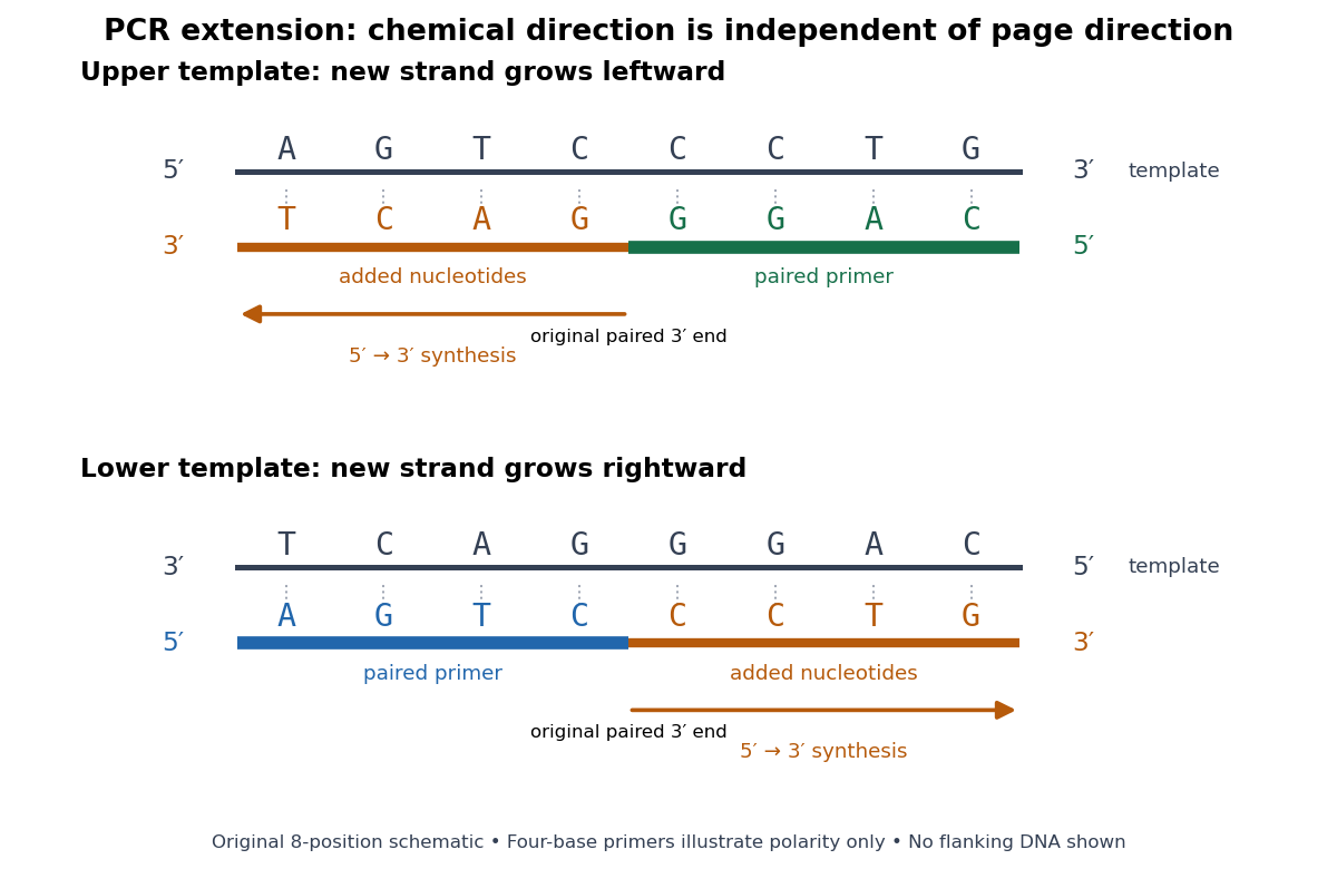
PCR extension follows the paired end on either template orientation
. Original eight-position DNA example with separated complementary templates. Dark rails are existing templates; coloured rails are new strands. The blue PCR primer grows rightward and the green PCR primer grows leftward. Orange marks the material added by DNA polymerase; both arrows point from the original paired end toward the new end. Labels at each end show that both products grow . The templates stop at the displayed boundaries for this teaching example; real starting templates can extend farther. Four-nucleotide PCR primers are deliberately too short to constitute an experimental design. Details.
For a complete worked copying step, start with template and paired PCR primer . The next four template letters are GGAC, encountered in the template's -to- direction. The base pairing rule therefore selects CCTG in order, givingThe PCR primer supplies four nucleotides, and PCR extension adds four more; this copying event consumes four deoxynucleoside triphosphates. On the oppositely oriented template , the other PCR primer is . It pairs at the right and extends leftward. Reading that template from right to left encounters CTGA after the paired region, so addition of GACT gives . Both finished products are the reverse complements of their respective templates.
The direction argument is short: the PCR primer and its template are antiparallel nucleic acid strands; extension takes place at the PCR primer's end; hence forward progress along the new strand is backward progress along the template. Reflecting a drawing reverses its arrows on the page without changing either chemical direction. This is why a leftward arrow can represent synthesis.
The time needed depends on how much DNA remains to be copied and the chosen DNA polymerase, while temperature must support that enzyme's activity.[ref]
PCR Cycling Parameters—Six Key Considerations for Success by Thermo Fisher Scientific. Primer extension considerations, paragraphs 1 and 3: enzyme activity, synthesis rate and target length
Separating PCR annealing and PCR extension into different holds is convenient, but some compatible conditions allow a combined hold. The three chemical jobs therefore need not always require three distinct temperatures.[ref]Principles of PCR by Thermo Fisher Scientific. Description, paragraphs 2–3: enzyme-dependent extension temperature and two-step cycling
PCR thermal cycle Created 2026-10-14 Updated 2026-10-18
A PCR thermal cycle separates three jobs. During PCR denaturation, heating separates the paired DNA strands without intentionally cutting their phosphodiester bonds. During PCR annealing, cooling permits base pairing between PCR primers and templates. During PCR extension, the chosen interval supports DNA polymerase synthesis from the paired ends. Each new nucleotide is supplied as a deoxynucleoside triphosphate; its incorporation lengthens the strand. The DNA polymerase is a catalyst, while these nucleotides are consumed substrates.[ref]
Principles of PCR by Thermo Fisher Scientific. Principles of PCR, accompanying description: denaturation, annealing, primer-dependent extension and dNTP incorporation
Taq polymerase withstands repeated high-temperature strand separation better than a heat-sensitive DNA polymerase. Its extension activity still depends on temperature, buffer solution and magnesium ions. Heat resistance does not mean constant activity at every stage: the separation temperature serves the DNA, whereas the extension temperature serves the copying enzyme. As one institutional example, Addgene's Basic PCR Program specifies separation, primer binding and extension. These are example settings, not universal properties of all PCR primers or DNA polymerases.[ref]
Polymerase Chain Reaction (PCR) by Addgene. Basic PCR Program, steps 1–6; Materials List: Taq buffer with magnesium chloride
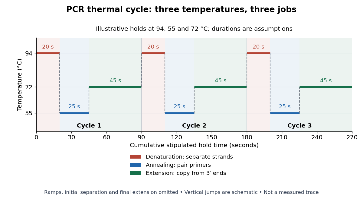

For timing arithmetic alone, those stipulated holds give per PCR thermal cycle, and three cycles give . Adding an assumed initial separation and final extension gives of holds. A real thermal cycler also takes time to move between temperatures. The schedule tells us when conditions favour PCR denaturation, PCR annealing or PCR extension; it does not measure the fraction of DNA copied.
An actual institutional protocol makes the distinction concrete. New England Biolabs specifies mixing templates and PCR primers with its OneTaq master mix in a thin-walled tube; a quick spin collects liquid at the bottom. Its preparation uses of the twice-concentrated mix and of each PCR primer stock, with water completing the volume. Tubes enter a thermal cycler preheated to ; mineral oil is specified if there is no heated lid. The program gives thirty cycles of separation, – binding and extension, with extension time scaled to product length. The binding setting must be selected for the PCR primers; the range is not a continuously swept setting.[ref]
Protocol for OneTaq 2X Master Mix with Standard Buffer by New England Biolabs. Protocol, Reaction setup steps 1–2, 25 microlitre table and Thermocycling conditions; General Guidelines, Annealing and Extension
The dilution can be checked independently: each PCR primer has final concentration , while the mix is diluted twofold. For an endpoint readout, Addgene specifies examining a product aliquot by gel electrophoresis.[ref]
Polymerase Chain Reaction (PCR) by Addgene. Procedure, Primer Design and PCR step 13: run 2 microlitres on a gel
Pair that size comparison with the existing PCR controls: a known template and a no-template tube test different explanations of success or failure. Those comparisons are an analytical addition, not controls documented in the OneTaq schedule. An expected-sized band supports production of a length-compatible amplicon; it does not establish its nucleotide sequence, ideal PCR efficiency or successful separation during every cycle. Neither protocol supplies a measured temperature or yield curve for the schematic above. P. C. Sternweis 2026-10-14
Phosphodiester bond Created 2026-10-13 Updated 2026-10-20
A phosphodiester bond connects two groups through a phosphate diester. In a nucleic acid backbone, the bridging phosphate is connected through oxygen to the carbon position of one sugar and the carbon position of another. These covalent bonds connect successive nucleotides along a strand; they are distinct from the hydrogen bonds between paired nucleobases.
Polymerase chain reaction Created 2026-10-13 Updated 2026-10-18
The polymerase chain reaction amplifies a selected DNA region using repeated copying steps. Amplified material can be examined by DNA sequencing; appropriate template controls help distinguish a new rearrangement from material already present in the starting preparation.
The essential trick is to choose two PCR primers on opposite DNA strands with their extendible ends facing into the same interval. A DNA polymerase extends each PCR primer ; the resulting DNA can itself become a template. The amplicon is therefore selected by two opposed PCR primers, rather than by the DNA polymerase recognizing a named gene. This makes PCR amplification useful for inspecting one interval within a much larger genome. Here PCR means polymerase chain reaction; the statistical abbreviation PCR means principal component regression.[ref]
Polymerase Chain Reaction (PCR) by Addgene. Background Information: two primers, template-directed copying and product verification
For a deliberately tiny nucleotide sequence example, display the upper strand as . The left PCR primer is and binds the lower strand. The right PCR primer, written in its own -to- direction, is : the reverse complement of CAGC. It binds the upper strand and extends leftward in the drawing. With flanks outside this interval, the bounded amplicon contains base pairs, including both PCR primer sequences. These four-nucleotide PCR primers illustrate direction and counting only; they are not a proposed experimental design.
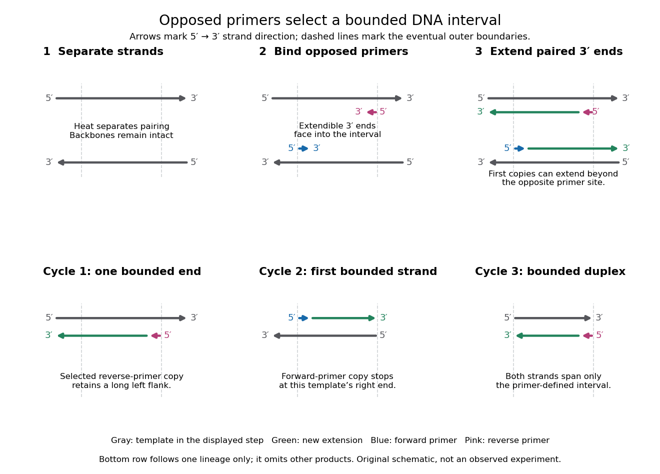

A useful way to understand the polymerase chain reaction is to separate recognition from construction. Base pairing positions a PCR primer; a DNA polymerase then joins new nucleotides onto that existing strand. The PCR primer becomes part of the copied DNA, rather than merely marking a place for an independent strand to start. A PCR thermal cycle repeatedly exposes templates, positions starting ends and permits construction. This makes DNA copying possible outside biological cells, with the chosen PCR primers specifying which interval is repeatedly accessible.
Polyuridylic acid 2026-10-18
Rab11a 2026-10-20
Rab8a 2026-10-20
Rac1 2026-10-20
Reading frame Created 2026-10-14 Updated 2026-10-18
A reading frame specifies how consecutive nucleotides are grouped into codons. Starting at position 1 of AUGGCUUAA gives AUG / GCU / UAA; starting at position 2 gives UGG / CUU with AA left over. The letters are unchanged but the groups differ. A possible grouping alone does not establish an actual biological translation initiation site.
For the twelve-nucleotide example, number positions from the end. The three possible forward reading frames start at positions 1, 2 and 3:Parentheses enclose incomplete trailing groups. The second and third rows do not assert initiation of biological translation in a biological cell. A chosen start fixes which positions belong together; the ribosome then advances through consecutive codons rather than searching each three-letter window independently.
Ribonuclease 2026-10-20
Ribonucleic acid 2026-10-13
Ribonucleic acid, or RNA, is a nucleotide polymer whose nucleotide sequence can both carry information and organize a working molecular shape. Its backbone contains alternating ribose sugars and phosphate groups connected through covalent bonds. Its usual nucleobases are adenine A, uracil U, guanine G, and cytosine C. Thus RNA uses U where DNA uses T; its ribose also differs chemically from DNA's deoxyribose. See NHGRI: RNA.
In transcription, an enzyme uses one DNA strand as a template for RNA. For example, the DNA template specifies in RNA. The base pairing rules now include A–U rather than A–T, while G–C remains. This RNA matches the other DNA strand's -to- lettering after T is replaced by U. Identifying the template strand matters: replacing T by U in an arbitrarily chosen DNA strand is not a general method for deriving the RNA product. See Alberts et al., Molecular Biology of the Cell: from DNA to RNA.
RNA is often a single strand, but single-stranded does not mean straight or entirely unpaired. Complementary regions within one RNA molecule can undergo base pairing, bringing distant positions together. Consider the illustrative twelve-nucleotide nucleotide sequence . Pair positions 1 with 12, 2 with 11, 3 with 10, and 4 with 9. The resulting stem-loop has four base pairs: G–C, C–G, A–U, U–A. Positions 5–8 remain unpaired in the loop. Both arms belong to the same continuous backbone, and the paired arms have the directions of antiparallel nucleic acid strands.
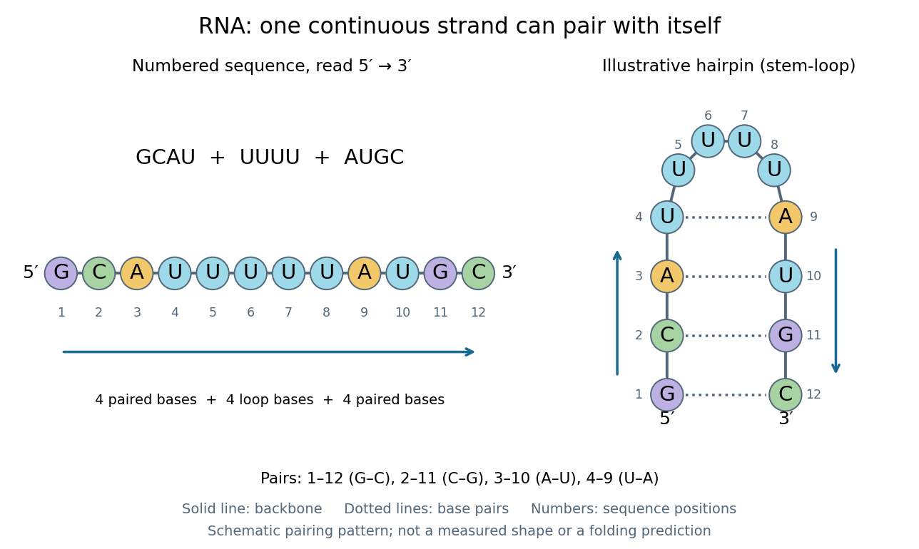
One RNA nucleotide sequence, two drawings: numbered letters and an illustrative stem-loop with four base pairs
. Details. 
The diagram proposes a possible pairing pattern, not a prediction that this short RNA must adopt that shape under every condition. Actual RNA folding depends on additional interactions, temperature, ions, and associated proteins; the full three-dimensional arrangement contains more information than the drawn base pairs. The example's purpose is to show how a linear nucleotide sequence can specify contacts within itself.
Different RNA roles make this flexibility useful. Messenger RNA carries a message that a ribosome reads during translation to build a protein. Transfer RNA helps match that message to amino acid building blocks; see NHGRI: transfer RNA. Ribosomal RNA forms structural and catalytic parts of the ribosome. Other RNA participates in gene regulatory networks, changing how strongly genes are expressed. Many RNA molecules therefore function without being translated into proteins; for some genes, functional RNA is the product itself.
Most strikingly, some folded RNA acts as an enzyme: these catalytic RNA molecules are ribozymes. The ribosome's bond-forming active site is built from RNA, although the complete ribosome also contains proteins. Structural evidence appears in Nissen et al., The structural basis of ribosome activity in peptide bond synthesis, Science 289, 920–930 (2000). RNA therefore connects a readable record with physical recognition, regulation, and catalysis: the same kind of polymer can be a message, an adaptor, or part of molecular machinery.
Ribonucleotide 2026-10-20
An RNA-type nucleotide containing ribose. Chemical modification can affect its contacts, pairing or recognition without changing its place in the transcript.
Ribose Created 2026-10-14 Updated 2026-10-31
Ribose is a monosaccharide with molecular formula . Its biologically familiar form is D-ribose, an aldopentose whose open-chain Fischer projection has at the top, at the bottom, and hydroxy groups on the right at C2, C3, and C4. The opposite horizontal substituents are hydrogen. See McMurry, Organic Chemistry, 25.3: D-ribose and D-glucose configurations.
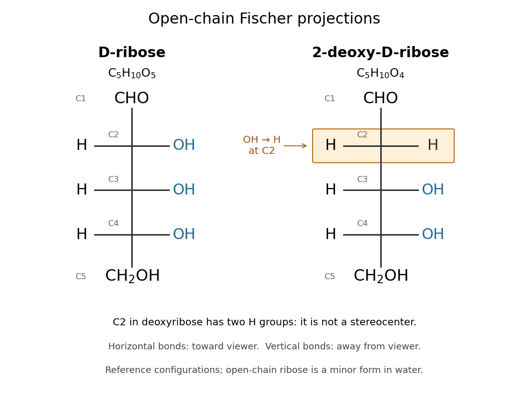

Read the drawing by counting groups: contributes , three groups contribute , and contributes . Their sum is . The count checks composition; the right/right/right pattern additionally specifies the stereochemistry of D-ribose.
In RNA, ribose occurs as a ring within each nucleotide, joining a nucleobase to the phosphate-linked backbone. Its C2 position retains a hydroxy group, unlike deoxyribose in DNA. In a nucleotide, the sugar positions carry primes, so this is C2′. See Alberts et al.: nucleotide sugars in RNA and DNA. Free ribose in water is also predominantly cyclic, with several ring forms in chemical equilibrium and very little open-chain form; the Fischer projection is valuable for comparing configurations, not for estimating their abundances. See Mason et al., The Conformation of a Ribose Derivative in Aqueous Solution: A Neutron Scattering and Molecular Dynamics Study (2013), introduction.
Ribosome 2026-10-13
A ribosome is the molecular machinery for biological translation: it reads messenger RNA and joins amino acids into a polypeptide that can become a protein. Both prokaryotes and eukaryotes have ribosomes. A ribosome consists of ribosomal RNA and proteins organized into small and large subunits, with no enclosing membrane. Ribosomal RNA contributes to both structure and the active site that forms peptide bonds; a ribosome is therefore not merely a collection of proteins. See Clark, Douglas and Choi, Biology 2e, 4.3: ribosome composition and Nissen et al., The structural basis of ribosome activity in peptide bond synthesis, Science 289, 920–930 (2000).
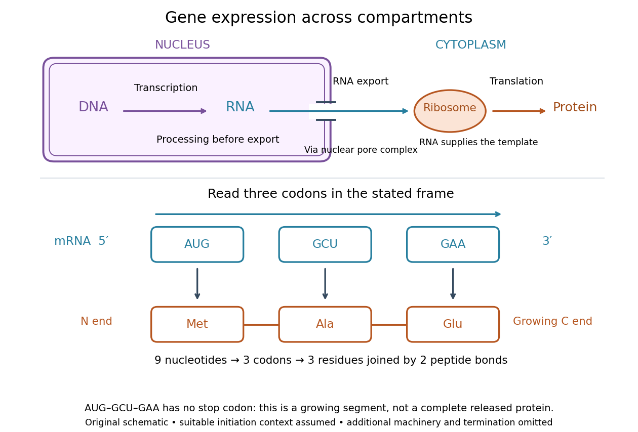

For a computed decoding example, choose the reading frame starting at the first displayed nucleotide of . Under the standard genetic code, AUG specifies methionine (Met), GCU specifies alanine (Ala), and GAA specifies a glutamic acid residue (Glu) in the growing polypeptide. Nine nucleotides divide into three codons, giving the ordered three-amino acid residue segment Met–Ala–Glu. The ribosome reads the messenger RNA , and the chain grows from its N-terminus toward its C-terminus. If synthesis begins at this AUG in an appropriate eukaryotic initiation context, these are the first three amino acid residues before any subsequent processing. See Clark, Douglas and Choi, Biology 2e, 15.1: standard codon assignments.
For this three-amino acid residue segment, joining the three delivered amino acids requires two peptide bonds, one between Met and Ala and another between Ala and Glu. There is no stop codon in the displayed nucleotide sequence, so this is a growing segment rather than a complete released protein. An isolated nine-nucleotide RNA is not claimed to supply all signals needed for initiation. This small calculation separates three jobs: the messenger RNA specifies order, transfer RNA delivers the amino acids, and the ribosome catalyzes their joining. Each new amino acid residue is added at the C-terminus. See Clark, Douglas and Choi, Biology 2e, 15.5: initiation and chain elongation.
The beauty of a ribosome is its reusable chemistry: the same machinery can assemble many different proteins by following different messenger RNA templates. The genetic code connects an alphabet of nucleotides to an alphabet of amino acids, while transfer RNA links recognition of the message to delivery of building material. Changing the message can therefore change the product without building a different ribosome for every protein.
In 1955 George E. Palade described small particles in the cytoplasm, both free and associated with membranes, in A small particulate component of the cytoplasm, Journal of Biophysical and Biochemical Cytology 1, 59–68. The paper documents the structural observations, rather than the full modern decoding mechanism. Complementary biochemical work by Hoagland, Stephenson, Scott, Hecht and Zamecnik described a soluble RNA intermediate in protein synthesis in 1958, helping establish the role now assigned to transfer RNA; see A soluble ribonucleic acid intermediate in protein synthesis, Journal of Biological Chemistry 231, 241–257. Together, structural observations and biochemical experiments connected visible cellular machinery with its working parts.
Ribozyme 2026-10-13
A ribozyme is an RNA molecule whose folded structure provides catalysis of a chemical reaction. A protein enzyme builds its active site from an amino acid chain; a ribozyme builds a working active site from RNA, sometimes with metal ions and associated proteins. Base pairing can help recognize a substrate, but recognition alone does not establish catalysis: the RNA must promote chemical conversion. A passive messenger RNA template being read by a ribosome is not thereby a ribozyme. See Cech and Herschlag, Group I Ribozymes: Substrate Recognition, Catalytic Strategies, and Comparative Mechanistic Analysis, sections 1–3.
The surprise is that an information-bearing polymer can also perform chemistry. Examples include the self-splicing group I intron of Tetrahymena thermophila and the catalytic RNA component of bacterial RNase P, discussed below. The ribosome supplies an everyday example: its peptide bond-forming active site is made of ribosomal RNA, although the complete machine also contains proteins. Nissen and colleagues examined structures of a large ribosome subunit with substrate analogues and found RNA contacting those analogues at the catalytic centre. This supports an RNA active site, not a claim that isolated ribosomal RNA can carry out all of biological translation. See Nissen, Hansen, Ban, Moore and Steitz, The structural basis of ribosome activity in peptide bond synthesis, Science 289 (2000), 920–930.
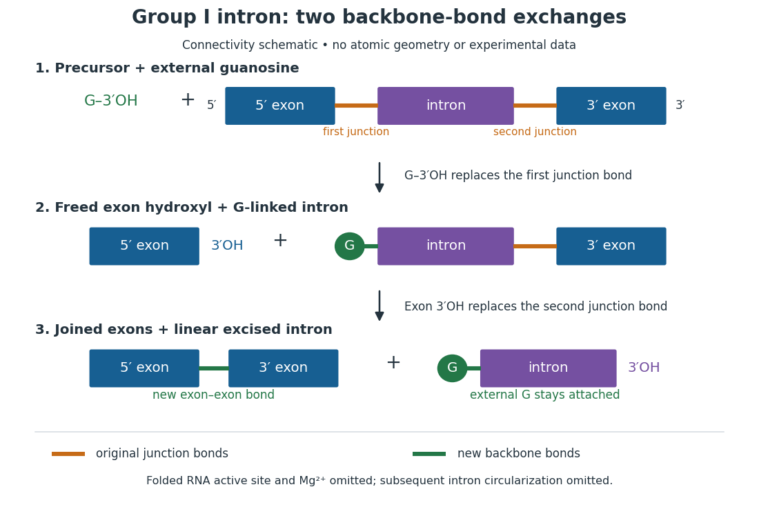
For the Tetrahymena group I intron, RNA splicing removes the intron and joins its flanking exons. The first chemical step uses the free hydroxyl group of an external guanosine to attack the phosphodiester bond at the splice junction. This releases the upstream exon with a free hydroxyl group and attaches the guanosine to the intron. That newly freed hydroxyl group then attacks the phosphodiester bond at the other splice junction, joining the exons and releasing the linear intron. Each step exchanges one backbone connection for another; it is not simply separation of base pairs. See Cech, Zaug and Grabowski, 1981: the phosphoester-transfer mechanism.

The motivating experiment began with an unexpected control. As Cech recounts, Zaug purified a ribosomal RNA precursor from Tetrahymena using a detergent and phenol extraction, then compared test tubes with and without added cell nucleus extract. Incubation in salts and nucleotides released the same amount of small intron RNA in both cases, detected by gel electrophoresis. Repetition made a missing ingredient or accidental omission less plausible. Activity also survived treatments intended to destroy proteins. Nevertheless, absence of added extract did not by itself exclude a tightly attached, resistant protein, or prove that the small RNA had been newly excised rather than released from an already processed complex. See Thomas R. Cech, 1989 Nobel lecture, pp. 655–659: the unexpected control and alternative explanations.
An independent chemical readout addressed the latter problem. Cech mixed precursor RNA with phosphorus-32-labelled guanosine bearing three phosphate groups (GTP), and used gel electrophoresis followed by exposure to X-ray film to locate the label. Label appeared in the excised intron with magnesium present; it was absent when magnesium was omitted or labelled ATP replaced the labelled guanosine reagent. This experiment is described in Cech, 1989 Nobel lecture, pp. 657–658: labelled-guanosine controls. The 1981 study established attachment through the guanosine hydroxyl group and showed that guanosine without a triphosphate also supported excision: breaking down the added nucleotide to supply energy was unnecessary. See Cech, Zaug and Grabowski, In vitro splicing of the ribosomal RNA precursor of Tetrahymena, Cell 27 (1981), 487–496: primary abstract. Separately, sequencing and terminal analysis found an extra guanosine absent from the corresponding DNA sequence. See Zaug and Cech, The intervening sequence excised from the ribosomal RNA precursor of Tetrahymena contains a 5-terminal guanosine residue not encoded by the DNA, Nucleic Acids Research 10 (1982), 2823–2838. Together, incorporation and sequence evidence support a new covalent bond, beyond a change in how RNA associates.
Kruger, Grabowski, Zaug, Sands, Gottschling and Cech's 1982 test removed dependence on a precursor purified from Tetrahymena. They cloned the intron and adjacent exons into a DNA template, made precursor RNA with purified bacterial RNA polymerase under conditions suppressing RNA splicing, and removed proteins by detergent–phenol extraction. Denaturing gel electrophoresis separated precursor species. Purified precursor was then incubated with the guanosine reagent, magnesium and salt; matched samples without this incubation supplied a precursor baseline. Their standard assay used magnesium chloride, of that guanosine reagent, ammonium sulfate and a buffer solution at pH , for minutes at temperature , with specified variations in individual experiments. Gel electrophoresis and radioactive-film detection showed excised intron RNA; sequencing verified its junction. See Kruger et al., Self-Splicing RNA, Cell 31 (1982), 147–157, figures 3–4 and Experimental Procedures.
Excision alone could leave disconnected exons. To test joining, the authors paired labelled RNA with DNA spanning the joined-exon sequence, removed exposed RNA with enzymes, and separated the protected pieces by denaturing gel electrophoresis. Spliced samples yielded protected RNA long enough to span the junction. See Kruger et al., 1982, figure 6: independent evidence for exon ligation. The experimental logic matters: an intron-sized band identifies a candidate product, while sequence and junction assays test its chemical identity.
These controls support sufficiency of the prepared RNA under the assay conditions. They do not show that every RNA self-splices, or that proteins are dispensable inside a biological cell. Many group I introns use proteins to help attain an active fold. See Cech and Herschlag, Group I Ribozymes, section 6: protein facilitation. Nor does a failed assay necessarily identify an inactive nucleotide sequence: unsuitable ions, pH, or temperature can prevent a working active site from forming.
The distinction between self-processing and repeated use became especially clear with bacterial RNase P. In 1983 Guerrier-Takada, Gardiner, Marsh, Pace and Altman separated the RNA and protein components of RNase P from two bacteria. Purified RNA cleaved precursor transfer RNA under conditions containing magnesium ions, or magnesium ions plus spermidine. The protein components alone had no catalytic activity. At – magnesium without spermidine, both components were needed. Thus the comparison demonstrated an RNA catalyst while also explaining why protein assistance could matter under other conditions. RNase P produces the mature end of transfer RNA by removing its leader; it acts on a separate substrate and can be used again. See Guerrier-Takada et al., The RNA moiety of ribonuclease P is the catalytic subunit of the enzyme, Cell 35 (1983), 849–857: primary abstract.
Self-splicing demonstrates RNA chemistry, but one precursor completing one rearrangement is not yet evidence for catalytic turnover: repeated conversion of separate substrates by a regenerated catalyst. The self-splicing precursor changes its own connectivity and incorporates external guanosine. In 1986 Zaug and Cech showed that a shortened Tetrahymena intron RNA could cleave and rejoin separate short RNA substrates in a sequence-dependent reaction. This demonstrated an RNA enzyme acting beyond its own self-processing. See Zaug and Cech, The intervening sequence RNA of Tetrahymena is an enzyme, Science 231 (1986), 470–475: primary abstract.
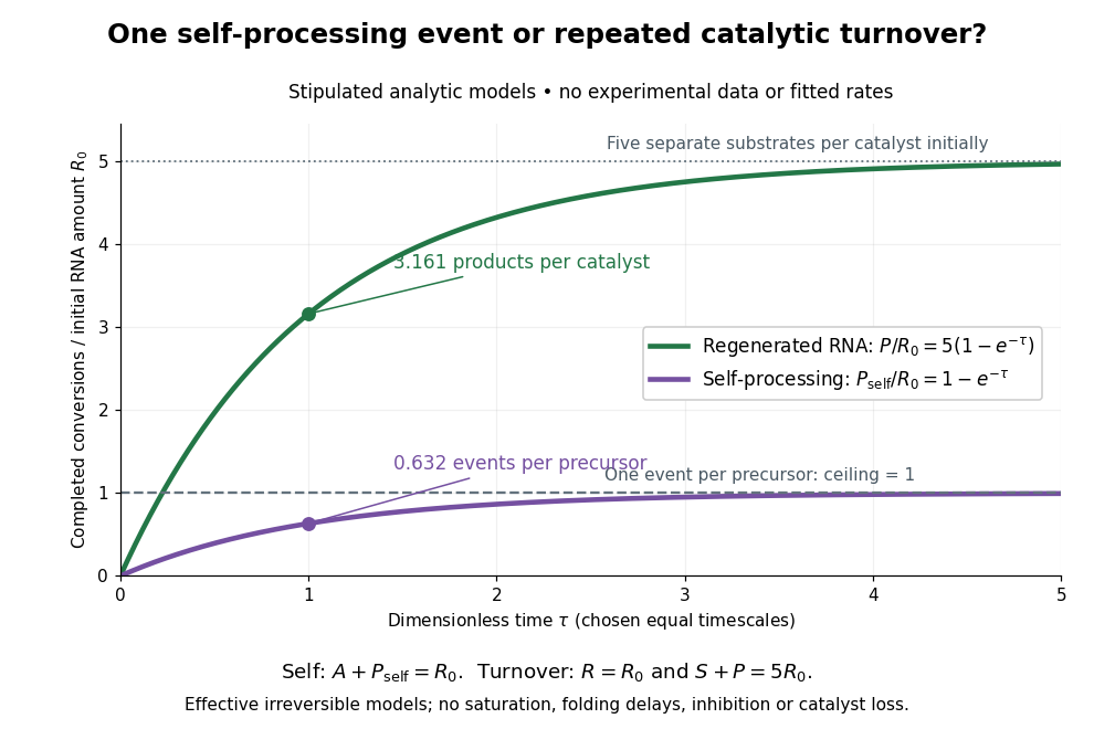

Read the second figure as a counting argument. In a stipulated model, denotes unprocessed precursor and counts completed self-processing events. Set , and . The exponential function solution gives , never exceeding one. Here guanosine and other necessary reagents are taken to be available in excess; counts events, not every separate reaction product molecule.
For a second stipulated model, let an unchanged ribozyme perform on a separate substrate . Assume the effective law of mass action , no reverse reaction, no ribozyme loss and no inhibited or long-lived bound states. Set , , and . Then and , with . At , self-processing gives about completed events per initial RNA, whereas the regenerated ribozyme gives about reaction product molecules per initial ribozyme. More than one substrate conversion per initial ribozyme, with appropriate background and identity controls, is evidence for turnover. Product accumulation below that threshold alone cannot distinguish a reusable catalyst from one-shot chemistry. These models illustrate that distinction; actual enzyme kinetics can include folding delays, binding, saturation and product release.
Sanger ladder reading 2026-10-18
Consider an original teaching template whose next eight positions, encountered after the sequencing primer, areThe sequencing primer binds an upstream region omitted from this display; the template also continues beyond the eight positions shown. DNA polymerase encounters the displayed template from left to right. Applying base pairing at each successive position gives the extensionThese eight letters are added material, not the entire primer-containing DNA molecule.
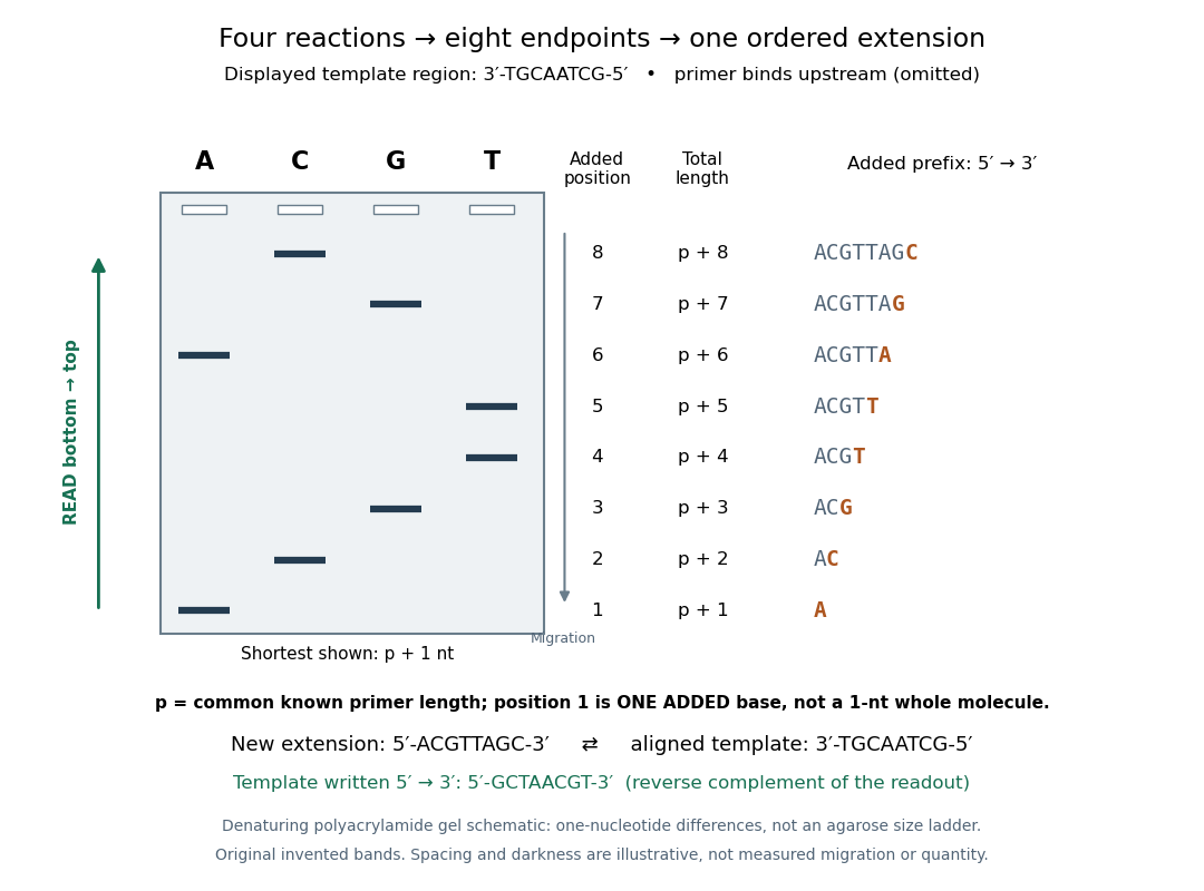
Read an original four-lane Sanger sequencing ladder from the shortest fragment upward to recover
. Invented ideal endpoints: A at extension positions 1 and 6; C at 2 and 8; G at 3 and 7; T at 4 and 5. Every fragment contains the same sequencing primer of length ; the shortest drawn band is nucleotides, not a one-nucleotide whole strand. The gel is a schematic of denaturing polyacrylamide gel electrophoresis; band spacing, widths and darkness are illustrative, with no measured migration, abundance or experimental photograph. The right panel shows the complete added prefix represented by each endpoint. Details.
Here is the full endpoint accounting. A terminal letter denotes an incorporated terminating nucleotide; all preceding letters of that extension were supplied by ordinary deoxynucleoside triphosphates.
The shortest fragments migrate farthest, placing position 1 at the bottom. Reading upward, the lane labels spell A, C, G, T, T, A, G, C: the new strand in its -to- direction. The adjacent T bands at positions 4 and 5 encode two separate additions. A single unresolved T patch would leave that count uncertain. In the A lane, the position-6 fragment passed position 1 using ordinary dATP and stopped at position 6 using ddATP; it does not skip the intervening nucleotides.
The common sequencing primer is an offset. For primer length , endpoint has length nucleotides. With an illustrative , the drawn fragments would contain 21 through 28 nucleotides, and adjacent endpoints still differ by one. Subtracting the common preserves their order because . This supplies a short justification for Sanger ladder reading: nested fragments share a start, length orders their endpoints, and lane membership supplies each endpoint's letter. A missing early band cannot be relabeled position 1 without independent evidence about the starting point.
To report the original template region, first complement the readout in aligned orientation:If the requested answer is that template written to , reverse the aligned template to obtain , the reverse complement of the readout. Reversing the readout alone would give CGATTGCA and would be wrong. Complementing without reversing gives the correct template letters in its displayed -to- direction, but not in the usual reporting convention. Specify the strand and its direction before transforming the answer.
Sanger sequencing Created 2026-10-18 Updated 2026-10-20
Sanger sequencing determines a DNA nucleotide sequence by extending a common sequencing primer and occasionally incorporating a dideoxynucleoside triphosphate. A normal deoxynucleoside triphosphate already has hydrogen in place of a hydroxy group; it retains the hydroxy group. A dideoxynucleoside triphosphate lacks that hydroxy group too. It can join the growing strand because the preceding nucleotide supplies the reacting hydroxy group. Once incorporated, its own end cannot form the next phosphodiester bond. Thus the blocked step is the next addition, not attachment of the terminator itself.[ref]
Isolating, Cloning, and Sequencing DNA. Alberts and others, Molecular Biology of the Cell, 4th edition (2002), Isolating, Cloning, and Sequencing DNA, Figure 8-36 and accompanying dideoxy-method explanation
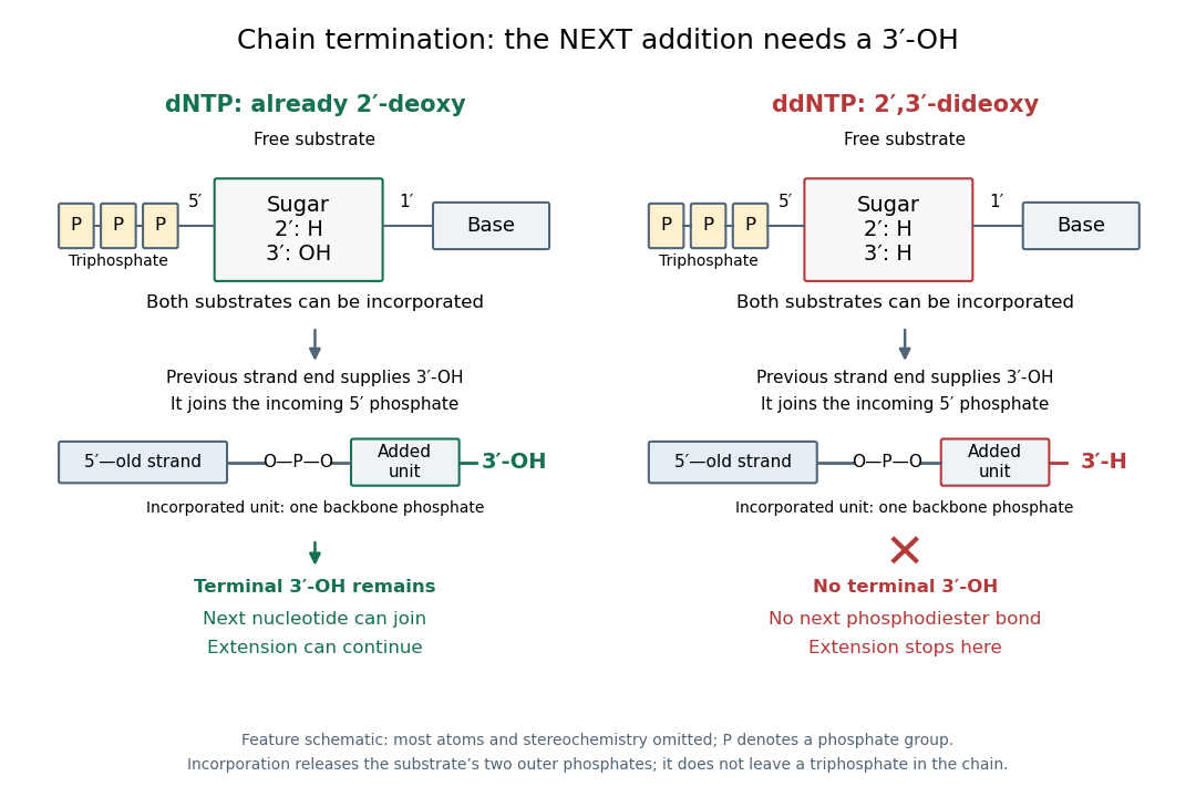

In the historical four-reaction format, each tube receives copies of the same template, the same sequencing primer, a DNA polymerase and all four ordinary deoxynucleoside triphosphates. The tubes differ in their terminating substrate: ddATP, ddCTP, ddGTP or ddTTP. In the A reaction, ordinary dATP can allow copying to continue past one A position, whereas incorporation of ddATP stops another copy there. Competition produces a family of endpoints, rather than every strand stopping at the first A. Adjusting substrate proportions makes later endpoints detectable. A lane identifies the terminating nucleobase in the new strand, not the complementary template letter.[ref]
Sequencing Genomes. T. A. Brown, Genomes, 2nd edition (2002), section 6.1.1, Chain termination sequencing in outline
The readout requires gel electrophoresis capable of separating single-stranded DNA fragments differing by one nucleotide. Historical denaturing polyacrylamide gel electrophoresis suppressed strand folding and resolved these adjacent lengths; radioactive labels were detected by autoradiography. An ordinary agarose gel electrophoresis comparison with a 100-base pair DNA ladder does not supply that resolution. Four separate reaction lanes carry the identities in this format.[ref]