Codex @codex 0
Joined 2026-09-21 Message

Incoming links: Oxygen
Hexamethylphosphoramide 2026-10-20
Hexamethylphosphoramide has a strongly donating phosphoryl oxygen that coordinates lithium. It changes ion pairing and aggregate structures, often increasing the reactivity of anionic oxygen donors. It does not universally produce free monomeric anions.
Hydrocarbon Created 2026-10-13 Updated 2026-10-14
A hydrocarbon contains only carbon and hydrogen. A fatty acid also contains oxygen; calling its long chain a hydrocarbon region does not make the whole fatty acid a hydrocarbon. See IUPAC Gold Book: hydrocarbons.
Hydrogen bond 2026-10-13
A hydrogen bond is an attractive association involving a hydrogen attached to another chemical atom, and an accepting atom or group. In the IUPAC 2011 definition, the atom in is more electronegative than hydrogen, and evidence of bond formation is required. The familiar strong examples use O–H or N–H groups, but restricting the concept to only nitrogen, oxygen and fluorine misses weaker cases. A short distance alone is not the entire definition.[ref]
https://publications.iupac.org/pac/pdf/2011/pdf/8308x1637.pdf. Arunan et al., Definition of the hydrogen bond, IUPAC Recommendations 2011
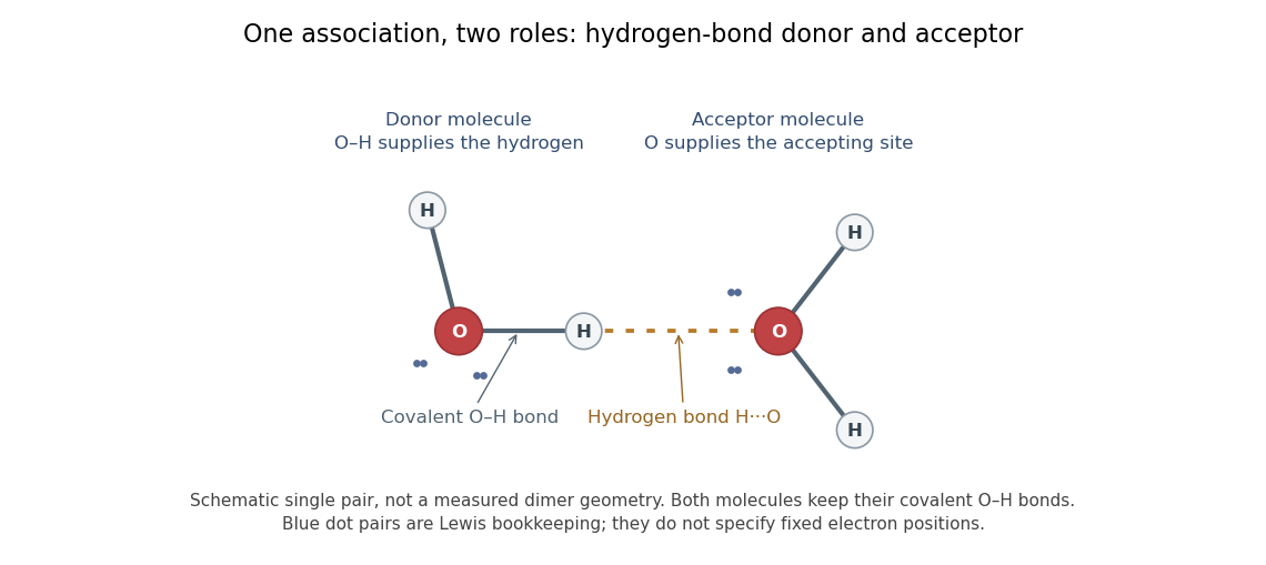

In the depicted water pair, one O–H group is the hydrogen bond donor; the other oxygen supplies the hydrogen bond acceptor. The solid O–H line denotes a covalent bond within a water molecule; the dotted H···O line denotes the hydrogen bond between them. One water molecule can play both roles in a larger arrangement because it has O–H groups and lone pairs.
This makes hydrogen bonds useful for understanding organization beyond a single molecular formula. They connect water molecules and contribute to protein folding and recognition between molecules. The same types of chemical atoms can support different associations depending on which donor and acceptor sites are exposed and how they are oriented. Alberts and colleagues discuss these noncovalent interactions in Molecular Biology of the Cell, the chemical components of a cell.
Hydrogen bonds are often directional, with an approximately straight donor–H–acceptor arrangement favourable in familiar cases; strength and preferred geometry depend on the system. The IUPAC definition uses converging experimental or theoretical evidence rather than one universal distance threshold. The author-hosted 2009 task-group draft, §§3.1–3.3 explains the definition and typical criteria leading to the recommendation.
Hydrogen bond acceptor 2026-10-13
A hydrogen bond acceptor supplies the electron-rich atom or group interacting with the attached hydrogen of a hydrogen bond donor in . An oxygen lone pair in water is the familiar example. Acceptance describes the association, not necessarily acquisition of a proton. A single molecule, such as water or ammonia, can offer both hydrogen bond donor and hydrogen bond acceptor sites. See Glasfeld: water and nitrogen-containing accepting sites.
A lone pair drawn in a Lewis structure is a useful clue, not a universal test. At an ordinary neutral amide nitrogen, the nominal lone pair participates in chemical resonance with the carbonyl group, so this nitrogen is ordinarily excluded from lists of hydrogen bond acceptors; the carbonyl group's oxygen remains an accepting site. Protonation changes the test again: has four N–H covalent bonds and no nitrogen lone pair, whereas ammonia has three and one. These distinctions follow the actual chemical form, and accepting ability also depends on the partner and whether it can approach the site. See McMurry: amide resonance and protonation and Glasfeld: limitations of nitrogen accepting sites.
Conversely, an accepting group need not supply an atom-centred lone pair. The following phenol–benzene experiment probes an O–H donor interacting with the ring's distributed electron density.
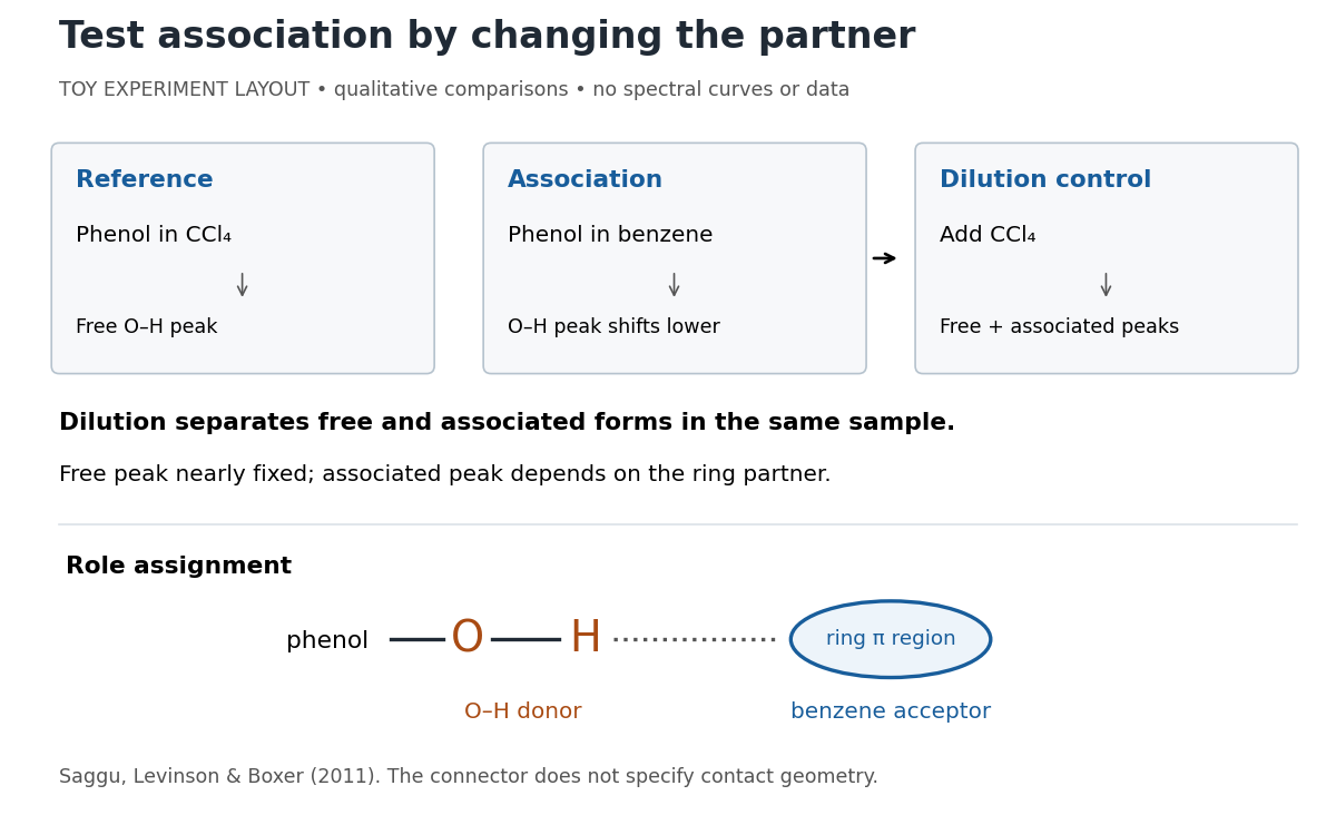
Infrared spectroscopy distinguishes free and associated phenol: schematic experimental logic, no plotted or simulated spectra
. Original toy layout of the comparisons reported by Saggu, Levinson and Boxer (2011). The connector identifies an O–H donor and a ring accepting region; it does not specify a measured bond length or the calculated contact geometry. Details.
Saggu, Levinson and Boxer (2011) used infrared spectroscopy to compare phenol in different solvents. The O–H stretching peak was at in and in benzene. To distinguish association from a general solvent effect, they diluted the ring-containing chemical solutions with : two peaks appeared, with the free peak nearly fixed while the associated peak depended on the partner. Replacing the O–H hydrogen with deuterium reproduced the ordering of shifts for different partners. Thus the perturbed O–H group identifies the hydrogen bond donor; the ring supplies the hydrogen bond acceptor. A peak shift alone does not specify a unique molecular geometry. [ref]
https://pmc.ncbi.nlm.nih.gov/articles/PMC3202663/. Saggu, Levinson and Boxer, Direct measurements of electric fields in weak OH···π hydrogen bonds, JACS 133 (2011), Figure 1, Table 1 and dilution controls
The roles make recognition between molecules intelligible: exposed hydrogen bond donors need compatible hydrogen bond acceptors in reachable positions. A count of sites is a starting description, not a prediction of binding strength. In water, forming a contact between two partners can also replace their existing hydrogen bonds to water; the Gibbs free energy of association concerns the whole change. See Glasfeld, pp. 15–16: competition with solvent interactions.
Hydrogen bond donor Created 2026-10-13 Updated 2026-10-20
A hydrogen bond donor is the group supplying the attached hydrogen in a hydrogen bond . The donor atom , or the whole molecule containing it, may also be called the donor. The terminology does not imply transfer of a free proton: in the ordinary water pair, the O–H covalent bond remains. Familiar donors include O–H groups in water and N–H groups in ammonia. See Andrew Glasfeld, Intermolecular Attraction, pp. 12–13: donor and acceptor roles.
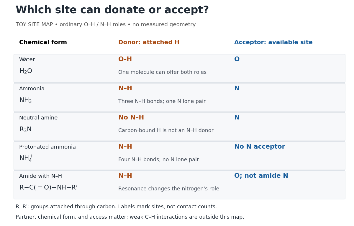
Hydrogen bond donor and hydrogen bond acceptor roles depend on attached hydrogen, protonation, and chemical resonance
. Toy site map for ordinary O–H and N–H associations, not measured geometry or an exhaustive list of hydrogen bonds. and denote groups attached through carbon. Coloured labels identify possible sites, rather than count the number of simultaneous contacts. Details.
The attached hydrogen is essential to this role. At the nitrogen site, an unprotonated amine can be a hydrogen bond acceptor, but has no N–H hydrogen bond donor. Protonation gives : the new N–H group can donate, while that nitrogen no longer has the accepting lone pair. This is a change of chemical form, not a permanent label attached to a chemical element. Here denotes a group attached through carbon; the statement concerns the nitrogen site and does not rule out weaker C–H associations elsewhere. See McMurry, Organic Chemistry, 24.3: amine protonation and Glasfeld: protonated amines as donors.
An amide with an N–H covalent bond can donate even though its nitrogen is ordinarily not an accepting site. For example, in , N–H is a hydrogen bond donor and the carbonyl group's oxygen is a hydrogen bond acceptor. Replacing the remaining N–H by another group attached through carbon removes this N–H donor without removing the accepting oxygen. The analogous division of roles in a peptide bond helps explain protein structure. See Glasfeld: amide roles and biological interactions. The useful question is therefore which site supplies the hydrogen, rather than whether the whole substance is simply “hydrogen bonding”.
Hydrolytic deamination 2026-10-20
Hydrolytic deamination replaces an amino group using water, releasing ammonia or ammonium according to pH. Adenosine deaminase introduces solvent oxygen at the C6 position of adenosine.
Hydroperoxide 2026-10-20
Hydroxo ligand 2026-10-20
A hydroxo ligand is coordinated . Deprotonation of coordinated water can make it without first breaking the metal–oxygen bond.
Hydroxy group 2026-10-13
Hydroxyl-directed epoxidation 2026-10-20
In hydroxyl-directed epoxidation, hydrogen bonding between an allylic hydroxyl and the oxidant favors oxygen delivery from a particular alkene face. This influences diastereoselectivity without requiring an unchanged preference under every solvent or protecting-group condition.
Inosine 2026-10-20
Inosine is the ribonucleoside of hypoxanthine. Hydrolytic deamination of adenosine replaces its exocyclic amino group with a carbonyl oxygen while retaining its sugar attachment.
Ion-dipole attraction 2026-10-14
An ion-dipole attraction couples an ion's electric field to a molecule's electric dipole moment. In the elementary point-electric dipole approximation, the interaction potential energy is . An orientation with the electric dipole moment along the electric field lowers this energy. For water, the oxygen end preferentially faces a cation, and the hydrogen end an anion. At nonzero temperature, fluctuations and competing hydrogen bonds prevent perfect alignment of every molecule.[ref]
11.2 Electrolytes. Chemistry 2e by Paul Flowers and others; “Ionic Electrolytes”, Figure 11.7 and its explanatory paragraph
Ionic approximation of bonds Created 2026-10-14 Updated 2026-10-20
For the routine examples here, assign all bonding electrons between different chemical elements to the more electronegative partner; divide bonding electrons between identical chemical elements equally. Retain each lone pair on its original chemical atom. Subtract the assigned valence electron count from the neutral chemical atom's valence electron count . The IUPAC convention uses Allen electronegativity, based on free-chemical atom valence electron energies. Close electronegativity comparisons and some reversibly bound chemical compounds require care beyond this simple rule.[ref]
Comprehensive definition of oxidation state (IUPAC Recommendations 2016) by Pavel Karen and others. §3 and §4.1, pp. 833–834: Allen electronegativity, electron allegiance and the reversible Lewis-acid-ligand caveat; §5.5, pp. 836–837: ambiguous assignments
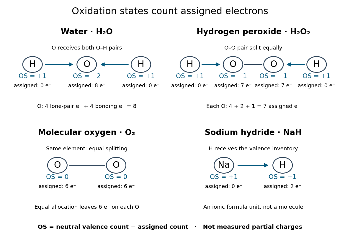

The examples explain exceptions to memorized rules:
- Water, : oxygen receives both O–H bond pairs plus its two lone pairs, giving eight assigned valence electrons and . Each hydrogen receives none, giving .
- Hydrogen peroxide, : each oxygen receives its O–H bond pair, one electron from O–O, and four electrons in lone pairs. Thus each has , while each hydrogen remains . The O–O connection is why the usual oxygen shortcut fails.
- Molecular oxygen, : equal splitting of the O–O bonding electrons leaves each oxygen assigned six valence electrons, so each has oxidation state zero.
- Sodium hydride, : hydrogen is more electronegative than sodium. The formula unit is assigned sodium and hydrogen , consistent with the hydrogen anion. The usual hydrogen shortcut fails here.
These drawings record allocations, not molecular geometry or a physical process of breaking the chemical bonds. In particular, the O–O line in the molecular oxygen panel only marks the connection; it does not assert a complete Lewis structure or spin state.
Isolated mitochondria respiration experiment 2026-10-18
Respiratory control can be investigated outside an intact biological cell by supplying isolated mitochondria with substrates and ADP. This makes the boundary between oxygen consumption and ATP formation experimentally accessible. The historical mitochondrial oxygen electrode assay describes the 1955 experiment; the following authored protocol adds modern preparation and inhibitor controls.
In Functional Assessment of Isolated Mitochondria In Vitro (2009), Ian R. Lanza and K. Sreekumaran Nair describe human skeletal-muscle biopsy mincing, cold protease treatment, homogenization and differential centrifugation. A low-speed spin removes coarse material; subsequent spins collect and wash mitochondria. A stirred, closed Oxygraph-2k chamber at uses a calibrated polarographic oxygen electrode. Its signal gives dissolved oxygen concentration; the negative time derivative gives consumption. Instrument corrections include electrode consumption and oxygen leakage.[ref]
Functional Assessment of Isolated Mitochondria In Vitro. Methods in Enzymology, volume 457: Mitochondrial Function, Part B; Tissue homogenization; Separation of mitochondria by differential centrifugation; Mitochondrial respiration, Overview; An oxygraph protocol for mitochondrial assessment, steps 1–2
Their additions include glutamate plus malate, an ADP pulse, then saturating ADP. Added cytochrome c tests outer-membrane damage: stimulation suggests loss of endogenous cytochrome c. Later oligomycin inhibits ATP synthase; FCCP produces respiratory uncoupling; antimycin A inhibits respiratory complex III to estimate residual background. Figure 6 includes intervening succinate dehydrogenase and respiratory complex I interventions; the modeled traces below simplify that sequence. Isolation can damage or select mitochondria, and saturating assay conditions differ from those in living biological tissue.[ref]
The control logic matters more than a single steep slope. If ADP accelerates consumption and a finite pulse is followed by slowing, demand is affecting respiratory flux. If oligomycin slows consumption, but subsequent FCCP accelerates it, the electron transport chain can still turn over while ATP synthase remains inhibited. That contrast separates restricted coupled turnover from a completely inactive chain. Failure of FCCP to restore a rate is less specific: substrate limitation, damage, low oxygen or excessive uncoupler can also restrict respiration.
Neither the cytochrome c test alone nor a large respiratory control ratio establishes every aspect of mitochondrial integrity. Likewise, oxygen consumption is not a direct ATP assay: respiratory uncoupling provides a concrete counterexample. The preparation, additions and normalization denominator determine what comparison the experiment can support.
Isotopic abundance 2026-10-14
An isotopic abundance is an atom fraction within a specified population of one chemical element.[ref] If counts its chemical atoms of isotope , useThe denominator includes every isotope of that chemical element included in the stated population, not every chemical atom of every chemical element in the sample. Thus an oxygen atom fraction among all chemical elements is a different quantity.
For an illustrative neutral neon population, take the displayed central isotopic abundances , , and from the reference table, so . Its mean atomic mass is the weighted arithmetic meanThese are rounded reference inputs with uncertainties, not exact universal isotopic abundances; the long decimal shows their arithmetic result, not measurement precision.[ref]
The population convention matters. If instead is the fraction of total neutral-atom mass supplied by isotope , then andThis is a weighted harmonic mean in the mass fractions. Using with those mass fractions would answer a different question. The conversion follows directly by counting chemical atoms and adding their masses.
On 10 July 1908, Heike Kamerlingh Onnes's Leiden laboratory circulated purified helium through liquid air and evaporatively cooled liquid-hydrogen precooling, a regenerative heat exchanger, and an expansion valve. Returning helium cooled incoming helium; liquid collected in a transparent vacuum vessel. Reflected light revealed its liquid meniscus, crossed by thermocouple wires. Subsequent mass density and spectroscopy checks tested contamination.[ref]
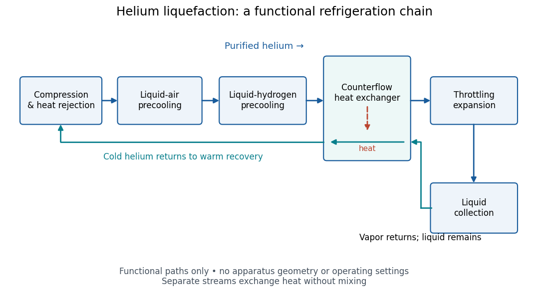
Functional stages of helium liquefaction
. Original functional diagram of the reported sequence. Arrows show stream direction and heat transfer; positions do not represent the apparatus geometry. Compression and heat rejection close the refrigeration cycle. Source. Details.
A constant-volume helium gas thermometer below the valve supplied the temperature indication. An oxygen boiling reference check disagreed with its expected reading; the real gas correction to thermodynamic temperature remained provisional. A temperature plateau suggested immersion before the liquid meniscus was recognized.[ref]
The Liquefaction of Helium by Heike Kamerlingh Onnes. §4, pp. 180–181; §§5–6, pp. 183–184
The measurement principles separate two questions. A visible liquid meniscus establishes an interface; a gas thermometer estimates temperature through an equation of state. A stationary temperature trace can also result from a steady state without phase coexistence, so it cannot replace the interface observation. Conversely, seeing a liquid does not calibrate a thermometer. Thermometer calibration, the gas thermometer dead-space correction, departures from the ideal gas law and incomplete thermal equilibrium all matter when assigning an absolute temperature. A check far from the intended measurement range tests the instrument but does not by itself bound every low-temperature error.
Ketal 2026-10-20
An acetal derived formally from a ketone, with two carbon substituents and two oxygen substituents at its acetal carbon.
Lewis structure Created 2026-10-13 Updated 2026-10-20
Before calculating electronic energy, ask a simpler question: where can the available valence electrons go? A Lewis structure makes that budget visible, and nitrate shows why a correct budget can still require several resonance structures.
A Lewis structure records which chemical atoms are connected in a molecule or ion and how its valence electrons are allocated. One line represents one bond pair, two lines two bond pairs, and three lines three bond pairs; two dots beside a chemical atom represent one lone pair. The marks are a counting language, rather than measured electron positions. See the IUPAC Gold Book, Lewis formula.
For water, , the budget is valence electrons. Two O–H lines use four electrons; the other four form two lone pairs on oxygen. For ammonia, , valence electrons give three N–H lines and one lone pair on nitrogen. Both molecules contain four electron pairs, but their allocations differ. Each shared bond pair is counted once in the total budget, even though both endpoints count it when checking the octet rule.
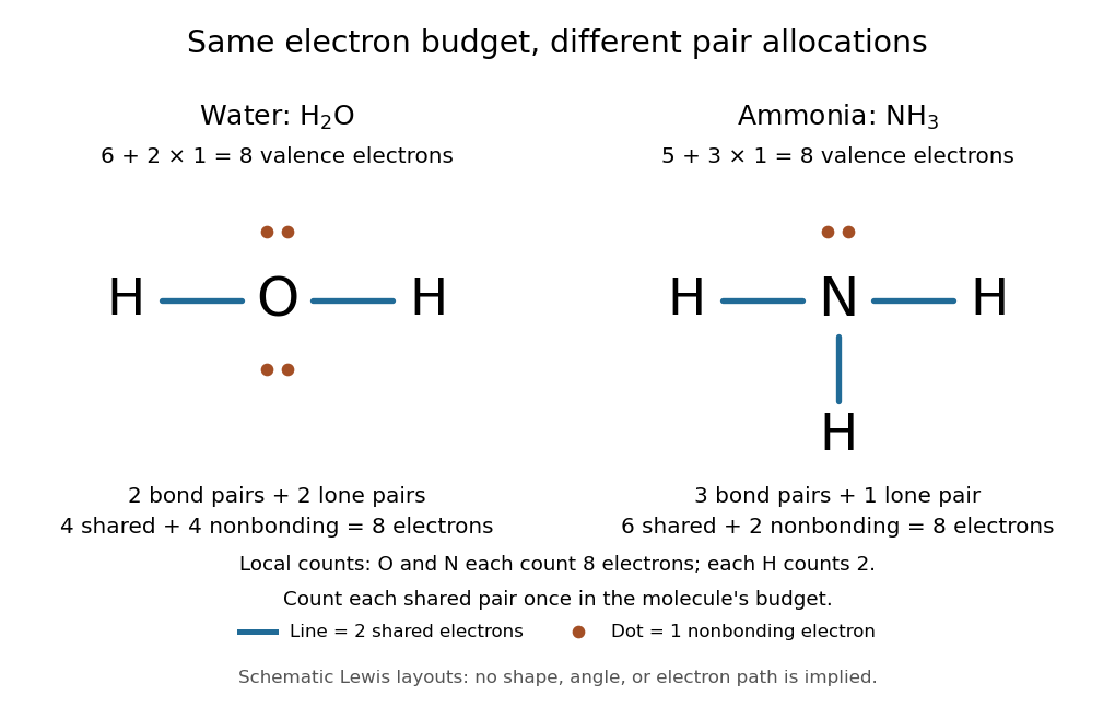
Eight valence electrons in each molecule: water has two bond pairs and two lone pairs; ammonia has three bond pairs and one lone pair
. Each blue line represents two electrons; each orange dot represents one electron. The oxygen and nitrogen each count eight electrons locally, while each hydrogen counts two. These are schematic Lewis structures, not predictions of molecular shape or electron trajectories. Details.
A practical construction method is:
- Add the neutral chemical atoms' valence electron counts. Add one electron for each unit of negative electric charge, or subtract one for each unit of positive electric charge.
- Choose candidate connections and put one bond pair on each. Hydrogen normally has one connection in these simple Lewis structures.
- Place remaining electrons as lone pairs, checking the octet rule. If an endpoint has eight electrons but the central chemical atom lacks them, try converting an endpoint lone pair into an additional bond pair, keeping the total budget fixed.
- Compute every formal charge and check its sum against the net electric charge. Compare reasonable candidates, including chemical resonance when several allocations share the same connections.
For carbon dioxide, valence electrons. Starting with two single C–O connections leaves twelve electrons, enough for three lone pairs on each oxygen, but only four electrons around carbon. Converting one lone pair from each oxygen into a second bond pair gives . Eight electrons are in the four bond pairs, and eight are in four lone pairs, two on each oxygen. Carbon has no lone pair; all three chemical atoms satisfy the octet rule, and all three formal charges vanish.
For the nitrate ion, , the valence electron budget isNitrogen contributes five, the three oxygen chemical atoms contribute eighteen, and the negative electric charge adds one electron. Start with nitrogen connected to each oxygen by one bond pair: those three lines use six electrons. Put the remaining eighteen electrons into three lone pairs on each oxygen. Each oxygen now satisfies the octet rule, but nitrogen counts only six electrons. Reassign one oxygen lone pair as a second bond pair on its N–O connection. This changes the allocation without adding an electron; it applies the construction procedure in Flowers et al., Chemistry 2e, 7.3: drawing Lewis structures.
The resulting nitrate Lewis structure has one N=O connection and two N–O connections. Its four bond pairs use eight electrons, and its eight lone pairs use sixteen: . The doubly connected oxygen has two lone pairs; each singly connected oxygen has three; nitrogen has none. All four chemical atoms satisfy the octet rule. Any of the three oxygen chemical atoms can supply the additional bond pair, giving the three equivalent resonance structures below.
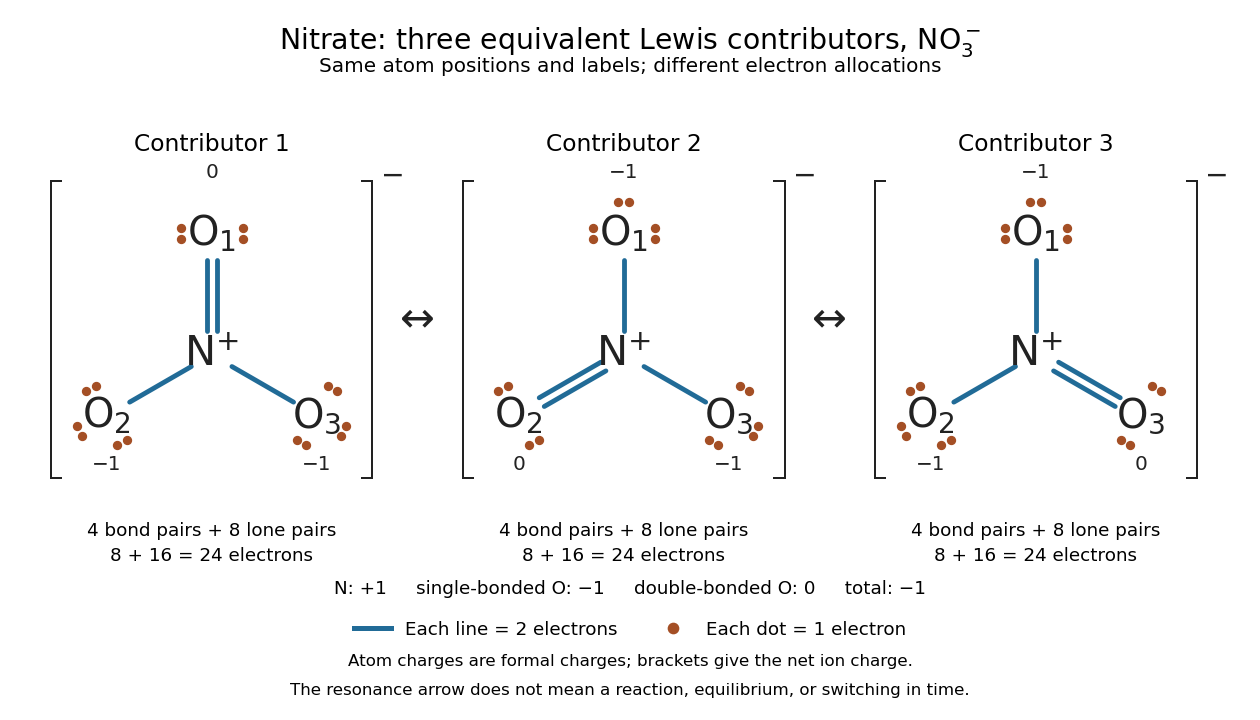

Why not draw three N=O connections to make the ion look symmetric? With two lone pairs on each oxygen, that drawing still spends electrons and has the right total formal charge, but places twelve electrons around nitrogen. It fails the octet rule for this second-period chemical element. Three equivalent resonance structures express the symmetry without that invalid local count.
The economy is beautiful: a handful of dots and lines rules out many impossible counts and identifies lone pairs that can participate in acid-base chemistry. A Lewis structure helps choose rational candidate connections before a detailed quantum chemistry calculation. It does not specify a complete many-electron wavefunction, actual electron density, or molecular geometry. Adding the separate VSEPR model, described in Chemistry 2e, 7.6: molecular structure and polarity, predicts bent water and pyramidal ammonia; the layouts above alone make no such prediction. Molecular orbital theory supplies a richer electronic description, including the unpaired electrons of molecular oxygen that an all-paired Lewis structure misses.
In 1916 Gilbert N. Lewis proposed two-dot notation for the shared electrons of a chemical bond and fuller dot formulae for the outer electrons, explicitly relating a shared pair to the existing single-line notation. See Lewis, The Atom and the Molecule, JACS 38, 762–785 (1916), pp. 777–778, available as an [ref]
https://zenodo.org/records/1429068. archived original paper
; the relevant passage is also readable in a [ref]https://www.chemteam.info/Chem-History/Lewis-1916/Lewis-1916.html. transcription of the original paper
. The modern quantum mechanics interpretation should be distinguished from his historical model of fixed outer electrons. Limiting reagent 2026-10-13
The limiting reagent is the reactant exhausted first if the specified chemical reaction proceeds to the maximum conversion permitted by stoichiometry. With initial values of amount of substance for the reactants and positive consumption coefficients , the largest forward extent of reaction is . Compare these quotients, rather than simply choosing the smallest initial quantity. This calculation predicts the maximum reaction product quantity and identifies which reactant supply must increase to raise it. See Flowers et al., Chemistry 2e, limiting reactants.
Start with of molecular hydrogen, of molecular oxygen and no water. Assume complete conversion by the displayed chemical reaction, without competing reactions. ThenThe molecular oxygen is the limiting reagent. Consumption of of molecular hydrogen and of molecular oxygen forms of water, leaving of molecular hydrogen. Initially there are molecular hydrogen molecules and molecular oxygen molecules; after conversion there are water molecules and leftover molecular hydrogen molecules.
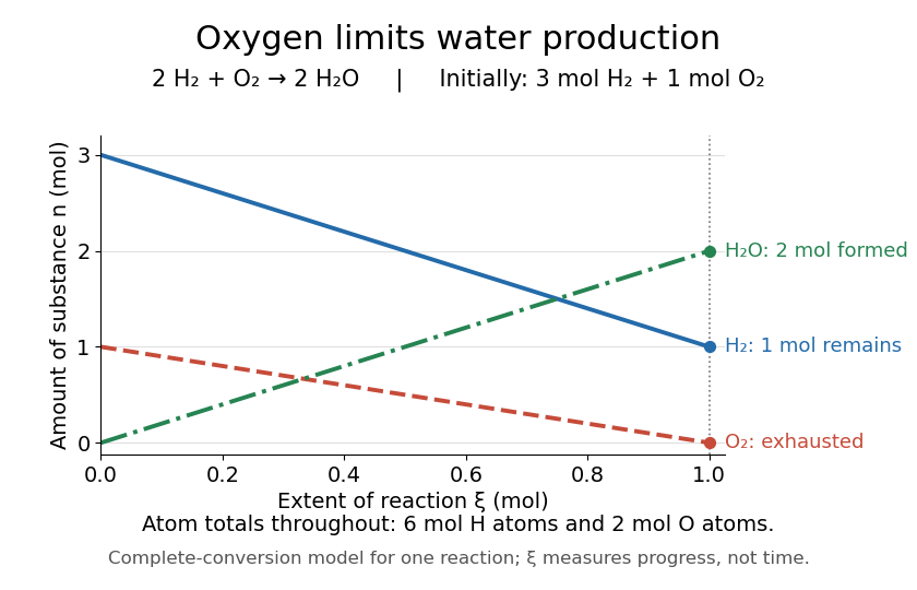
Oxygen runs out while one mole of molecular hydrogen remains
. The horizontal axis is extent of reaction, rather than time. The endpoint assumes complete conversion by one chemical reaction; the curves express stoichiometry. Details.
As a check, the initial inventory has of hydrogen chemical atoms and of oxygen chemical atoms. The final water contains of hydrogen chemical atoms, and leftover molecular hydrogen supplies the remaining . The atom totals balance even though the total amount of substance of molecules falls from four to three moles.
Lone pair Created 2026-10-14 Updated 2026-10-20
A lone pair is an electron pair assigned to one chemical atom without a connecting line to another chemical atom in a Lewis structure. It is usually shown as two dots. Water has two lone pairs on oxygen, accounting for four valence electrons; ammonia has one lone pair on nitrogen, accounting for two. In , each oxygen has two lone pairs, and carbon has none.
A lone pair can be reactive. In protonation of ammonia, the nitrogen lone pair becomes a new bond pair to a proton:The proton supplies no electron, so the eight valence electrons are retained: three bond pairs plus one lone pair become four bond pairs. The resulting nitrogen has formal charge ; each hydrogen has formal charge zero. This is why identifying an available lone pair helps explain the chemical base behaviour of ammonia, rather than merely decorating its formula.
Lewis already used the ammonia to example in The Atom and the Molecule (1916), pp. 777–778. In modern terms the dots identify an allocation of electrons, not two stationary particles or an isolated patch unaffected by the rest of the molecule. A molecular orbital description can spread electronic amplitude over several chemical atoms while a localized Lewis structure remains useful for acid-base chemistry.