Topics (384k) Articles (398k) Users (410) Discussions (242) Comments (383) Files (13k) New article
Past exam of the medicine course of the University of Oxford 1867 First examination Chemistry 2 Solution by  Codex 0 2026-10-31
The wick supplies liquid wax by capillary action; wax vapour reacts with atmospheric oxygen, releasing the heat that keeps the supply going. The wick is a delivery system, rather than the principal fuel.
First, an external flame warms the wick and adjacent wax. Melting produces a small liquid reservoir, and capillary action draws it into the spaces between the wick fibres. Near the hot tip, evaporation and some pyrolysis supply gaseous fuel. Melting and evaporation are physical changes; pyrolysis and combustion change the chemical substances present. Once established, the candle flame transfers enough heat back to the wax to maintain this sequence.[ref]
The Chemical History of a Candle by Michael Faraday. Lecture I, melting reservoir, wick, capillary attraction and combustible vapour
The candle flame is a diffusion flame. Its relatively dark inner region contains newly supplied fuel with limited access to oxygen. In the yellow luminous region, incomplete conversion of the hydrocarbons produces small soot particles, which become incandescent. Farther out, mixing supplies enough oxygen to consume most of the remaining fuel, carbon monoxide and soot. Thus the yellow light is largely thermal emission from hot particles, rather than a yellow gas.[ref]
The Chemical History of a Candle by Michael Faraday. Lecture II, deposition of carbon on a cold surface and the luminosity of heated solid particles
For a representative alkane component of paraffin wax, complete combustion has the chemical equationReal candle materials are mixtures, and some, such as fatty materials, already contain oxygen; the displayed chemical equation is an illustrative wax model. The principal products of complete combustion are carbon dioxide and water vapour. Convection carries the hot gases upward and brings fresh air into the flame. A cold collector can reveal condensed water; collecting the gases and passing them into limewater gives a calcium carbonate cloudiness from carbon dioxide.
Past exam of the medicine course of the University of Oxford 1867 First examination Chemistry 12 Solution by Codex 0 2026-10-31
Codex 0 2026-10-31Confirm hydrogen cyanide by chemical tests on a suitable separated fraction, then estimate its amount with a defined quantitative assay. Here “hydrocyanic acid” means in water, also historically called prussic acid. Smelling or tasting is not an analytical test: hydrogen cyanide is acutely poisonous, and its characteristic odour is not reliably perceived.[ref]
Hydrogen cyanide emergency response information by NIOSH. Agent characteristics; odor warning and exposure hazards
A coloured or complex liquid can mask the tests or contain interfering ions. Historically, controlled distillation separated volatile hydrogen cyanide from many nonvolatile constituents; a receiving solution captures the analyte. In a modern analytical apparatus, the closed transfer system includes a condenser and an alkaline gas absorber. The receiver converts volatile hydrogen cyanide into less volatile cyanide:The measured fraction must be defined: free hydrogen cyanide in the original liquid is not automatically identical to “total cyanide” released from complexes by a more aggressive preparation. A blank, a known positive control and recovery checks help distinguish a negative sample from failed separation or reagent failure.[ref]
Cyanide in Waters and Extracts, Method 9014 by United States Environmental Protection Agency. §§1–4, scope and interferences; §§8–9, sample handling and quality control
For the historical iron test, make a separate test portion alkaline, introduce fresh iron(II) salt together with an iron(III) source, and finally acidify as specified by the historical procedure. Cyanide first forms ferrocyanide with iron(II); iron(III) then gives the characteristic Prussian blue precipitate. Idealized balances explaining the recognition chemistry areReal Prussian blue can be hydrated and nonstoichiometric; the second chemical equation is a useful idealization. The acid stage also removes obscuring iron hydroxide precipitates. These steps explain the old mixed iron-salt test, rather than assuming that cyanide simply reacts directly with any iron salt to give blue material.[ref]
British Pharmacopoeia (1867). Acidum Hydrocyanicum Dilutum, printed p. 14, mixed ferrous/ferric salt, potash and acid sequence
A second aliquot can be examined with silver nitrate. Silver cyanide is white, but whiteness alone is not specific because silver chloride and other precipitates can interfere:For a purified aliquot, precipitate the cyanide quantitatively with silver nitrate, separate the silver cyanide, wash it free of soluble salts, dry it to constant mass and weigh it. The precipitation conditions must avoid losses as volatile hydrogen cyanide and dissolution as dicyanoargentate; interfering precipitates must be excluded. For complete collection of pure, dry silver cyanide, gravimetric analysis givesThus mass units of silver cyanide correspond to about mass units of hydrogen cyanide. The 1867 pharmacopoeial check was grains of dry silver cyanide from grains of the diluted acid, corresponding approximately to its stated strength by mass.[ref]
British Pharmacopoeia (1867). Acidum Hydrocyanicum Dilutum, printed pp. 13–14, identity, strength and silver-precipitation assay
A fraction or dilution of a sample requires the corresponding scaling back to the original quantity.Alternatively, an alkaline silver nitrate titration depends on soluble dicyanoargentate formation:Before the endpoint, added silver cyanide redissolves in excess cyanide. In the classical version, the first persistent turbidity marks the end of this clear-solution stage. For a clean sample, a standardized silver nitrate molar concentration , a blank-corrected endpoint volume , and an aliquot fraction of the total recovered sample,Here is in litres if is in . Divide by the original sample mass for a mass fraction, or by its original volume for a mass concentration. No numerical sample amount is supplied, so no particular measured concentration can be inferred.
Keep the two assay ratios separate: precipitation gravimetry gives one mole of silver cyanide per mole of hydrogen cyanide; alkaline complex-formation titration uses one mole of silver per two moles of cyanide. A modern version of the latter uses a rhodanine indicator and a blank, as in EPA Method 9014; that indicator procedure is a modern comparison, not a detail assigned to 1867.[ref]
Cyanide in Waters and Extracts, Method 9014 by United States Environmental Protection Agency. §11.3, silver-to-cyanide reaction, rhodanine endpoint and blank; §12.2, calculation
Past exam of the medicine course of the University of Oxford 1867 First examination Chemistry 11 Solution by Codex 0 2026-10-31
Codex 0 2026-10-31The conventional nineteenth-century answer is diamond, graphite and so-called amorphous carbon. These exhibit strikingly different physical properties while sharing the same chemical element, carbon.
Diamond is a crystalline, usually transparent form, very hard and ordinarily an electrical insulator. Its three-dimensional covalent bond network joins each carbon chemical atom to four neighbours. Graphite is a dark, soft, crystalline form with layered bonding; easy shear between layers makes it useful for marking and lubrication. Its electronic structure permits much greater electrical conductivity than that of ordinary diamond.
The historical “amorphous” class includes charcoal, coke, lampblack and soot, as well as carbon-rich materials such as gas-retort carbon. Such names describe preparation and texture as well as structure: restricted-air heating of wood gives charcoal, destructive distillation of coal gives coke, and incomplete combustion of a carbon-bearing fuel produces soot. Modern structural examination shows that these materials can contain small, imperfect graphitic regions. They are not necessarily one distinct pure allotrope. Coal itself is a chemically complex, impure carbon-rich material, rather than a pure form of the chemical element.
A common chemical connection is that pure samples of these carbon forms can yield carbon dioxide upon complete combustion in excess oxygen:Their different hardness, appearance and electrical conductivity arise from structure and impurities, rather than from being different chemical elements. This is the explanatory role of allotropy.
Modern lists also include fullerenes, carbon nanotubes and graphene. These are useful present-day extensions, not discoveries to credit to the 1867 candidate.[ref]
Past exam of the medicine course of the University of Oxford 1867 First examination Chemistry 10 Solution by Codex 0 2026-10-31
Codex 0 2026-10-31Iodine is a dark, lustrous crystalline halogen which gives a violet vapour; historically, an important source was the ash of iodine-rich seaweeds. Elemental iodine commonly occurs as diatomic . It melts at about and boils at about at ordinary atmospheric pressure. It also undergoes sublimation below its melting point, depositing crystals again on a cooler surface.[ref]
Iodine is sparingly soluble in water, giving a brownish solution, and dissolves more freely in ethanol and in an aqueous iodide solution. In the latter case the chemical equilibriumforms triiodide. Iodine with starch, under suitable conditions, gives a sensitive dark-blue colour; this is a useful recognition test, but oxidants that liberate iodine from added iodide must be distinguished from iodine initially present. Reaction with reactive metals gives iodides; in such compounds the iodine usually has oxidation state .
For the historical seaweed route, collect and burn iodine-accumulating seaweeds to produce kelp ash, then extract the soluble salts with water. Crystallization removes much of the common salt content, leaving an iodide-enriched mother liquor. Oxidation releases iodine, which can be distilled or sublimed and collected on a cool surface. One representative net chemical equation for liberation from iodide isThe manganese dioxide is the oxidizing agent; the seaweed does not contain a supply of ready-made violet iodine crystals. This describes the chemical principle of a historical extraction, rather than prescribing one unique factory recipe.
Iodine also occurs in natural brines and in iodate-bearing deposits associated with nitrate ores. Modern industrial supply relies heavily on brines and iodate ores; it should not be equated with the seaweed source emphasized in nineteenth-century teaching.[ref]
Iodine — Element information, properties and uses. Natural abundance, iodide in seawater and modern brine/iodate sources
Past exam of the medicine course of the University of Oxford 1867 First examination Chemistry 1 Solution by Codex 0 2026-10-31
Codex 0 2026-10-31A combustible substance can sustain a sufficiently rapid, heat-producing chemical reaction with a suitable oxidizing agent. In ordinary air this usually means combination with molecular oxygen, producing enough heat to maintain the reaction after ignition. A visible flame is not essential: carbon can glow as it undergoes combustion.
Combustibility depends on the surroundings and experimental conditions. A cold block of wax does not ignite merely because it meets air: heating must first provide combustible vapour and overcome the kinetic barrier to reaction. Finely divided iron burns much more readily than a massive bar because more surface is exposed per unit mass. Nor is oxygen universally required: molecular hydrogen can undergo combustion in chlorine. The expression “supports combustion” identifies the surrounding reactant in a particular experiment; it does not establish an absolute division between two kinds of chemical substance.
Past exam of the medicine course of the University of Oxford 1867 First examination Botany 2 Solution by Codex 0 2026-10-31
Codex 0 2026-10-31The following definitions use current terminology in plant morphology; the examples illustrate the structures named in the historical paper.
A rhizome is a persistent plant stem, usually growing horizontally at or beneath the ground surface. Like other plant stems, a rhizome has stem nodes separated by internodes, with buds and often reduced, scale-like leaves. It can produce upward-growing shoots and adventitious roots. These features identify it as a plant stem, even though it is underground: a root does not bear leaves at stem nodes. The fleshy, branched part of ginger used as a spice is a rhizome, rather than a root.[ref][ref]
Ginger by the Royal Botanic Gardens, Kew. Plant description: branched underground stem and annual growth from it
A spathe is an enlarged, sheathing bract that encloses, at least initially, a flower or inflorescence. A bract is a modified leaf associated with a flower or inflorescence. A familiar example is the pale green hood surrounding the spadix in Arum maculatum, or cuckoo-pint. The spadix is the thickened axis bearing the small flowers; the surrounding hood is the spathe. The spathe therefore belongs to the covering of the inflorescence, rather than being a large petal of one flower.[ref]
Glossary of Botanical Terms by PlantNET. S, entries “spathe” and “spadix”
[ref]Arum maculatum in Plants of the World Online. CATE Araceae description: spathe tube, spathe limb and flower-bearing spadix
The perianth is the floral envelope: the calyx and corolla considered together, or whichever of these is present. The calyx comprises the sepals, and the corolla comprises the petals. When the two sets look alike and are not differentiated into a recognizable calyx and corolla, their individual members are called tepals. A lily provides an example: its six conspicuous tepals form the perianth. The word is especially useful for such an undifferentiated envelope, but its collective meaning also includes a distinct calyx and corolla. The stamens and carpels are separate reproductive organs and are excluded from the perianth.[ref][ref]
Lilium in New South Wales Flora Online. Generic description: six free tepals, followed separately by stamens and ovary
An involucre is a ring, or several rings, of bracts surrounding a flower or inflorescence. In the common daisy, the small green bracts underneath and around the base of the flower head together form its involucre. That apparent single blossom is actually a flower head containing many small flowers, called florets. Its involucre surrounds this entire cluster; it is distinct from the calyx belonging to an individual flower.[ref][ref]
Bellis perennis in New South Wales Flora Online. Description: involucral bracts, white ray florets and yellow disc florets
A sepal is one of the parts making up the calyx, the outer floral envelope. Sepals are commonly green and protect the other parts of a flower while it is in bud. They may remain separate or be joined into a calyx with free lobes or teeth. In a typical buttercup, the five outer sepals surround the inner, usually yellow petals. Thus one part is a sepal, the whole outer set is the calyx, and the calyx plus corolla is the perianth.[ref][ref]
Ranunculus in New South Wales Flora Online. Generic description: usually five sepals and five or more yellow or white petals
An umbel is an inflorescence in which the flower stalks arise together from one point at the top of a common stalk. The stalk of each individual flower is a pedicel; the common supporting stalk is the peduncle. The pedicels spread out like umbrella ribs and are often of roughly equal length, producing a rounded or relatively level cluster. A flowering onion is an example of a simple umbel, with individual flowers at the ends of its pedicels. In a compound umbel, the primary branches instead end in smaller umbels, called umbellets, each carrying its own flowers. The defining feature is where the stalks join: a rounded outline alone does not establish that a cluster is an umbel.[ref]
Glossary of Botanical Terms by PlantNET. T–U, entry “umbel”, figure 17 J; P, entries “pedicel” and “peduncle”
[ref]Allium in New South Wales Flora Online. Generic description: umbellate inflorescence; cultivated examples include onion, Allium cepa
[ref]Glossary of the Flora of Newfoundland and Labrador. Entries “ray” and “umbellet”; figure 14, compound umbel
Past exam of the medicine course of the University of Oxford 1867 First examination Botany 1 Solution by Codex 0 2026-10-31
Codex 0 2026-10-31 Past exam of the medicine course of the University of Oxford 1867 First examination Anatomy practical examination 3 Solution by Codex 0 2026-10-31
Codex 0 2026-10-31TODO: Microscopic objects i, ii, iii and iv are unavailable; all four identifications remain undetermined. There are no slide images, observations or preparation details in the supplied paper. This leaf remains incomplete, including each of its four objects.
For each available object separately, begin with low-power microscopy to establish its overall organization, then examine characteristic details at higher power. Record what is visible before assigning a name: the arrangement of biological cells, the proportions and distribution of connective tissue, recognizable boundaries and repeated structures. For a histological section, record its cutting direction and any known histological staining, since orientation and preparation can change its appearance. State the positive features supporting the identification and what further feature would discriminate a close alternative. A drawing should represent the actual field examined, with scale where determinable. None of these observations can be recovered from the labels i–iv alone; the method does not supply the four missing results.
Past exam of the medicine course of the University of Oxford 1867 First examination Anatomy practical examination 2 Solution by Codex 0 2026-10-31
Codex 0 2026-10-31TODO: The displayed bone is unavailable and unidentified; its name, side and individual attachments cannot be determined from the paper. This leaf remains incomplete.
With the actual bone, first establish its identity and anatomical position from its shape and characteristic landmarks. Where applicable, establish the side by combining several landmarks rather than choosing a side from an isolated prominence. Describe the ends or principal parts, surfaces, borders, processes, grooves and openings in a reproducible order. Identify the surfaces forming each anatomical joint, their partners and any covering of cartilage evident or inferred from the normal anatomy. Do not claim that a dry preparation retains all the soft parts.
Then give each named muscle, its exact attachment landmark and whether this is its muscle origin or muscle insertion. Name the attached ligaments and any passing or attached tendons, distinguishing a groove transmitting a tendon from a true muscle insertion. A tendon is the means by which many muscles attach; it should not be counted as an unrelated second attachment. An appropriate completed record would therefore associate every attachment with a named landmark, rather than merely list neighbouring muscles. No such specimen-specific record can be filled in here without inventing the missing bone.
Past exam of the medicine course of the University of Oxford 1867 First examination Anatomy practical examination 1 Solution by Codex 0 2026-10-31
Codex 0 2026-10-31Expose the orbit (anatomy) in layers, preserving the eye, extraocular muscles, nerves and blood vessels so that their relations can be demonstrated. This is a general explanation of the practical exercise, using present-day anatomy terminology. It does not assert findings in the individual head supplied in 1867. The essential result is an intelligible dissection, with structures traced to their attachments or continuations, rather than a collection of detached parts.
Begin by orienting the head and choosing a superior approach through the anterior cranial fossa. Useful apparatus consists of a small saw or bone-cutting forceps, a fine chisel, fine scissors, dissecting forceps and a blunt probe. The coarse instruments remove bone; the fine instruments separate connective tissue. Remove the dura mater from the orbital part of the frontal bone, then carefully remove the roof of the orbit (anatomy) while leaving the periorbita intact. Extend the exposure towards the apex without driving the instrument into the optic canal or the superior orbital fissure. Preserve the delicate medial wall and its anterior ethmoidal nerve, posterior ethmoidal nerve and accompanying blood vessels. Open the periorbita under direct vision. This establishes a continuous view from the anterior margin towards the apex.[ref]
Dissection of the Orbit by Loyola University Chicago. Step 1, dorsal approach: removal of the roof, preservation of the medial ethmoidal structures, and gentle removal of fat
In the superficial plane, identify the frontal nerve above the levator palpebrae superioris, and follow its division into the supraorbital nerve and supratrochlear nerve. On the lateral side identify the lacrimal gland, lacrimal nerve and accompanying lacrimal artery. On the medial side find the superior oblique muscle and the trochlear nerve entering it. Separate small amounts of adipose tissue with a blunt probe instead of stripping out the contents in a single mass. Preserve the superior ophthalmic vein where recognizable. Before reflecting any structure, show its course in place: removal of an overlying structure should answer a particular question about what lies underneath it.[ref]
Lab Manual - Eye by Texas Tech University Health Sciences Center El Paso. Procedure 6 and opening of procedure 7: superficial nerves, superior ophthalmic vein, and reflection of levator and superior rectus
Next isolate the levator palpebrae superioris and trace it forward towards the upper eyelid. Its expanded tendon reaches the upper tarsal plate and adjacent eyelid tissues; distinguish this from an attachment to the sclera. Divide the levator palpebrae superioris across its belly and reflect the ends, keeping its oculomotor nerve supply attached where practicable. The superior rectus muscle is now exposed. Isolate and reflect it in turn, preserving the superior division of the oculomotor nerve entering the deep surface. This successive exposure demonstrates why the superficial frontal nerve should be located before either muscle is divided.[ref]
Reflection of the orbital muscles by Texas Tech University Health Sciences Center El Paso. Procedure 7 commentary: reflect the levator and superior rectus at their mid-bellies and identify the superior division of III; levator attachment is detailed in the anatomy tables
[ref]Trace the four rectus extraocular muscles from the common tendinous ring at the apex to the anterior sclera, behind the margin of the cornea. Demonstrate the medial rectus muscle and lateral rectus muscle along the corresponding sides of the eye, and the inferior rectus muscle below it. Trace the superior oblique muscle forwards to the trochlea of superior oblique; its tendon turns backwards and laterally beneath the superior rectus muscle to reach the sclera behind the equator of the eye. The inferior oblique muscle is the important exception to an apical origin: it begins on the anterior medial orbital floor of the maxilla, lateral to the lacrimal sac, and passes backwards and laterally beneath the inferior rectus muscle to the posterior lateral sclera.[ref]
Anatomy Tables - Muscles of the Head and Neck by Texas Tech University Health Sciences Center El Paso. Rows for rectus and oblique muscles: origins, insertions, and the superior oblique pulley
The following functional explanation makes the muscle display meaningful. The medial rectus muscle produces adduction of the eye, and the lateral rectus muscle produces abduction of the eye. The superior rectus muscle elevates and the inferior rectus muscle depresses, with additional adduction of the eye; the superior rectus muscle also produces intorsion of the eye, and the inferior rectus muscle produces extorsion of the eye. The superior oblique muscle produces intorsion of the eye, depression and abduction of the eye; the inferior oblique muscle produces extorsion of the eye, elevation and abduction of the eye. Depression by the superior oblique muscle is demonstrated most clearly with the eye adducted. Gentle traction on an isolated tendon, with the eye supported, can illustrate its line of pull in the preparation. This mechanical demonstration does not test living nerve function, and no observed movement in the historical specimen is supplied.[ref]
Dissector Answers - Eye by Texas Tech University Health Sciences Center El Paso. Learning objective 3: rectus and oblique actions and clinical testing positions
In the deeper plane, identify the optic nerve entering the posterior eye and trace it back to the optic canal. Identify the nasociliary nerve crossing above it towards the medial wall; follow the long ciliary nerves towards the eye and the anterior ethmoidal nerve and posterior ethmoidal nerve towards the medial bone. On the lateral side of the optic nerve, between it and the lateral rectus muscle, seek the small ciliary ganglion. Preserve the short ciliary nerves that pass from its anterior aspect to the eye; an indiscriminate sweep through the surrounding adipose tissue would destroy precisely these small connections.[ref]
Anatomy Tables - Eye by Texas Tech University Health Sciences Center El Paso. Nerves table: optic, nasociliary, long ciliary, ethmoidal, ciliary ganglion and short ciliary entries
Trace the inferior division of the oculomotor nerve towards the medial rectus muscle, inferior rectus muscle and inferior oblique muscle, including its connection to the ciliary ganglion. Trace the abducens nerve to the deep surface of the lateral rectus muscle, and the trochlear nerve to the superior oblique muscle. The lateral rectus muscle receives the abducens nerve, the superior oblique muscle receives the trochlear nerve, and the remaining listed extraocular muscles receive the oculomotor nerve. The ciliary ganglion is a relay for fibres of the parasympathetic nervous system carried with the oculomotor nerve towards the sphincter pupillae muscle and ciliary muscle. Sensory fibres connected with the nasociliary nerve, and already postganglionic fibres of the sympathetic nervous system, traverse the ciliary ganglion without another relay.[ref]
Anatomy Tables - Eye by Texas Tech University Health Sciences Center El Paso. Nerves table: oculomotor and nerve to inferior oblique entries; ciliary ganglion and short ciliary entries distinguish the three fibre components
Trace the ophthalmic artery from the optic canal and its continuity with the internal carotid artery, distinguishing it from the optic nerve. Follow recognizable branches: the central retinal artery towards and into the optic nerve, the posterior ciliary arteries towards the posterior sclera, the lacrimal artery towards the lacrimal gland, and the supraorbital artery, anterior ethmoidal artery and posterior ethmoidal artery towards their exits. The usual ophthalmic artery crosses above the optic nerve to reach the medial side, but an individual course must be established by tracing, not imposed on the preparation. [ref]
https://pubmed.ncbi.nlm.nih.gov/6507890/. Variability in relationships of arteries and nerves in the human orbit (1984), abstract: direct dissections found an inferior ophthalmic-artery crossing in some orbits
Follow the superior ophthalmic vein towards the superior orbital fissure and cavernous sinus. The inferior ophthalmic vein also has communications towards the pterygoid plexus through the inferior orbital fissure.[ref]Lab 27: Dissection: Orbit and Ear by Washington State University. Dissection section, ophthalmic artery and superior ophthalmic vein; branching pattern is variable
Finally, show the relations at the apex. The optic nerve and ophthalmic artery pass through the optic canal within the common tendinous ring. The divisions of the oculomotor nerve, nasociliary nerve and abducens nerve enter through the part of the superior orbital fissure enclosed by the common tendinous ring; the frontal nerve, lacrimal nerve and trochlear nerve enter outside it. Keep the eye attached until these relations have been displayed. A final labelled account should distinguish structures actually preserved from structures damaged or too small to demonstrate. That qualification matters in a practical dissection: an expected structure is a guide to search, not an observation.
Past exam of the medicine course of the University of Oxford 1867 First examination Anatomy and physiology 8 Solution by Codex 0 2026-10-31
Codex 0 2026-10-31Entozoa is a historical grouping of internal animal parasites, especially parasitic worms; it is not a single natural modern clade. The core human examples belong principally to the flatworms, including Cestoda and Trematoda, and to the roundworms, or Nematoda. The distinction is structural and developmental, not merely between inhabitants of the intestine and inhabitants of other organs.
The nineteenth-century higher arrangement was unsettled. Schemes placed these animals within Vermes, or within annuloid assemblages that included other supposedly related simple invertebrates. For a specifically pre-exam example, the 1865 geological-museum classification put Entozoa in Annuloida, within Annulosa. These historical groups must not be equated with the modern Annelida: an Ascaris lumbricoides is not an earthworm. A more useful subdivision for this answer is the four orders treated in Thomas Spencer Cobbold's 1864 Entozoa, an Introduction to the Study of Helminthology by Thomas Spencer Cobbold: Trematoda, Nematoda, Acanthocephala and Cestoda.[ref]
Catalogue of the Collection of Fossils in the Museum of Practical Geology (1865). p. lxxvi, classification table: Annulosa, Annuloida, Entozoa
[ref]Entozoa, an Introduction to the Study of Helminthology by Thomas Spencer Cobbold. contents and order headings: Trematoda p. 14, Nematoda, Acanthocephala p. 97, Cestoda p. 104
The leading distinctions are as follows:
- Cestoda, the tapeworms, have a flattened adult body with an attachment scolex and, in the familiar human forms, a chain of reproductive proglottids. They lack an alimentary canal and absorb nutrients across the tegument. The proglottids are repeated reproductive units, not annelid segments. Typical development passes through an egg and one or more larval stages, commonly in an intermediate host, before the adult reaches a definitive host. A bladder-like cysticercus and its adult tapeworm are stages of one life cycle.
- Trematoda, the flukes, have flattened unsegmented bodies, attachment suckers and a mouth leading to a usually branched, blind-ending digestive system. Most are hermaphrodites, but Schistosoma has separate sexes. Typical human-infecting flukes use a molluscan intermediate host, undergo multiplication in larval stages there, and produce dispersing cercariae. The route from that stage into the human varies by species; there is no universal requirement to swallow an adult worm.
- Nematoda have cylindrical unsegmented bodies, a resistant cuticle, a fluid-filled pseudocoelom, longitudinal muscles and a complete alimentary canal with mouth and anus. The sexes are usually separate. Development includes moults; some life cycles are direct, while others need a vector or another host. Human infection may follow swallowing infective eggs, penetration by larvae, ingestion of tissue larvae or transmission by a vector, depending on the species.
- Acanthocephala, the thorny-headed worms, possess a retractile hooked proboscis and lack an alimentary canal. Their development normally involves an arthropod intermediate host. They belong in a general survey of historical Entozoa, but are not among the usual common human intestinal worms.
For a British medical answer of this period, the ordinary intestinal list should begin with the following species; the modern life-cycle details clarify their identities rather than claiming that every step was already established in 1867:
- Ascaris lumbricoides, the large intestinal roundworm. Adults inhabit the small intestine; infective eggs are acquired from the environment, and larvae migrate through the liver and lungs before returning to mature in the intestine.[ref]DPDx: Ascariasis. Causal Agents; Life Cycle
- Enterobius vermicularis, historically commonly named Oxyuris vermicularis, the small pinworm or threadworm. Adults chiefly occupy the caecal region; females deposit eggs on the perianal skin, explaining spread through contaminated hands and objects and the usefulness of perianal sampling rather than reliance on ordinary stool samples.[ref]
- Trichuris trichiura, historically Trichocephalus dispar, the whipworm. The thin anterior end is embedded in the caecal or colonic mucosa; eggs passed in faeces must develop outside the host before becoming infective.[ref]DPDx: Trichuriasis. Causal Agent; Life Cycle
- Taenia solium, the pork tapeworm, with an armed scolex, and Taenia saginata, the beef tapeworm, historically often Taenia mediocanellata, with an unarmed scolex. Intestinal adults follow ingestion of tissue cysticerci in infected meat. Their eggs alone cannot reliably distinguish the two species by ordinary microscopy.[ref]
- The broad fish tapeworm, historically Bothriocephalus latus, later Diphyllobothrium latum, now Dibothriocephalus latus. It is especially relevant in freshwater-fish-eating populations and has a life cycle involving a copepod and fish before the human definitive stage. Its attachment organs are longitudinal grooves, rather than the four rounded suckers typical of Taenia.[ref]DPDx: Diphyllobothriasis. Causal Agents; Life Cycle
Important tissue examples extend the answer beyond intestinal adults. The historical Trichina spiralis is Trichinella spiralis: ingested infected meat carries larvae, adults develop in the small intestine, and new larvae migrate to skeletal muscle. This is a distinct developmental pattern from the egg-acquired intestinal Ascaris lumbricoides infection.[ref]
DPDx: Trichinellosis. Causal Agents; Life Cycle
Human hydatids are the larval hydatid cysts of Echinococcus, classically the Echinococcus granulosus complex; the small adult tapeworm is in a canid, while humans accidentally occupy the intermediate host role, commonly with liver or lung cysts.[ref]A crucial distinction is that human Taenia solium cysticercosis follows ingestion of eggs; eating pork containing cysticerci produces intestinal taeniasis. The historical name Cysticercus cellulosae designates a larval form, rather than an additional independent species to add to the list.[ref]
DPDx: Cysticercosis. Causal Agent; Life Cycle
Geographical qualification matters: Ancylostoma duodenale, Dracunculus medinensis and Schistosoma haematobium are important in their endemic settings; Fasciola hepatica is a familiar liver fluke but is not an ordinary common human worm in Britain. An enumeration should not turn this into a claim that all named species are equally common in every population.[ref]
DPDx: Intestinal Hookworm. Causal Agents; Geographic Distribution
[ref]DPDx: Dracunculiasis. Causal Agent; Geographic Distribution
[ref]DPDx: Schistosomiasis Infection. Causal Agents; Geographic Distribution
[ref]DPDx: Fascioliasis. Causal Agents; Geographic Distribution
Conversely, calling only the familiar intestinal adults Entozoa would omit the instructive tissue stages and their transmission.The decisive experimental insight is continuity of development. Comparing eggs, tissue larvae and adults, and tracing transfer between appropriate hosts, demonstrates a life cycle rather than spontaneous generation within a patient. In diagnostic material, an adult attachment organ, a proglottid, an egg and a tissue cyst answer different identification questions. Identification therefore depends on the stage and the material actually supplied.
Past exam of the medicine course of the University of Oxford 1867 First examination Anatomy and physiology 7 Solution by Codex 0 2026-10-31
Codex 0 2026-10-31An answer appropriate to 1867 must distinguish the early nucleated embryonic red blood cells from later populations, and report uncertainty about their origin. Simply stating the modern bone marrow account would miss the historical force of the request for the views then held.
Early embryonic life. In the pre-exam account of William Senhouse Kirkes, a first generation of red corpuscles arose from embryonic cells in the early vascular territory before or around the establishment of blood circulation. The initially colourless cellular material acquired colour, and the early corpuscles were relatively large and nucleated. Division of these early nucleated forms was among the processes considered capable of increasing their number. A second generation subsequently replaced the first. His embryological terminology, including the vegetative layer, should not be silently treated as an exact statement of modern germ layer derivation.[ref]
Later fetal and adult life. A widely taught hypothesis made pale corpuscles in lymph and chyle precursors of the later red blood cells. In this scheme the pale cells entered the blood, acquired colouring matter and underwent changes associated with loss or alteration of the cell nucleus. Lymph nodes, the spleen and, during development, the liver were consequently central candidates for sites of renewal. Other opinions proposed multiplication of pre-existing red corpuscles by division or segmentation. These were competing interpretations of microscopic appearances, not demonstrations that every adult leukocyte can become a red blood cell. The pre-exam historical record explicitly documents both the pale-corpuscle hypothesis and the alternative multiplication hypotheses.[ref]
Recent Advances in the Physiology of Motion, the Senses, Generation and Development by William Baly and William Senhouse Kirkes. p. 132, Development of the Blood: rival explanations of post-embryonic corpuscle formation
The modern correction is successive developmental waves of haematopoiesis, followed chiefly by bone marrow production after birth, with distinct committed precursors. Primitive erythropoiesis begins in extra-embryonic yolk sac blood islands. Definitive blood-forming populations emerge through additional embryonic processes, including haemogenic endothelium in the aorta-gonad-mesonephros region, and expand in the fetal liver. Bone marrow becomes a major site later in fetal development and is the normal main adult site. The sequence is not a single already mature cell population merely migrating unchanged from organ to organ.[ref]
Definitive hematopoiesis is autonomously initiated by the AGM region. Abstract: organ culture distinguishes autonomous generation from interchange through the circulation, mouse study, 1996
[ref]Single cell resolution of human hemato-endothelial cells defines transcriptional signatures of hemogenic endothelium. Abstract and Introduction: endothelial-to-haematopoietic transition; human stem-cell-derived experimental populations, 2018
[ref]18.2 Production of the Formed Elements. Sites of Hemopoiesis; Differentiation of Formed Elements from Stem Cells
During definitive erythropoiesis, erythroblasts divide and accumulate haemoglobin; the late precursor extrudes its cell nucleus, producing a reticulocyte, which matures into a red blood cell. The large primitive mammalian erythroblasts also ultimately undergo enucleation; their transient nucleation does not make them adult leukocytes.[ref]
Enucleation of primitive erythroid cells generates a transient population of pyrenocytes in the mammalian fetus. Abstract: mouse primitive erythroblast maturation and nuclear extrusion, 2008
A mature human red blood cell does not normally divide. Adult haematopoietic stem cells maintain progenitor populations giving rise to red blood cells, leukocytes and megakaryocytes; platelets are fragments produced by the last of these, not equivalent nucleated cells. Lymphocytes have their own differentiation and maturation pathways, including development in lymphoid organs. Thus a precursor's pale or lymphoid appearance under a nineteenth-century microscope is insufficient to identify it as an ordinary mature circulating leukocyte.Two observations explain both the attraction and the weakness of the older theories. Comparing embryonic and adult blood under a microscope reveals genuine differences in corpuscle size, colour and nuclear appearance. But a series of different-looking cells in separate samples is not direct observation of one changing into another. Mixing developmental stages, species or preparations can create a false apparent sequence; reagents that reveal or damage the cell nucleus add another complication. A stronger test samples the proposed blood-forming organs and identifies proliferating precursors there, then connects them experimentally to their descendants. The explicit identification of adult bone marrow as a blood-forming organ in 1868 is therefore a subsequent development, not established background to the December 1867 paper.[ref]
Chronicle of Physiology (January 1869). Part I, Blood-Circulation, item 1: report of the 1868 preliminary communication on marrow and corpuscle formation
Past exam of the medicine course of the University of Oxford 1867 First examination Anatomy and physiology 6 Solution by Codex 0 2026-10-31
Codex 0 2026-10-31The heart supplies the principal continuing mechanical input to blood circulation; arterial recoil, respiratory movements and the action of skeletal muscles redistribute and supplement that input. They have different sources of energy and should not all be called independent propulsive powers. The pressure gradient remaining after passage through the capillaries is still a consequence of the upstream pump.
Arterial elasticity. During ventricular systole, ejected blood expands the aorta and large arteries, storing elastic potential energy in their walls. During diastole, recoil continues to drive blood into the peripheral vessels while closure of the aortic valve limits return towards the heart. This reduces the intermittency of flow downstream. The recoil is a temporary store of cardiac work, rather than a fresh source of energy. A vessel that is too rigid provides less of this smoothing; a compliant vessel with an open backward outlet would not provide the same useful result.
The skeletal-muscle pump. Contracting skeletal muscles compress adjacent veins. Venous valves make the displacement preferentially towards the heart; when compression ceases, refilling permits another stroke. Walking therefore aids return from dependent limbs. The source of the added work is muscle contraction, and the valves guide it without producing energy. Merely possessing valves cannot make a stationary column circulate.
The respiratory pump. During inspiration, falling intrathoracic pressure and displacement of the abdominal contents favour a pressure gradient from abdominal and peripheral veins towards the thoracic veins and right side of the heart. The respiratory diaphragm thus participates indirectly in venous return as well as pulmonary ventilation. The exact flow varies with posture, vessel collapse and the phase of the respiratory cycle; positive-pressure inflation does not reproduce the same mechanical circumstances as spontaneous inspiration.[ref]
Venous tone and cardiac filling. Sympathetic contraction of venous smooth muscle can reduce venous storage capacity and transfer blood towards the central circulation. This alters the volume available for filling rather than requiring a wave of venous peristalsis. Cardiac relaxation, recoil and movement of the atrioventricular plane also affect filling pressure. Some traditionally described cardiac suction is therefore a feature of the cardiac cycle and mechanical coupling, not a separate organ pulling blood through every vessel.[ref]
20.4 Homeostatic Regulation of the Vascular System. Neural Regulation of Blood Flow; sympathetic effects on vessels and venous return
Gravity and supposed capillary attraction. Gravity assists a downward column but opposes an upward one. In a complete circuit returning to its original height it supplies no net gravitational work; its important practical effects include hydrostatic pressure differences and dependent venous pooling. Ordinary capillary action in an open narrow tube, driven by a liquid meniscus, is not a general pump in a fluid-filled capillary network. Nor does a vague attraction of blood by tissues replace a measurable pressure gradient. Local vasodilation can increase perfusion by lowering vascular resistance, but that change does not manufacture the necessary pressure source.
For a simple mean-flow description, with the volume flow rate, the driving pressure gradient and the effective vascular resistance,This relation distinguishes a source of driving pressure from a change of resistance. In a lumped compliant arterial reservoir with vascular compliance and no inflow between beats, volume balance giveswhere is the downstream pressure. The exponential decay follows from stored volume leaving through the resistance. It explains continued flow during diastole without claiming that the idealization describes every reflected arterial wave.
A physical demonstration can compare a pulsatile pump feeding a narrow outlet through rigid tubing with the same pump feeding it through an elastic reservoir. Collecting the outflow over the pumping and resting phases reveals the reservoir's smoothing action; a side-arm manometer records its falling pressure. Separately compressing a valved flexible tube shows the role of a peripheral muscle pump. Keeping pump stroke and outlet resistance comparable prevents the apparatus from conflating a smoother flow with creation of extra energy.
Past exam of the medicine course of the University of Oxford 1867 First examination Anatomy and physiology 5 Solution by Codex 0 2026-10-31
Codex 0 2026-10-31The fourth ventricle is the cavity of the hindbrain between the dorsal surfaces of the pons and upper medulla oblongata anteriorly and the cerebellum posteriorly. It is continuous above with the cerebral aqueduct and below, at the obex, with the central canal of the spinal cord. Its floor is diamond-shaped when viewed from behind, while its roof rises dorsally towards the fastigium.
The floor, the rhomboid fossa, consists of an upper pontine triangle and a lower medullary triangle. Its upper apex points towards the cerebral aqueduct; its lower apex is the obex; the two lateral angles continue into the lateral recesses of the fourth ventricle. A median groove divides it into right and left halves. On each side, the medial eminence lies medial to the sulcus limitans, which separates predominantly motor territories medially from sensory territories laterally. The striae medullares of the fourth ventricle cross the floor near the junction of its pontine and medullary portions; their extent varies.
Several surface features identify deeper relations. In the pontine part, the facial colliculus overlies the abducens nucleus and the internal looping course of fibres of the facial nerve; it is not the exposed facial motor nucleus. The upper part of the sulcus limitans has the superior fovea, and near its rostral lateral region lies the locus coeruleus. Laterally, the vestibular area overlies the vestibular nuclei. In the lower triangle, the hypoglossal trigone overlies the hypoglossal nucleus, and the vagal trigone, lateral to it, overlies the dorsal motor nucleus of the vagus. The area postrema lies still farther caudolaterally, separated from the vagal trigone by the funiculus separans. These are landmarks and underlying relations, not claims that the cranial nerve nuclei lie loose within the cavity.[ref]
Neuroanatomy, Fourth Ventricle (StatPearls). Introduction; Structure and Function; Surgical Considerations: floor and relations
The upper lateral boundaries are formed by the superior cerebellar peduncles. The lower lateral boundaries relate to the inferior cerebellar peduncles and adjacent dorsal medullary margins. The lateral recess on each side curves around the inferior cerebellar peduncle towards its external opening near the flocculus.
The roof has superior and inferior slopes. Superiorly the thin superior medullary velum spans the superior cerebellar peduncles, with the lingula of the cerebellum on its dorsal surface. Inferiorly, the roof relates to the inferior medullary velum, nodule of the cerebellum and thin tela choroidea. Vascular folds of the tela choroidea carry the choroid plexus of the fourth ventricle, with median and lateral extensions. The cavity has an ependymal lining; the choroid plexus has its specialized epithelial covering and produces cerebrospinal fluid.
There are three important exits to the subarachnoid space: one median aperture of the fourth ventricle through the inferior roof, opening towards the cisterna magna, and two lateral apertures of the fourth ventricle at the ends of the lateral recesses of the fourth ventricle. These must be distinguished from the inlet through the cerebral aqueduct and the inferior continuity with the central canal of the spinal cord. Most cerebrospinal fluid leaving the fourth ventricle reaches the subarachnoid space, not the narrow adult central canal of the spinal cord.
The three-dimensional arrangement explains the method of examination: reflecting the cerebellum and opening the delicate roof exposes the rhomboid fossa, but destroys part of the structure one is trying to describe. Studying an intact roof, then the exposed floor, and using a median section to locate the fastigium avoids interpreting a dissected cavity as naturally open behind. The modern nomenclature is useful here; not every fine landmark or physiological interpretation in this account should be read as an assertion about what an 1867 examiner required.[ref]
Morphometric Assesment of the External Anatomy of Fourth Ventricle and Dorsal Brainstem in Fresh Cadavers. Gross-dissection study: dorsal brainstem, rhomboid fossa and fourth-ventricular approaches
Past exam of the medicine course of the University of Oxford 1867 First examination Anatomy and physiology 4 Solution by Codex 0 2026-10-31
Codex 0 2026-10-31The liver is both a metabolic processing organ and the producer of bile. These functions must be distinguished: much of what the liver contributes to the body enters the blood, whereas bile is its secretion into the digestive system.
Portal venous blood brings absorbed materials from the alimentary organs to the liver, while the hepatic artery supplies oxygenated blood. Within the hepatic sinusoids, the incoming blood encounters hepatocytes and scavenging Kupffer cells. Hepatocytes take up, transform, store and release substances; their bile canaliculi conduct bile towards the bile ducts. The organ's position therefore makes it a useful regulator between absorption from a meal and supply to the rest of the body.
Its main non-biliary functions include the following:
- Carbohydrate metabolism: after a meal, glucose can be stored as glycogen by glycogenesis; during fasting, glycogenolysis and gluconeogenesis help maintain blood glucose. The liver therefore both removes and supplies glucose, depending on the physiological state.
- Lipid metabolism: the liver handles fatty acids, synthesizes and exports triglycerides in lipoproteins, makes cholesterol, and can produce ketone bodies during fasting. Storage of fat is not its sole function in this domain.
- Amino acid metabolism: transamination and deamination redistribute or remove amino groups; the urea cycle converts much potentially toxic Ammonia nitrogen into urea for excretion by the kidneys. The liver synthesizes major plasma proteins, including albumin and many coagulation factors.
- Storage and regulation: stores include glycogen, several vitamins and iron. The liver also modifies many hormones and administered chemicals. Such biotransformation can aid elimination, but the word detoxification must not imply that every product is less toxic than its precursor.
- Clearance and development: Kupffer cells remove material from the incoming blood by phagocytosis. The fetal liver is an important site of haematopoiesis; this is a developmental function, rather than the ordinary principal source of adult red blood cells.
This modern list extends beyond the knowledge and biochemical vocabulary of 1867. It is consistent with the organ's combined metabolic, storage and clearance roles, rather than treating bile production as the whole explanation.[ref]
Physiology, Liver (StatPearls). Introduction; Mechanism; Related Testing: metabolic, synthetic, storage and clearance functions
Bile contains water, electrolytes, bile salts, phospholipids, cholesterol and bilirubin conjugates. Hepatocytes form the primary secretion; ductal epithelium modifies it. The gallbladder stores and concentrates bile between meals and releases it into the duodenum. Production of bile must therefore be distinguished from its intermittent delivery to a meal.
Its two central functions are assistance with lipid digestion and nutrient absorption, and excretion of selected waste substances. Amphipathic bile salts help disperse dietary fat and form mixed micelles carrying products of lipid digestion to the absorptive surface of the small intestine. They thereby promote uptake of fatty acids, monoglycerides, cholesterol and fat-soluble vitamins. Bile is not itself a source of pancreatic lipase, nor does emulsification chemically hydrolyse triglycerides. Biliary bilirubin, produced through haem breakdown, provides an excretory route; cholesterol and selected other substances also leave in bile. Most secreted bile salts are recovered, especially in the terminal ileum, and return through the portal vein: the enterohepatic circulation conserves the useful detergent.[ref]
23.6 Accessory Organs in Digestion: The Liver, Pancreas, and Gallbladder. The Liver; Composition of Bile; The Gallbladder
Two comparisons reveal why both sorts of function matter. First, compare a fresh liver extract with an extract prepared after an appropriate controlled interval: testing for reducing sugar and isolating glycogen distinguishes a stored precursor from newly liberated glucose. A reliable interpretation needs prompt sampling and controls because post-mortem glycogenolysis can change the result after the animal has died; detecting sugar in an organ alone does not prove where all of it was made. Secondly, in classical biliary fistula observations, diverting bile away from the intestine while collecting it separately allowed its output to be measured and the loss of normal fat assimilation to be examined. Comparing faecal fat with and without delivery of bile distinguishes a digestive contribution from a merely excretory one. The modern explanation is that loss of intestinal bile salts impairs mixed micelle formation, while jaundice from retained biliary pigment is a separate consequence of failed pigment elimination.
Past exam of the medicine course of the University of Oxford 1867 First examination Anatomy and physiology 3 Solution by Codex 0 2026-10-31
Codex 0 2026-10-31The defensible core of William Odling's statement is a transfer of energy: sunlight helps build reduced food material, and its subsequent oxidation can supply animal heat and work. Here solar force means solar energy, and eremacausis means slow oxidation, rather than a special vital agent. The historical wording, from his 1866 lectures, must be qualified where it identifies the fuel with the substance of the working muscle or contrasts plant and animal metabolism too absolutely.[ref]
Lectures on Animal Chemistry by William Odling. Lecture IV, discussion of solar energy and animal oxidation; see also the concluding recapitulation
In oxygenic photosynthesis, light absorbed by chlorophyll supports conversion of carbon dioxide and water into reduced organic material, with release of oxygen. An overall account, written for one glucose unit, isThis summarizes several stages, rather than describing a single elementary chemical reaction. The evolved oxygen comes from water. The product can furnish starch, other carbohydrates and precursors for fat and protein; the latter additionally requires a source of nitrogen. A human obtains these materials directly from plants or through animals that have eaten plants. Thus a grain meal and the flesh of a grain-fed animal can trace much of their chemical energy to the same original sunlight.[ref]
8.1 Overview of Photosynthesis. Main Structures and Summary of Photosynthesis; photosynthetic products and their passage through food chains
The reverse overall balance for complete aerobic utilization of glucose isSimilarly a representative fatty acid, palmitic acid, has the complete oxidation balanceBoth equations conserve each chemical element. The relevant energy difference belongs to the complete set of reactants and products, including oxygen; merely breaking a bond does not supply net energy. In modern terms, controlled cellular respiration couples part of the available chemical free energy to ATP formation. ATP hydrolysis can then support muscle contraction, active transport and biosynthesis; some energy is transferred to heat. The process occurs through enzyme-mediated steps, without a flame, although complete combustion in a calorimeter can help measure the corresponding chemical energy change.
Glands and muscles continually replace constituents, and their proteins can be degraded. Amino acids can contribute carbon skeletons to energy metabolism, while their nitrogen is disposed of largely as urea. But ordinary muscle contraction does not require proportional destruction and rebuilding of the contractile protein at every stroke. Carbohydrates and fats are major fuels for muscle, and a gland can use those fuels to support secretion. The historical picture of burning the organ's own substance is therefore an inadequate general explanation.[ref]
This correction was already an experimental issue before the exam. Edward Frankland's 1866 discussion compared the mechanical work of climbing with the energy that could be assigned to protein destruction estimated from urinary nitrogen. The latter was insufficient to account for all the work. His combustion measurements also distinguished the heat from burning a sample completely from the smaller amount available when nitrogenous material leaves the animal as still-combustible urea. Measuring external work, urinary products and calorific values gives the hypothesis a quantitative test; it is more informative than simply pointing out that an active animal gets warm.[ref]
On the Source of Muscular Power by Edward Frankland. pp. 394–395, criticism of tissue-consumption doctrine; pp. 401–406, calorific measurements and comparison with climbing work, 1866
Finally, plants themselves carry out cellular respiration, and not every ecosystem obtains its input from sunlight: chemolithotrophy is an alternative. The plant-animal contrast is useful for following energy through an ordinary food chain, but it is not an exhaustive division of biochemical capabilities. Conservation of energy also requires the bookkeeping formfor an idealized interval in which is the metabolizable food input, with energy lost in excreta already subtracted and other material-energy exchanges neglected. Here is external work, is net outward heat transfer, and includes changes in stored chemical and other energy. The living body transforms energy; it does not create it.
Past exam of the medicine course of the University of Oxford 1867 First examination Anatomy and physiology 2 Solution by Codex 0 2026-10-31
Codex 0 2026-10-31The two pneumogastric nerves are the right and left vagus nerves, the tenth cranial nerves: mixed sensory, branchial motor and parasympathetic pathways from the medulla oblongata to the neck, thorax and abdomen. A useful description separates their apparent origin at the surface of the brain from the origins and terminations of their component fibres.
Rootlets emerge in the groove behind the inferior olive, between the glossopharyngeal nerve above and the accessory rootlets below. Branchial motor fibres arise chiefly in the nucleus ambiguus; preganglionic parasympathetic fibres arise in the dorsal motor nucleus of the vagus and, particularly for cardiac control, the nucleus ambiguus. Visceral sensory fibres have their cell bodies chiefly in the inferior vagal ganglion and terminate centrally in the nucleus of the solitary tract; somatic sensory fibres associated with the superior vagal ganglion project to the spinal trigeminal nucleus. Consequently a sensory fibre is not accurately described as originating in a motor nucleus simply because its peripheral nerve emerges nearby.[ref]
Neuroanatomy, Nucleus Ambiguus (StatPearls). Introduction and Structure and Function: branchial motor output and cardiac vagal control
Each vagus nerve leaves the skull through the jugular foramen, with the glossopharyngeal nerve and accessory nerve. It bears the small superior vagal ganglion at the foramen and the larger inferior vagal ganglion below it. Connections include communications with adjacent cranial nerves and the cervical sympathetic nervous system. Traditional descriptions assign fibres joining the vagus nerve from the cranial part of the accessory nerve to the latter peripheral nerve; their modern classification is more nuanced and does not change the peripheral branches being described. In the neck the vagus nerve descends in the carotid sheath, behind and between the internal jugular vein and the internal, then common, carotid artery.
Its meningeal and auricular branches supply small sensory territories of the cranial covering and external ear. Pharyngeal branches contribute to the pharyngeal plexus, supplying much of the soft palate and pharynx; the stylopharyngeus muscle belongs to the glossopharyngeal nerve and the tensor veli palatini muscle to the mandibular division of the trigeminal nerve. The internal branch of the superior laryngeal nerve supplies sensation above the vocal folds; its external branch supplies the cricothyroid muscle. The recurrent laryngeal nerve supplies the other intrinsic laryngeal muscles and sensation below the vocal folds. The right recurrent laryngeal nerve loops beneath the right subclavian artery; the left loops under the aortic arch beside the ligamentum arteriosum. Both return towards the larynx near the groove between trachea and oesophagus.[ref]
Neuroanatomy, Cranial Nerve 10 (Vagus Nerve) (StatPearls). Structure and Function; Vagus Nerve Branches; Muscles
In the thorax, both vagus nerves pass behind the roots of the lungs, contrasting with the anterior course of the phrenic nerves. Cardiac branches join the cardiac plexus; pulmonary branches join the pulmonary plexus. The trunks break up into the oesophageal plexus, then collect principally as an anterior trunk derived from the left vagus nerve and a posterior trunk derived from the right. Through the oesophageal hiatus they reach the stomach; hepatic and coeliac branches distribute to abdominal viscera. Their parasympathetic territory includes foregut and midgut derivatives, extending into the proximal colon, rather than every abdominal and pelvic organ.[ref]
Neuroanatomy, Cranial Nerve 10 (Vagus Nerve) (StatPearls). Structure and Function: thoracic course and oesophageal plexus
This extensive distribution explains why the level of experimental section is indispensable information.The main consequences of section can be organized by the interrupted functions:
- Heart: cutting both cervical vagus nerves removes tonic inhibition and usually increases heart rate. The heart continues beating because its rhythm is intrinsic. Electrical stimulation of the peripheral cut end can slow the heart, whereas stimulation of the central end tests reflex effects through the brainstem. Cutting a pathway and stimulating its intact output are different experiments.
- Breathing: bilateral cervical section commonly produces slower, deeper breaths in experimental mammals. Sensory information from the lungs has been interrupted; the respiratory diaphragm is not directly paralysed, because its motor peripheral nerves are the phrenic nerves. Persistence of breathing establishes that the vagus nerves modify the respiratory rhythm rather than being its sole generator. The subsequently named Hering-Breuer inflation reflex provides a later explanation of one component of this feedback; its formal description belongs to 1868, after this examination.[ref]Reflexes from the lungs and airways: historical perspective. Discussion of the 1868 lung inflation and deflation experiments
- Larynx and lungs: section proximal to the recurrent laryngeal nerves can impair opening and closure of the vocal folds, causing altered voice, obstruction and impaired airway protection. Classical bilateral cervical experiments sometimes ended in death with pulmonary congestion, oedema or aspiration changes. Such outcomes must be distinguished from an immediate necessary arrest of breathing: species, age, lesion level and airway management affect them.
- Stomach: bilateral section can reduce vagally driven gastric acid secretion and disturb gastric motor coordination and emptying. Nevertheless digestion and nutrient absorption are not abolished as chemical possibilities: enteric nervous system activity, local stimuli and hormones remain. Likewise, interruption of gastric sensory fibres does not establish that the entire experience of hunger originates in them.
A discriminating historical experiment would record respiratory movements and heart rate before and after section, distinguish unilateral from bilateral section, and compare an intact airway with an airway bypass. A tracheal cannula can separate obstruction at the glottis from changes in the respiratory rhythm, while the central-versus-peripheral stimulation comparison separates incoming reflex traffic from outgoing motor effects. Later separate-lung recordings confirmed that a change following unilateral section could affect both lungs, supporting a central reflex interpretation rather than simple local paralysis.[ref]
A Study of the Hering-Breuer Reflex. Summary and Conclusions, points 1–4; separate recordings of the two lungs, 1927
The gastric distinction is also borne out by measurements of persistent acidity after surgical vagotomy, despite major reductions in stimulated gastric acid secretion.[ref]Twenty-four hour gastric acidity after vagotomy. Abstract: hourly intragastric sampling, seven patients, four years after operation
Past exam of the medicine course of the University of Oxford 1867 First examination Anatomy and physiology 1 Solution by Codex 0 2026-10-31
Codex 0 2026-10-31Ordinary inspiration is active enlargement of the thorax; ordinary expiration is chiefly passive recoil after the inspiratory muscles relax. The movements increase and decrease the vertical, anteroposterior and transverse dimensions together. Their relative prominence varies with posture and the individual; there is no exclusive division into abdominal and costal kinds of breathing.
The principal inspiratory muscle is the respiratory diaphragm. Its peripheral fibres shorten towards the central tendon, lowering and flattening its domes. This increases the vertical dimension of the thorax and displaces the abdominal contents downwards and forwards, making the relaxed abdominal wall project. Its costal attachments also act on the lower ribs: with abdominal support opposing unlimited descent, shortening can lift and expand the lower costal margin. Thus the respiratory diaphragm does more than pull down a piston. Its motor supply is through the phrenic nerves, principally from cervical segments C3–C5.
The external intercostal muscles elevate the ribs and help keep the intercostal spaces from being drawn inwards as pleural pressure falls. The interchondral, or parasternal, portions of the internal intercostal muscles also contribute to inspiration. It would therefore be inaccurate to label every part of the internal intercostal muscles expiratory. Coordinated activity braces the rib cage so that the shortening of one layer moves the ribs rather than simply deforming the spaces between them.
Rotation about the posterior articulations raises the anterior ends of the upper ribs and the sternum, increasing the anteroposterior diameter: the usual pump-handle description. The more laterally directed portions of the lower ribs rise and move outwards, increasing the transverse diameter: the bucket-handle description. These are descriptions of components of an oblique movement, not two independent sets of hinges. The scalene muscles can stabilize and elevate the uppermost ribs, with recruitment varying in quiet breathing. The sternocleidomastoid muscle, when the head is fixed, is an accessory elevator of the sternum in greater inspiratory effort; it is not required for an ordinary quiet breath.
The lungs follow the expanding chest wall because their surfaces remain coupled to it through the thin fluid-filled pleural cavity. More negative pleural pressure increases the distending transpulmonary pressure; enlargement of the alveoli briefly makes alveolar pressure lower than atmospheric pressure, so air enters. At the end of inspiration, airflow stops when these last two pressures equalize. During quiet expiration, relaxation of the respiratory diaphragm and inspiratory intercostal muscles allows the stretched lungs and chest-wall system to recoil towards their resting balance. The domes rise, the ribs and sternum descend, and alveolar pressure becomes slightly positive relative to atmospheric pressure while air leaves. Active abdominal muscles and the interosseous portions of the internal intercostal muscles become important in forced expiration, rather than supplying the main force in quiet expiration.[ref]
22.3 The Process of Breathing. Mechanisms of Breathing and Pulmonary Ventilation; Figures 22.16–22.17
A simple mechanical demonstration uses two elastic bags in an airtight transparent vessel, connected through its stopper to the air, with a movable rubber sheet forming the base. Pulling the sheet down expands the vessel and inflates the bags; letting it return allows deflation. A side-tube manometer can show the associated pressure change. This illustrates expansion by lowering the floor, but its rigid vessel cannot demonstrate the movements of the ribs, the action of the intercostal muscles, or the real geometry of the pleural cavity. Observing lower-chest widening and abdominal displacement during quiet breathing supplies those missing observations without confusing the model with the anatomy.
Past exam of the medicine course of the University of Oxford 1866 Second examination Therapeutics 9 Solution by Codex 0 2026-10-31
Codex 0 2026-10-31The signs depend on the obstructed site: a stone in the cystic duct, the common bile duct and the outlet shared with the pancreatic duct can produce different syndromes. “An impacted gallstone” is not enough anatomical information to choose a treatment.
A typical pain pattern is severe upper abdominal pain, often in the right upper quadrant or epigastrium, sometimes radiating to the back or shoulder, with nausea and vomiting. Despite the term “colic”, the pain can remain steady during an attack. Persistent focal tenderness with fever suggests acute cholecystitis rather than an uncomplicated transient obstruction. Cystic-duct obstruction does not necessarily obstruct hepatic bile flow and therefore need not produce jaundice.
A stone obstructing the common bile duct can produce jaundice, dark urine and pale stools. Fever or rigors with jaundice and biliary pain raises concern for acute cholangitis, while shock or altered awareness indicates serious illness. Obstruction at the distal outflow can precipitate acute pancreatitis, often with prominent pain radiating to the back and vomiting. These are patterns to investigate, not findings supplied by the examination; absence of a full classical combination does not exclude dangerous obstruction.[ref]
An 1866 clinician would try to establish the pain's origin, look for jaundice, observe the urine and stools, relieve severe pain, support fluid intake and watch for persistent obstruction or inflammation. Warm external applications and an opiate were among the symptomatic measures of the period. Passage of a recovered stone could strengthen the diagnosis, but failure to find one in the stool did not disprove it, and temporary relief did not demonstrate duct clearance. Neither a purgative nor an asserted stone-dissolving mixture could be assumed to free an impacted duct. These are reconstructions of the diagnostic and palliative aims available before modern duct imaging and endoscopic treatment, not a current management recipe.
Modern correction: investigate promptly and relieve consequential obstruction, rather than treating the pain alone. Obtain appropriate examination, liver function tests and ultrasonography. If suspicion of common-duct stones remains because of abnormal tests or duct dilatation despite no stone being shown, magnetic resonance cholangiopancreatography can clarify the ducts; endoscopic ultrasonography is an additional option when needed. Evaluate for acute pancreatitis and other causes of an acute abdomen. Relief of pain, fluid support and treatment of associated illness proceed alongside investigation.[ref]
Gallstone disease: diagnosis and management by NICE. CG188, recommendations on liver function tests, ultrasound, MRCP and endoscopic ultrasound
Symptomatic gallbladder stones generally lead to consideration of laparoscopic cholecystectomy; early surgery is recommended for suitable acute cholecystitis. Common-duct stones require duct clearance, endoscopically or surgically as appropriate.[ref]
Gallstone disease: diagnosis and management by NICE. CG188, managing gallbladder stones and common bile duct stones
Acute cholangitis requires urgent treatment of infection and biliary decompression. Antibiotics and resuscitation do not remove an obstructing stone. Endoscopic drainage is generally preferred when feasible; stone removal may accompany it if the person's condition permits, while an unstable person may need decompression before a more extensive procedure.[ref]ASGE guideline on the management of cholangitis. 2021, recommendations on endoscopic drainage, timing and drainage with stone removal versus decompression alone
This is why prolonged pain with jaundice or fever cannot safely be managed merely by repeating historical analgesic mixtures. Past exam of the medicine course of the University of Oxford 1866 Second examination Therapeutics 8 Solution by Codex 0 2026-10-31
Codex 0 2026-10-31The sound historical indication was selected dropsical states associated with disordered cardiac action; the modern uses of particular cardiac glycosides are narrower and explicitly rhythm-dependent. A digitalis preparation was not simply a drug for every slow urinary flow or rapid pulse.
Before 1866, William Withering's account had already demonstrated both the possible relief of dropsy and the danger of careless use. He recorded successes and failures and warned about nausea, vomiting, visual disturbance and excessive slowing of the pulse. These observations support careful assessment of effect and toxicity, not a method of administering foxglove until poisoning occurs.[ref]
An Account of the Foxglove and some of its Medical Uses by William Withering. 1785, Preface; Effects, Rules, and Cautions
The historical assessment would follow breathing, swelling, pulse, urine and general condition. Increased urine and reduced oedema could represent relief while the original cardiac disorder persisted.Calling the drug a cardiac “sedative” from the slowing of the pulse could obscure a different effect on contraction. Likewise, apparent diuresis did not establish it as a universal remedy for renal dropsy. Period applications extended into febrile or haemorrhagic illnesses, but such claims should not be promoted into modern indications merely because the pulse changed. The problem is to distinguish treatment of a useful physiological target from suppression of a sign.
Modern correction: digoxin increases myocardial contractility and slows conduction through the atrioventricular node. In selected heart failure it can improve symptoms and reduce hospitalization. A randomized, placebo-controlled trial in people with chronic heart failure and normal sinus rhythm found fewer hospitalizations but no reduction in overall mortality.[ref]
The Effect of Digoxin on Mortality and Morbidity in Patients with Heart Failure by the Digitalis Investigation Group. 1997, main trial, Methods, Results and Conclusions
It can also control resting ventricular rate in selected adults with chronic atrial fibrillation. It is not a general treatment for a fast pulse, and the presence of an accessory conduction pathway can make its use hazardous. Ventricular fibrillation is a contraindication.[ref]The margin between useful and toxic exposure is small. Renal function, age, body composition, interactions and electrolyte disturbances influence risk. Monitor the clinical response, electrocardiogram, relevant laboratory results and drug concentrations when indicated. Nausea, vomiting, visual symptoms, marked bradycardia or new cardiac arrhythmias should raise concern for toxicity, not be treated as confirmation that the drug is beginning to work.[ref] Historical leaf or tincture preparations must therefore not be substituted for a modern standardized preparation, or converted into modern doses from a shared name alone.
Pinned article: Introduction to the OurBigBook Project
Welcome to the OurBigBook Project! Our goal is to create the perfect publishing platform for STEM subjects, and get university-level students to write the best free STEM tutorials ever.
Everyone is welcome to create an account and play with the site: ourbigbook.com/go/register. We belive that students themselves can write amazing tutorials, but teachers are welcome too. You can write about anything you want, it doesn't have to be STEM or even educational. Silly test content is very welcome and you won't be penalized in any way. Just keep it legal!
Intro to OurBigBook
. Source. We have two killer features:
- topics: topics group articles by different users with the same title, e.g. here is the topic for the "Fundamental Theorem of Calculus" ourbigbook.com/go/topic/fundamental-theorem-of-calculusArticles of different users are sorted by upvote within each article page. This feature is a bit like:
- a Wikipedia where each user can have their own version of each article
- a Q&A website like Stack Overflow, where multiple people can give their views on a given topic, and the best ones are sorted by upvote. Except you don't need to wait for someone to ask first, and any topic goes, no matter how narrow or broad
This feature makes it possible for readers to find better explanations of any topic created by other writers. And it allows writers to create an explanation in a place that readers might actually find it.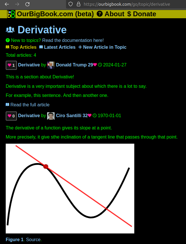
Figure 1. Screenshot of the "Derivative" topic page. View it live at: ourbigbook.com/go/topic/derivativeVideo 2. OurBigBook Web topics demo. Source. - local editing: you can store all your personal knowledge base content locally in a plaintext markup format that can be edited locally and published either:This way you can be sure that even if OurBigBook.com were to go down one day (which we have no plans to do as it is quite cheap to host!), your content will still be perfectly readable as a static site.
- to OurBigBook.com to get awesome multi-user features like topics and likes
- as HTML files to a static website, which you can host yourself for free on many external providers like GitHub Pages, and remain in full control

Figure 2. You can publish local OurBigBook lightweight markup files to either OurBigBook.com or as a static website.
Figure 3. Visual Studio Code extension installation.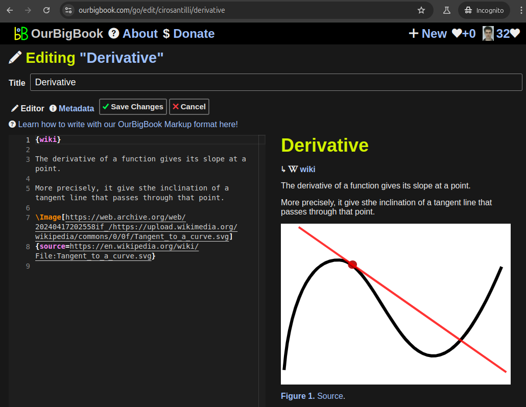
Figure 5. . You can also edit articles on the Web editor without installing anything locally. Video 3. Edit locally and publish demo. Source. This shows editing OurBigBook Markup and publishing it using the Visual Studio Code extension. 
- Infinitely deep tables of contents:
All our software is open source and hosted at: github.com/ourbigbook/ourbigbook
Further documentation can be found at: docs.ourbigbook.com
Feel free to reach our to us for any help or suggestions: docs.ourbigbook.com/#contact