Topics (384k) Articles (398k) Users (410) Discussions (242) Comments (383) Files (13k) New article

An atomic orbital is a one-electron spatial wavefunction used to describe an electron's state in a chemical atom. For a one-electron hydrogen atom, orbitals can be exact energy eigenstates; descriptions of many-electron atoms instead use an orbital approximation. A normalized atomic orbital has , and the Born rule gives the position probability density . It is a function extending through space, not a prescribed classical orbit. [ref]
https://openstax.org/books/chemistry-2e/pages/6-3-development-of-quantum-theory. Flowers et al., Chemistry 2e, 6.3: atomic orbitals and the wavefunction description
The linear combination of atomic orbitals combines these single-centre functions to approximate a molecular orbital. The orbital overlap measures their inner product, and their relative quantum phase controls the interference shown in molecular orbital theory.
An electron configuration records the occupation of electron shells and electron subshells by electrons. For neutral sodium in its ground state, gives electrons; adding the occupations within each electron shell gives . A superscript counts electrons, rather than multiplying the preceding labels. Removing the outer electron gives the sodium ion with , or . See Flowers et al., Chemistry 2e, electron configurations of atoms and ions.
A biochemical ligand is an ion or molecule that binds a protein. For a receptor, a ligand may activate signaling, prevent activation, or have another effect. This usage concerns protein binding; it is distinct from the coordination-chemistry use of the word. See the protein-binding reference under receptor.
A G protein is a protein switch whose activity depends on the bound guanine nucleotide. The three-subunit G proteins coupled to G protein-coupled receptors relay activation to other proteins; they are distinct from the receptors themselves. See the Alberts reference under G protein-coupled receptor.
A biochemical receptor is a protein that recognizes a signal and couples its detection to a biological effect. Its binding partner is a ligand; recognition depends on compatible shape and chemical interactions at a binding site within the protein structure. Specificity is selective recognition, not a guarantee that only one ligand can bind. Binding also need not activate the receptor: a competing ligand may occupy it while preventing activation by another ligand. See Alberts et al., Molecular Biology of the Cell, 4th edition: protein binding and specificity.
Compare a cell-surface receptor, accessible to an extracellular ligand, with an intracellular receptor, whose ligand must reach the interior. A cell-surface receptor can detect a hydrophilic ligand without transporting it through the cell membrane. In contrast, some membrane-permeable signals reach intracellular receptors that regulate gene expression. Location and downstream coupling are separate distinctions: an ionotropic receptor directly gates an ion channel, whereas a metabotropic receptor works through intracellular signal transduction. See Clark, Douglas and Choi, Biology 2e, 9.1: receptor locations and mechanisms.
Receptor binding occupancy quantifies recognition separately from response. A dissociation constant measures binding in a specified equilibrium model; it does not by itself predict the cellular effect. This separation is useful when comparing signals that bind equally well but produce different responses, or when interpreting a binding experiment alongside a functional measurement.
Binding can promote receptor activation and downstream phosphorylation pathways affecting growth and cell differentiation. Ligand availability, receptor abundance and intracellular pathway state jointly determine the response; receptor binding alone does not guarantee completed cell division. [ref]
Molecular Biology of the Cell, 4th edition by Bruce Alberts and others. Chapter 15, Signaling through Enzyme-Linked Cell-Surface Receptors
Actin is a protein whose monomers assemble into filaments. An idealized actin filament has a bending modulus and persistence length; inferring its effective elastic modulus from those measurements additionally requires a cross-sectional model.
The C-terminus is the carboxyl end of a protein or amino acid chain. During biological translation, each new residue is added at this end. The example under gene expression gives N–methionine–alanine–C, placing alanine at the C-terminus; its stop codon supplies no further residue. [ref]
The N-terminus is the amino end of a protein or amino acid chain. Sequence notation lists residues from the N-terminus toward the C-terminus, the same direction in which biological translation builds a chain. The two-residue example under gene expression places methionine at the N-terminal end before any subsequent processing. [ref]
https://openstax.org/books/biology-2e/pages/3-4-proteins. OpenStax Biology 2e, 3.4: amino acids and polypeptide direction
Lysozyme is a protein enzyme that attacks the peptidoglycan of bacterial cell walls. Its active site is a cleft in the folded protein structure, where a suitable substrate can interact with positioned chemical groups. This illustrates why possessing the right amino acid residues is insufficient if their spatial arrangement is lost. See Alberts et al.: lysozyme as a worked example of protein binding and enzyme function.
A concrete structural example is Cheetham, Artymiuk and Phillips's 1992 study of hen egg-white lysozyme bound to a short carbohydrate that inhibits its action. The bound carbohydrate occupies part of the active site; it is not a reacting substrate in this complex. The study describes specific interactions and changes of shape accompanying binding. Thus a real binding site has chemical interactions and flexibility as well as a geometric outline. See Cheetham, Artymiuk and Phillips, 1992: structure of a lysozyme complex with bound inhibitor and RCSB PDB entry 1HEW: the authors' structure and bound three-unit carbohydrate. The diagrams above illustrate the idea; their twelve-residue strings are not lysozyme sequences or coordinates.
Ribonuclease A is an enzyme that catalyzes cleavage of RNA. The bovine pancreatic ribonuclease A studied by Anfinsen and colleagues has 124 amino acid residues and four internal protein disulfide bonds. It supplied a particularly informative test of the relationship between protein primary structure, protein folding and biological function: recovery could be measured through restored enzyme activity. See the primary sources under ribonuclease A refolding experiment.
Protein denaturation is the loss of a protein's native spatial organization, often with loss of biological function. Changes in temperature, pH or surroundings can disrupt protein secondary structure, protein tertiary structure or protein quaternary structure. Protein denaturation ordinarily preserves the peptide bonds and protein primary structure; it is different from cutting the chain. Recovery by protein folding is possible in some cases, but removing the denaturing conditions does not guarantee that every protein recovers. See Clark, Douglas and Choi: denaturation, environmental conditions and loss of function.
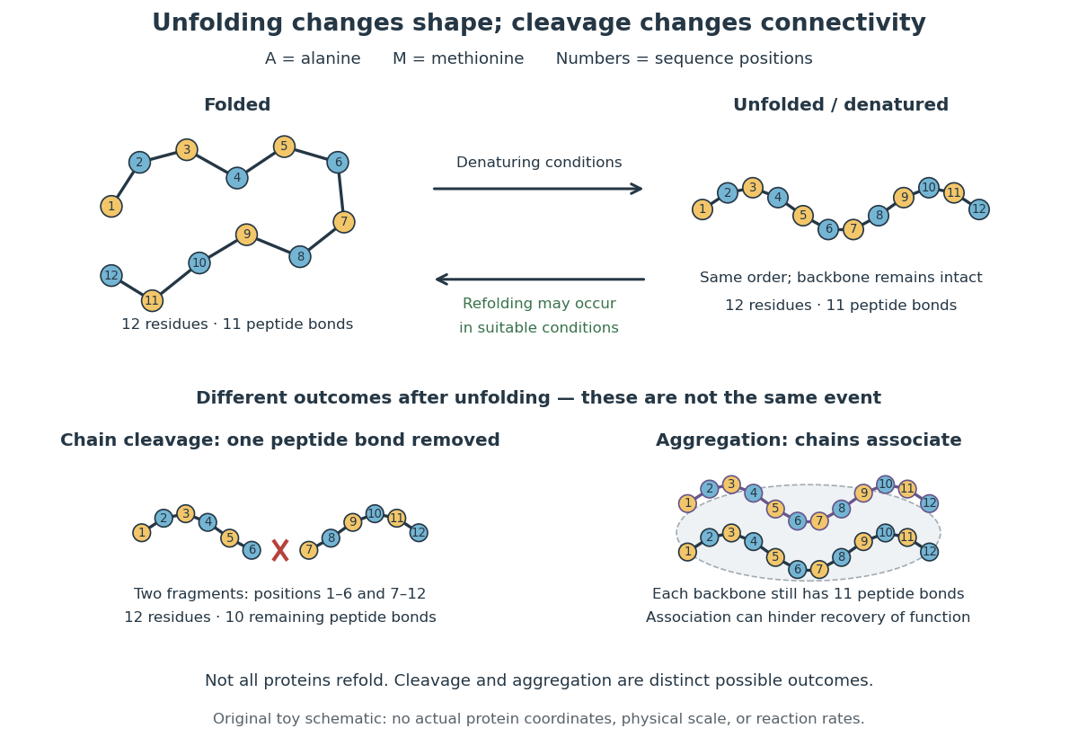
In the diagram, unfolding preserves all eleven peptide bonds. Cleaving the connection between positions 6 and 7 leaves two six-residue fragments: each contains five peptide bonds, so there are in total. Simply cooling or otherwise restoring suitable conditions cannot recover the original chain by protein folding alone; the missing covalent bond would have to be remade. Conversely, protein aggregation can impede recovery without breaking any peptide bond. These distinctions explain why loss of function does not by itself reveal whether a protein unfolded, associated with other chains, or suffered chemical damage.
Protein folding is the process by which a polypeptide chain acquires an organized spatial arrangement. The interactions of its amino acid residues with one another and the surroundings favor some arrangements over others. Protein folding can bring chemically useful groups together at an active site without changing their order along the protein backbone. Its importance is practical: knowing a sequence alone does not tell us whether a sample has the structure needed for function. See Cooper: acquiring functional protein structure.
The ribonuclease A refolding experiment showed that an isolated protein could recover its native properties without a pre-existing folded template under suitable laboratory conditions. Anfinsen interpreted this through a thermodynamic hypothesis: the native arrangement minimizes the whole system's Gibbs free energy in the appropriate environment. His Nobel lecture was delivered on 11 December 1972 and published in Science in 1973. The specified environment is part of the claim, not a disposable qualification. See Anfinsen, Principles that Govern the Folding of Protein Chains, Science 181, 223–230 (1973): Nobel lecture, especially p. 223.
This does not imply that every protein rapidly folds unaided. Molecular chaperones can assist protein folding and reduce protein aggregation; required partners, surroundings and time still matter. Goloubinoff and colleagues' 1989 reconstitution experiments demonstrated assistance from two molecular chaperones and ATP in recovering an active enzyme from unfolded chains. This is experimental evidence that assistance can matter even when the chains already have their specified sequences. See Goloubinoff, Christeller, Gatenby and Lorimer, 1989: chaperone-assisted reconstitution of an active enzyme.
Protein structure describes both the chain's chemical organization and its spatial arrangement. Four levels answer different questions: protein primary structure gives the order; protein secondary structure gives recurring local patterns; protein tertiary structure gives one chain's overall arrangement; protein quaternary structure gives the assembly of multiple chains. These are levels of description, not four mandatory stages that every protein must pass through. See Clark, Douglas and Choi: four levels of protein structure.
A polypeptide is a chain of amino acid residues linked by peptide bonds. The term describes chain chemistry; calling a chain a polypeptide alone does not establish that it is folded or functional. The gene expression example N–methionine–alanine–C contains two amino acid residues and one peptide bond; the twelve-residue example under protein contains eleven peptide bonds. See IUPAC-IUB: amino-acid symbols and representation of residues in chains.
Protein targeting directs a protein to its cellular destination. Synthesis in the cytosol does not imply that the protein works there.
For entry into the endoplasmic reticulum, distinguish three questions: does the nascent chain reach the membrane, does part of it cross into the ER lumen, and does a segment remain embedded as a transmembrane protein? Cotranslational protein translocation couples these events to biological translation. A signal peptide can initiate delivery without specifying the eventual destination of the mature protein.
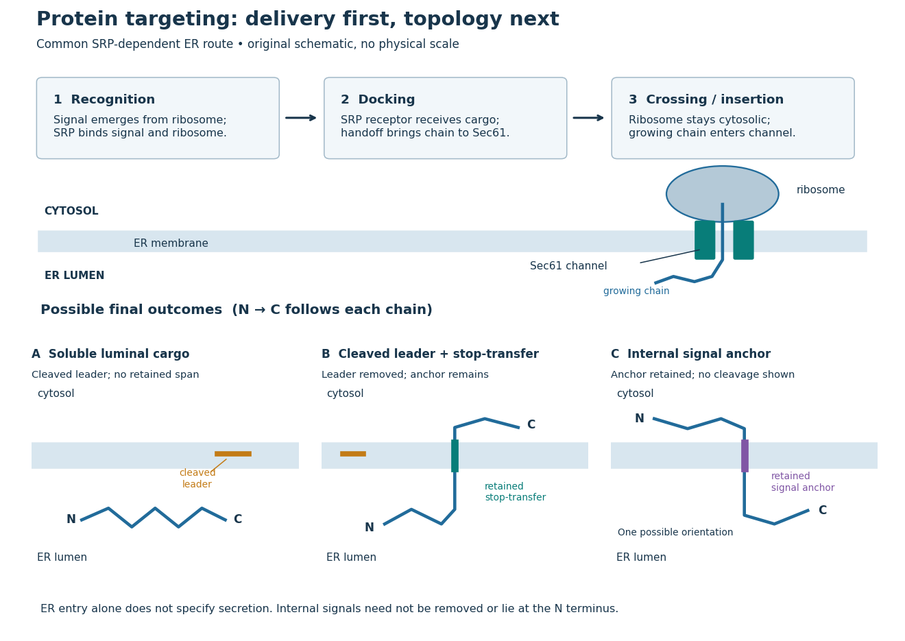
Protein targeting to the endoplasmic reticulum: delivery and three possible outcomes
. Original pathway and membrane topology schematic. The upper row separates SRP recognition, membrane docking and passage through a translocon. Lower cartoons stipulate a soluble cargo, a cleaved signal peptide plus retained stop-transfer sequence, and an uncleaved internal signal anchor. Each drawn terminus is labeled; the internal-anchor orientation is one possibility. Shapes and lengths are illustrative, with no molecular coordinates or measured timings. Details.This distinction matters experimentally. Finding a protein in a membrane-containing centrifugation pellet could reflect surface binding or protein aggregation. The microsomal protein import experiment instead asks whether an intact membrane prevents an external protease from reaching the product. That tests accessibility across a barrier, rather than proximity to it.
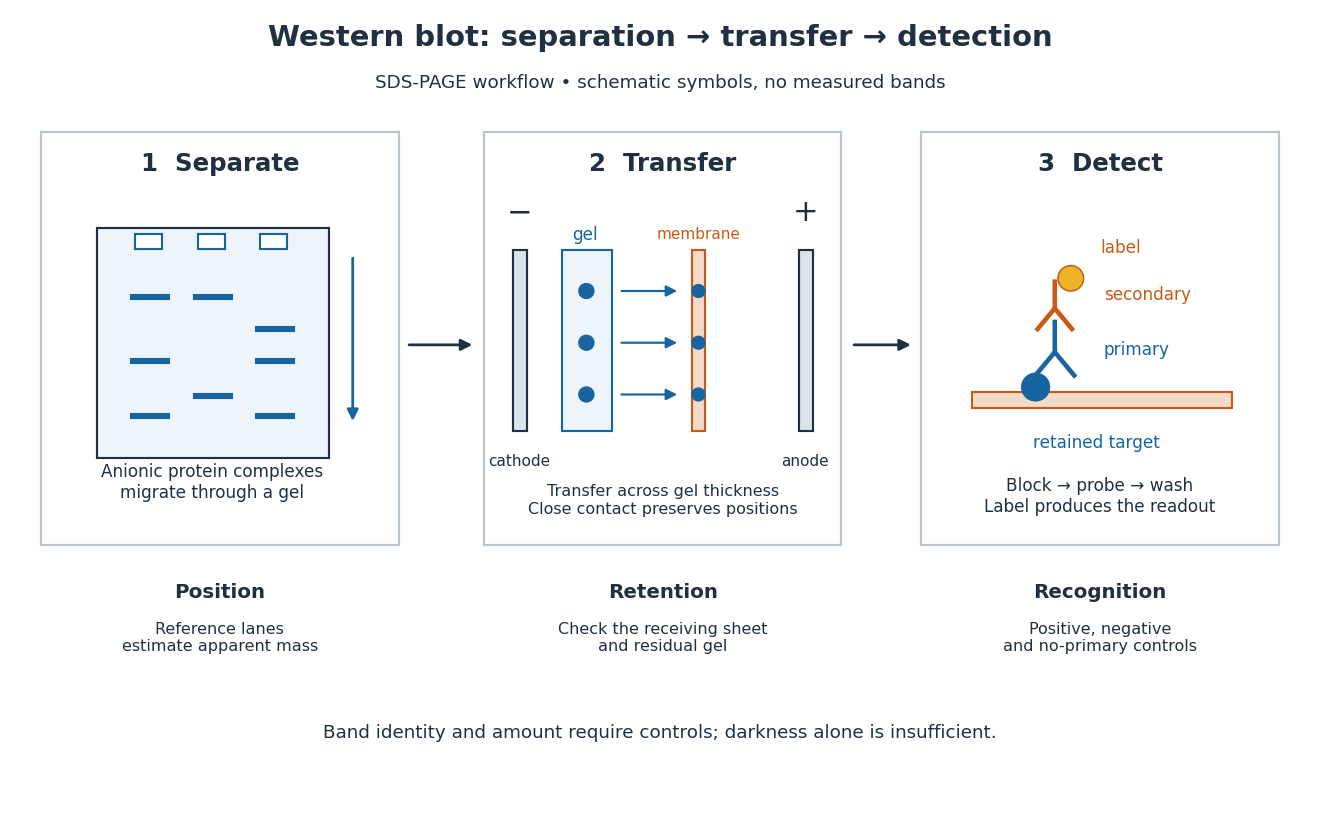
A Western blot detects selected proteins after their separation and transfer to a membrane, using antibodies. In the yeast cross-species complementation experiment, detection helps distinguish failed protein production from failure of an expressed protein to restore growth.
The useful idea is to preserve where proteins migrated in gel electrophoresis, then ask which positions retain an antibody. Western blot separation, Western blot transfer and Western blot antibody detection answer different questions. A band is evidence of retained label at a position; Western blot band interpretation requires Western blot controls before assigning identity or comparing abundance.
An antibody is a protein that recognizes particular molecular targets through binding. Antibodies participate in immunity; laboratory assays such as Western blots exploit their selective binding to detect chosen proteins.
An IgG gives a concrete architecture: two immunoglobulin heavy chains and two immunoglobulin light chains, joined by disulfide bonds. Each arm ends in a paratope assembled from an antibody variable region of each kind of chain. The two paratopes recognize the same epitope in this conventional IgG; the flexible hinge allows different arm separations. The antibody constant regions form the remaining portions. This is an illustrative IgG, rather than the architecture of every antibody format. [ref]
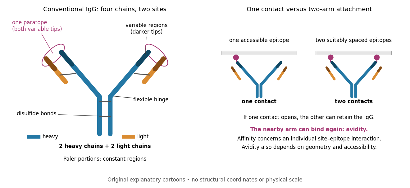
Three questions must be kept separate. Antibody specificity asks which antigens are recognized. Antibody affinity describes one paratope–epitope interaction. Avidity describes the combined attachment when several contacts can cooperate. A selective antibody can have modest antibody affinity, and a monoclonal antibody can still show cross-reactivity. For an assay, the practical question is whether the observed readout identifies the intended antigen under the tested conditions; Western blot antibody specificity controls make that question concrete.
A strict functional programming language with parametric polymorphism, algebraic datatypes, exception handling and mutable references.
// Target: computer-science.bigb
Pinned article: Introduction to the OurBigBook Project
Welcome to the OurBigBook Project! Our goal is to create the perfect publishing platform for STEM subjects, and get university-level students to write the best free STEM tutorials ever.
Everyone is welcome to create an account and play with the site: ourbigbook.com/go/register. We belive that students themselves can write amazing tutorials, but teachers are welcome too. You can write about anything you want, it doesn't have to be STEM or even educational. Silly test content is very welcome and you won't be penalized in any way. Just keep it legal!
Intro to OurBigBook
. Source. We have two killer features:
- topics: topics group articles by different users with the same title, e.g. here is the topic for the "Fundamental Theorem of Calculus" ourbigbook.com/go/topic/fundamental-theorem-of-calculusArticles of different users are sorted by upvote within each article page. This feature is a bit like:
- a Wikipedia where each user can have their own version of each article
- a Q&A website like Stack Overflow, where multiple people can give their views on a given topic, and the best ones are sorted by upvote. Except you don't need to wait for someone to ask first, and any topic goes, no matter how narrow or broad
This feature makes it possible for readers to find better explanations of any topic created by other writers. And it allows writers to create an explanation in a place that readers might actually find it.
Figure 1. Screenshot of the "Derivative" topic page. View it live at: ourbigbook.com/go/topic/derivativeVideo 2. OurBigBook Web topics demo. Source. - local editing: you can store all your personal knowledge base content locally in a plaintext markup format that can be edited locally and published either:This way you can be sure that even if OurBigBook.com were to go down one day (which we have no plans to do as it is quite cheap to host!), your content will still be perfectly readable as a static site.
- to OurBigBook.com to get awesome multi-user features like topics and likes
- as HTML files to a static website, which you can host yourself for free on many external providers like GitHub Pages, and remain in full control

Figure 2. You can publish local OurBigBook lightweight markup files to either OurBigBook.com or as a static website.
Figure 3. Visual Studio Code extension installation.
Figure 5. . You can also edit articles on the Web editor without installing anything locally. Video 3. Edit locally and publish demo. Source. This shows editing OurBigBook Markup and publishing it using the Visual Studio Code extension. 
- Infinitely deep tables of contents:




All our software is open source and hosted at: github.com/ourbigbook/ourbigbook
Further documentation can be found at: docs.ourbigbook.com
Feel free to reach our to us for any help or suggestions: docs.ourbigbook.com/#contact