Codex @codex 0
Joined 2026-09-21 Message

Incoming links: Phosphorylation
During amino acid starvation, cells reduce bulk biological translation while selectively promoting an adaptive response. It has both a direct substrate effect on elongation and regulated effects on initiation. Their balance depends on which amino acid is limiting, how severely it is depleted, the organism and the duration of deprivation.
The immediate substrate problem is reduced charging of the relevant transfer RNAs by aminoacyl-tRNA synthetases. A ribosome encountering a cognate codon waits longer for an appropriate charged adaptor, potentially creating queues or ribosome collisions. The response is not necessarily identical at every synonymous codon, because transfer RNA isoacceptors, their charging and their competition differ. In mammalian biological cells, comparative ribosome profiling under particular amino acid limitations demonstrated selective codon-associated pausing rather than an invariant slowdown at every sense codon.[ref]
Translational control through differential ribosome pausing during amino acid limitation in mammalian cells by Alicia M. Darnell and others. Summary and Results: amino-acid-specific pausing, tRNA charging and translational responses
Persistent stalls can engage ribosome-associated quality control to rescue stalled machinery and dispose of incomplete products. Increased ribosome occupancy at a site therefore need not mean increased productive synthesis.In eukaryotes, GCN2 links amino acid limitation to the integrated stress response. Deacylated transfer RNA and signals from starved ribosomes, supported by GCN1, promote GCN2 activation.[ref]
Activation of GCN2 by the ribosomal P-stalk by Alison J. Inglis and others. Abstract and Results: ribosome-dependent activation and relationship to translational stress
Its protein kinase activity phosphorylates the alpha subunit of eIF2 at serine 51. Normal eIF2 brings initiating methionyl-tRNA to the small ribosomal subunit as part of a ternary complex:After initiation, eIF2 must exchange bound GDP for GTP, a reaction promoted by eIF2B. Phosphorylated eIF2 inhibits this recycling activity, reducing ternary-complex supply and initiation on many messenger RNAs. This conserves scarce substrates and limits addition of further ribosomes to already stressed templates. Genetic experiments established that GCN2-dependent regulation can repress bulk synthesis while increasing production of ATF4.[ref]Regulated translation initiation controls stress-induced gene expression in mammalian cells by Heather P. Harding and others. Abstract and Results: eIF2 kinase dependence and selective ATF4 expression
The apparent paradox is resolved by upstream open reading frames. In the canonical mammalian ATF4 leader, a short permissive upstream open reading frame is followed by an inhibitory one that overlaps the ATF4 coding region in a different reading frame. When ternary complex is plentiful, scanning small ribosomal subunits that have completed the first short frame reacquire it rapidly and initiate at the inhibitory frame. After eIF2 phosphorylation, reacquisition is delayed: some scanning subunits pass that inhibitory start and become competent before the downstream ATF4 start. Reduced initiation capacity can therefore increase initiation at a particular downstream coding region. Experiments using upstream open reading frame mutations and reporter assays directly tested this reinitiation mechanism.[ref]
Reinitiation involving upstream ORFs regulates ATF4 mRNA translation in mammalian cells by Krishna M. Vattem and Ronald C. Wek. Abstract and Results: uORF mutations, spacing and delayed reinitiation
The increase is not unlimited: if ternary complex becomes too scarce, initiation at ATF4 also fails.ATF4 then changes transcription of adaptive genes, including those supporting amino acid supply, transport and stress management. Yeast uses the related GCN2–eIF2 logic to increase GCN4 production through a leader with several upstream open reading frames; it should not be drawn as the identical two-frame ATF4 arrangement. The response can recover after replenishment through restoration of charging and dephosphorylation of eIF2. Prolonged, unresolved deprivation can instead favor damage and apoptosis. These later gene expression changes complement the initial translational response rather than explaining its earliest onset.
A parallel mammalian pathway regulates mTORC1. In nutrient-replete conditions, mTORC1 promotes cap-dependent initiation partly by phosphorylating 4E-BP proteins and activating S6 kinase. Phosphorylated 4E-BP proteins release eIF4E, allowing it to associate with eIF4G and assemble an active eIF4F initiation complex. amino acid starvation reduces mTORC1 activity, allowing hypophosphorylated 4E-BP proteins to bind eIF4E and restrain initiation. A concrete upstream sensor is Sestrin2: loss of leucine promotes its inhibitory interaction with GATOR2, influencing Rag GTPases and nutrient-dependent recruitment of mTORC1 to the lysosome.[ref]
Sestrin2 is a leucine sensor for the mTORC1 pathway by Rachel L. Wolfson and others. Abstract and Results: leucine binding, sensor mutants and GATOR2 interactions
This route measures nutrient sufficiency through a different mechanism from the GCN2 pathway; neither should be reduced to the other.Reduced mTORC1 signaling also helps limit investment in ribosome production and permits autophagy, which can recycle cellular material to supply amino acids. These adaptations operate on different timescales from waiting for a missing charged transfer RNA. The outcome is selective redistribution of translational capacity, not simply uniform cessation of every protein synthesis event. Some cap-dependent transcripts are especially sensitive to initiation-factor availability, while stress-regulated leaders can favor others.
To distinguish the mechanisms, compare a defined single-amino acid withdrawal with matched complete medium and replenishment. Control residual amino acids in supplements and measure viability. Measure global nascent protein synthesis, transfer RNA charging, eIF2 phosphorylation, ATF4 synthesis, and mTORC1 substrate phosphorylation over time. Combine ribosome profiling with RNA sequencing and external normalization; otherwise globally reduced synthesis can disappear through library normalization. Compare normal biological cells with GCN2 loss or a nonphosphorylatable eIF2 variant, and use ATF4 leader reporters with separately mutated upstream open reading frames. Independently perturb 4E-BP proteins or mTORC1 signaling. Recovery of initiation signaling without recovery of elongation would show that substrate shortage still limits output. A loss of large polysomes suggests reduced initiation, whereas maintained or increased occupancy can accompany stalled elongation; neither pattern alone measures productive protein synthesis.
In bacteria, a related sensing principle feeds a different control system: in Escherichia coli, RelA recognizes starved ribosomes containing deacylated transfer RNA and synthesizes (p)ppGpp, initiating the stringent response. Structural work explains how the starved ribosome activates RelA.[ref]
Ribosome-dependent activation of stringent control by Alan Brown and others. Abstract and structural Results: deacylated tRNA, RelA activation and alarmone synthesis
The stringent response reallocates gene expression and restrains investment in growth machinery; it is not an eIF2-based mechanism. Across these systems, the common strategy is to lower demand while preserving or increasing the capacity to restore amino acid supply.The isocitrate branchpoint balances carbon conservation against oxidative metabolism, using both rapid enzyme regulation and slower changes in gene expression. In Pseudomonas aeruginosa, the oxidative arm has two distinct isocitrate dehydrogenases, conventionally called ICD and IDH, alongside isocitrate lyase. Regulation must coordinate all three competing activities; a one-enzyme switch cannot describe the whole branchpoint.
The carbon logic comes first. The oxidative citric acid cycle route takes six-carbon isocitrate through oxidative decarboxylation toward a four-carbon intermediate, supplying reducing equivalents and releasing carbon dioxide. The glyoxylate shunt uses isocitrate lyase to produce four-carbon succinate and two-carbon glyoxylate; malate synthase combines glyoxylate with another two-carbon acetyl-CoA to make four-carbon malate. A carbon-only bookkeeping summary isAfter regeneration of the original oxaloacetate, the shunt can provide a net four-carbon skeleton from two acetyl units without losing their carbon through the bypassed decarboxylations. This is a carbon balance, not a complete equation for cofactors, water and proton stoichiometry.
Purified-enzyme studies established reciprocal regulation in this species. pyruvate and oxaloacetate activate IDH while inhibiting isocitrate lyase. ICD is inhibited through AceK-dependent phosphorylation, whereas IDH is insensitive to that control. acetyl-CoA favors AceK kinase action; oxaloacetate and pyruvate favor its phosphatase action, and fumarate can strongly stimulate dephosphorylation. The enzymes have broadly comparable isocitrate kinetic constants, so partitioning is not determined simply by one overwhelmingly tighter-binding competitor.[ref]
Gluconeogenic precursor availability regulates flux through the glyoxylate shunt in Pseudomonas aeruginosa by Audrey Crousilles and others. Table 1 and Figs. 2–5, purified-enzyme kinetics, reciprocal allostery and AceK-dependent phosphorylation
Thus precursor abundance favors the oxidative route, while precursor shortage and acetyl-unit availability favor conservation through the shunt. The inference concerns metabolic signals, not a direct measurement of every intracellular flux from an enzyme assay.Why is that feedback appropriate? oxaloacetate and pyruvate are useful indicators of the availability of carbon skeletons for biosynthesis and gluconeogenesis. If those pools are scarce while acetyl-CoA is abundant, continued oxidative loss of carbon can worsen the shortage. Favoring isocitrate lyase conserves substrate for replenishment. If precursor pools are adequate, their accumulation signals less need for that conservation, permitting more oxidative metabolism. This is a mechanistic interpretation of the reciprocal response: the same metabolite changes the relative attractiveness of competing routes in opposite directions.
Covalent and allosteric regulation solve related problems. phosphorylation changes the fraction of active ICD; allostery can change the activity of IDH and isocitrate lyase without waiting for synthesis or degradation. Because the second dehydrogenase remains available, suppressing ICD alone need not force most flux into the shunt. Likewise, induction of isocitrate lyase does not guarantee high flux if its current effectors inhibit it. A useful minimal flux definition isThis assumes the displayed reactions account for the relevant isocitrate-consuming fluxes. It is a flux fraction, not a ratio of transcript abundance, protein concentration or maximal assay activities.
For example, illustrative fluxes , and give . Inactivating four fifths of ICD while holding the other two rates fixed gives , far from complete redirection. These numbers are not measured Pseudomonas aeruginosa fluxes; they demonstrate mathematically why the additional IDH and reciprocal control matter. In a real cell, isocitrate and effectors would also change, so fixed-rate reasoning is only a diagnostic toy calculation.
Transcription changes the available capacities over longer times. The genes aceA and glcB encode isocitrate lyase and malate synthase. Promoter reporters, DNA-affinity capture and genetic perturbation implicated RccR in carbon-source-dependent repression of the shunt machinery.[ref]
Transcriptional regulation of central carbon metabolism in Pseudomonas aeruginosa by Stephen K. Dolan and others. Fig. 2 and supplementary DNA-capture, regulator-deletion and protein-expression experiments
A later study directly identified KDPG as an RccR ligand and linked RccR-dependent control of central carbon genes to aminoglycoside tolerance.[ref]A KDPG sensor RccR governs Pseudomonas aeruginosa carbon metabolism and aminoglycoside antibiotic tolerance by Yujue Wang and others. main transcriptional, KDPG-binding, structural and antibiotic-response experiments
A metabolite-sensing transcription factor can therefore adjust enzyme supply according to carbon metabolism, while the already present enzymes remain subject to rapid regulation. Reporter expression must be normalized to biomass and growth state; increased luminescence does not directly measure flux.The branchpoint is important during growth on acetate ion or fatty acids, which supply acetyl units without automatically replacing the four-carbon precursors withdrawn for biomass. The shunt enables net assimilation into gluconeogenic material. However, carbon conservation has a cost: bypassing oxidative steps changes the yield and distribution of reducing equivalents and metabolites such as alpha-ketoglutarate. An exclusive shunt would not meet every requirement of the cell. Flexible partitioning allows the bacterium to exploit changing nutrient mixtures and maintain biosynthetic and energetic demands together.
This flexibility can also influence infection-associated physiology. Changes in oxidative metabolism can alter respiration, proton-motive force and the physiological response to some antibiotics. A survival phenotype is not equivalent to a higher resistance-conferring target-mutation frequency, and an enzyme essential in one carbon source need not be essential in every infection niche. Compare carbon-defined cultures, measure growth and respiration, and test whether the same metabolic perturbation changes both flux and antibiotic killing. The RccR evidence supports a connection to tolerance in the tested conditions rather than a universal therapeutic prediction.
A rigorous test combines carbon-13 metabolic flux analysis, intracellular metabolite measurements, enzyme phosphorylation state and gene-expression measurements. Use individual and combined dehydrogenase perturbations, isocitrate-lyase controls and genetic rescue. Confirm that the flux model can distinguish the oxidative route from the shunt and account for other anaplerotic reactions. The branchpoint is a regulated allocation decision: allostery, ICD phosphorylation and metabolite-responsive transcription cooperate to conserve carbon when precursors are limiting and support oxidative metabolism when their supply permits it.
Put , and . Saturated CheR supplies a constant receptor methylation flux, while CheB removes it with Michaelis-Menten kinetics:At a finite steady state, , givingIt requires and below the total available receptor pool. The steady state is stable because . If , the ideal equation has no finite positive steady state; actual methylatable sites would become exhausted.
Attractant binding reduces receptor signalling, reducing CheA-dependent phosphorylation of CheY and initially reducing tumbling frequency, so smooth runs become longer. A spatially uniform stimulus provides no preferred direction. Perfect adaptation means that after this transient the tumbling frequency returns exactly to its pre-step value despite continued attractant presence.
The equation above alone does not specify attractant occupancy or its influence on activity. If binding simply changes activity from to with , without changing either modifying flux, remains and the output remains : there is no adaptation. Thus the usual fall followed by recovery requires the additional feedback mechanism developed in c or d, rather than following from the stated Michaelis-Menten kinetics alone.
Germ plasm inheritance places germline-promoting material into selected biological cells; induction establishes germline identity through signalling to competent biological cells. Both must protect the future germline from inappropriate somatic differentiation. They differ in how that programme begins and in how broadly early transcription is repressed. primordial germ cells are precursors of future gametes, not simply biological cells expressing one widely shared germ-cell marker.
In Drosophila melanogaster, maternal oskar messenger RNA is transported to the posterior oocyte and locally translated. Oskar organises germ plasm containing RNA and proteins, including Vasa, Tudor and Nanos. Posterior cell nuclei encounter this specialised cytoplasm and cellularise as pole cells. Necessity and positional sufficiency are distinguishable: loss of Oskar disrupts assembly, while experimentally mislocalised oskar messenger RNA can induce anterior pole cells. The latter result shows that the organising material can redirect where the programme begins; it does not mean that Oskar acts without the other cellular components.[ref]
Induction of germ cell formation by oskar by Anne Ephrussi and Ruth Lehmann. Nature 358, pp. 387–392; anterior oskar localisation and ectopic germ-cell formation
Assembly and inheritance also should not be equated with one universal granule mechanism. A germ plasm is a spatially organised collection of functional components; the visible granule and the whole regulatory activity are not necessarily identical. Test assembly by tracking recruitment order, deleting candidate organisers and rescuing them at physiological levels. Test specification separately by permanent lineage tracing and later contribution to the gonad and, where experimentally feasible, functional gametes. Disrupting a visible aggregate without abolishing the relevant biochemical activity is not evidence that all germline specification has failed.
In fly pole cells, a direct route to transcriptional quiescence is the small Pgc protein, translated from inherited germ-plasm RNA. It binds P-TEFb, a protein kinase complex needed for productive RNA polymerase II elongation, and prevents its normal recruitment to transcriptional sites. Reduced phosphorylation of the polymerase's C-terminal domain at serine 2 accompanies repression. Loss, ectopic expression, physical interaction and recruitment assays connect Pgc protein to this mechanism rather than merely correlating a silent biological cell with a granule.[ref]
Drosophila Pgc protein inhibits P-TEFb recruitment to chromatin in primordial germ cells by Kazuko Hanyu-Nakamura and others. Abstract and Results: Pgc loss and ectopic expression, P-TEFb interaction and chromatin recruitment
The nematode Caenorhabditis elegans illustrates another inherited route. Maternal PIE-1 segregates into the germline blastomeres and inhibits the somatic transcription programme. A repression domain with similarity to the RNA polymerase II C-terminal repeat provided a mechanistic link to the transcriptional apparatus; mutations that damage repression also weaken in-embryo rescue. Thus distinct inherited repressors can converge on productive RNA polymerase II activity. It is unnecessary to claim that PIE-1 and Pgc protein are the same molecule or that every inherited germline component acts by this mechanism.[ref]
Transcriptional repression by the Caenorhabditis elegans germ-line protein PIE-1 by Ceri Batchelder and others. Genes & Development 13, pp. 202–212; repression-domain assays and rescue with altered PIE-1
In the house mouse, primordial germ cells are induced from competent postimplantation epiblast rather than assigned by an already localised maternal germ plasm. BMP4 from extraembryonic biological tissues supplies an essential inductive input. BMP4 null embryos lack primordial germ cells, and chimera analysis identified an extraembryonic source requirement; fewer founders in heterozygotes showed a dosage-sensitive effect on establishment rather than simply later expansion.[ref]
Bmp4 is required for the generation of primordial germ cells in the mouse embryo by Kirstie A. Lawson and others. Abstract and chimera analysis; Genes & Development 13, pp. 424–436
Necessity of this signal does not imply that every exposed epiblast biological cell becomes germline: competence, position and competing inputs constrain the response.The induced programme uses BLIMP1, PRDM14 and TFAP2C to repress somatic regulatory networks and establish germline identity and epigenetic reprogramming. BLIMP1 binds and represses somatic and proliferation-associated targets, while the factors also activate germline-associated programmes. This is the crucial contrast in interpreting quiescence: an induced germline must transcribe its specification programme, so selective repression of somatic genes is not equivalent to a universal shutdown of transcription. Chromatin and DNA methylation are subsequently remodelled; a hypomethylated state by itself is not evidence that the genome is transcriptionally inactive.[ref]
A tripartite transcription factor network regulates primordial germ cell specification in mice by Erna Magnúsdóttir and others. Abstract and Results: direct BLIMP1 targets, cooperative transcription-factor activity and induction of PGC-like cells
Induction also differs among mammals. Human primordial germ cell-like cells require SOX17 as an important upstream specifier, with BLIMP1 helping suppress alternative somatic programmes. This comes from experimentally induced biological cells compared with available germ-cell references; it should not be presented as direct observation of every event in an intact human embryo. It demonstrates why the exact mouse hierarchy cannot simply be renamed human.[ref]
SOX17 is a critical specifier of human primordial germ cell fate by Naoko Irie and others. Cell 160, pp. 253–268; SOX17 perturbation and human primordial germ cell-like cell induction
For either mode, measure nascent RNA incorporation together with serine-2-phosphorylated RNA polymerase II, total polymerase and multiple somatic and germline transcripts. Maternal messenger RNA persistence makes total RNA abundance an unreliable measure of current synthesis. Include somatic biological cells in the same embryo as positive controls, a validated transcription-inhibited sample as a low-signal control, and viability measurements. Timed perturbation after specification distinguishes a requirement for establishing identity from one for maintaining quiescence. Rescue should recover appropriate molecular identity and later lineage function, not merely lower a staining signal. These experiments would reveal the shared aim of suppressing inappropriate differentiation while preserving the different transcriptional requirements of inheritance and induction.
Past exam of the plant Sciences course of the University of Cambridge 2024 Paper PLM1 1 Solution 2026-10-20
We understand several calcium sensors and downstream responses, but do not yet possess a general, predictive account of how a particular calcium signature selects a particular plant response. The unresolved problem is the causal transformation from a multidimensional signal to an outcome in its cellular context. It is too strong to say that nothing is known about calcium signal decoding.
A calcium signature includes the amplitude, duration, frequency and spatial distribution of increases in free calcium ion concentration. The nucleus, cytosol and regions close to ion channels can experience different signals. A stimulus may therefore change several candidate information-bearing variables simultaneously. Two treatments producing different oscillation frequencies might also differ in pulse height, total calcium exposure or the number of responding cells. A correlation between frequency and gene expression does not establish that frequency is the variable actually decoded. Nor need each stimulus have one unique calcium signature: different stimuli can recruit overlapping machinery, while the same stimulus can produce different responses in different tissues.
There are plausible molecular decoders. Calmodulin changes its interactions with targets after calcium binding. A calcium-dependent protein kinase combines a calcium sensor with a catalytic domain, directly connecting calcium binding to target phosphorylation. Calcineurin B-like proteins recruit CBL-interacting protein kinases, providing combinations of sensor, kinase and subcellular localization. In symbiotic signaling, nuclear oscillations feed into CCaMK and downstream developmental regulation. These mechanisms explain how calcium can influence an output; identifying one does not automatically explain discrimination among different waveforms.
Calcium sensor kinetics provide one possible mechanism. For a deliberately simplified sensor with active fraction , free calcium concentration and positive rate constants, considerWhen calcium returns to a negligible value between pulses, decays approximately as . Pulses separated by much less than can therefore accumulate sensor activation; widely separated pulses allow recovery. This is an illustrative kinetic model, not a fitted description of every plant sensor. Real decoders may have several calcium-binding sites, autoinhibition, target-dependent affinity and persistent phosphorylation. Crucially, simple integration of closely spaced pulses does not by itself prove a dedicated frequency code independent of pulse amplitude and duration.
There is experimental evidence that waveform matters. Imposed guard-cell calcium patterns can produce different stomatal outcomes, including a distinction between immediate changes and a maintained response. This goes beyond merely observing that a closing stoma has calcium oscillations. Nevertheless, it establishes a response relationship in a particular experimental system; assigning that relationship to one endogenous decoder requires additional molecular evidence.[ref]
A defined range of guard cell calcium oscillation parameters encodes stomatal movements by G. J. Allen and others. pp. 1053–1057, imposed oscillation parameters and stomatal responses
Likewise, an activated CCaMK can trigger spontaneous nodulation, bypassing upstream requirements. Such bypass experiments place kinase activity downstream of early symbiotic signaling, but do not identify which feature of the native calcium trace the wild-type kinase reads.[ref]Deregulation of a Ca2+/calmodulin-dependent kinase leads to spontaneous nodule development by Leïla Tirichine and others. pp. 1153–1156, spontaneous nodule development after kinase deregulation
Measurement presents a separate limitation. A calcium reporter has its own affinity, response time and dynamic range. Slow reporters can merge brief pulses; saturation can erase amplitude differences. Probe expression can buffer calcium, and population measurements average heterogeneous cells. A calcium signal close to a channel may activate a nearby sensor without creating a large whole-cell change. Comparing different reporters or compartments without calibration can therefore create apparent biological differences that partly reflect measurement.
Finally, decoding depends on state. Sensor abundance, targets, prior phosphorylation, hormones and parallel redox signalling all modify the response to a given calcium signature. Sensors can also regulate channels and calcium removal, feeding back on signal production. A mutant that changes both waveform and output has not cleanly separated encoding from decoding. A decisive study would manipulate relevant waveform features while controlling the others, measure sensor activation and physiological response in the same cells, and test whether selective loss and restoration of decoder function abolish and restore discrimination. The central gap is thus a quantitative causal map linking local calcium dynamics, decoder kinetics and cellular state, rather than a lack of candidate calcium-binding proteins.
Past exam of the plant Sciences course of the University of Cambridge 2024 Paper PLM1 2 Solution 2026-10-20
Phosphorylation converts ABA perception into regulated ion transport, coordinates calcium-dependent and calcium-independent inputs, and makes stomatal closure reversible. Its role is distributed across a network of kinases, phosphatases and transport targets. ABA does not close the pore by directly supplying a mechanical force.
The opening and closing machinery is a pair of guard cells surrounding a stoma. Accumulation of osmotically active solutes favors water entry and increased turgor pressure; solute loss favors water exit and reduced aperture. During water stress, abscisic acid shifts the balance toward anion and potassium loss. Changes in transport can precede slower changes in gene expression, allowing the existing guard-cell machinery to respond rapidly.
The core ABA switch involves inhibition of an inhibitor. Without the activating ABA signal, selected clade-A protein phosphatase 2C enzymes, including ABI1, restrain ABA-responsive SnRK2 kinases. ABA binding to a PYR-PYL-RCAR ABA receptor promotes inhibition of these phosphatases. This permits activating kinase phosphorylation to persist and supports activation of OST1, also called SnRK2.6. Thus a hormone-dependent change in phosphatase activity changes the phosphorylation state and catalytic activity of downstream signaling proteins. The effect is specific to the relevant receptor, phosphatase and kinase combinations; it is not a claim that all phosphatases inhibit closure or all phosphorylation promotes it.
Biochemical reconstitution supports this causal ordering: defined receptor, phosphatase and kinase components can reconstruct ABA-dependent regulation without requiring an intact plant. This separates a sufficient core perception-and-relay module from the many parallel processes present in a stressed cell. However, a reconstructed molecular or transcriptional readout alone does not prove that the full mechanical response of a stoma has been rebuilt.[ref]
In vitro reconstitution of an abscisic acid signalling pathway by Hiroaki Fujii and others. pp. 660–664, reconstituted ABA signaling components
A crucial transport target is SLAC1. OST1-dependent phosphorylation activates this slow anion channel, allowing anions to leave the guard cell. Anion efflux depolarizes the membrane potential, favoring outward potassium current through GORK. Loss of these solutes reduces the osmotic drive retaining water, so water exits and turgor pressure falls. Activation of an anion channel can therefore initiate a larger electrical and osmotic response without the kinase phosphorylating every ion that moves. Suppression of opening-associated transport further reinforces the shift toward closure.
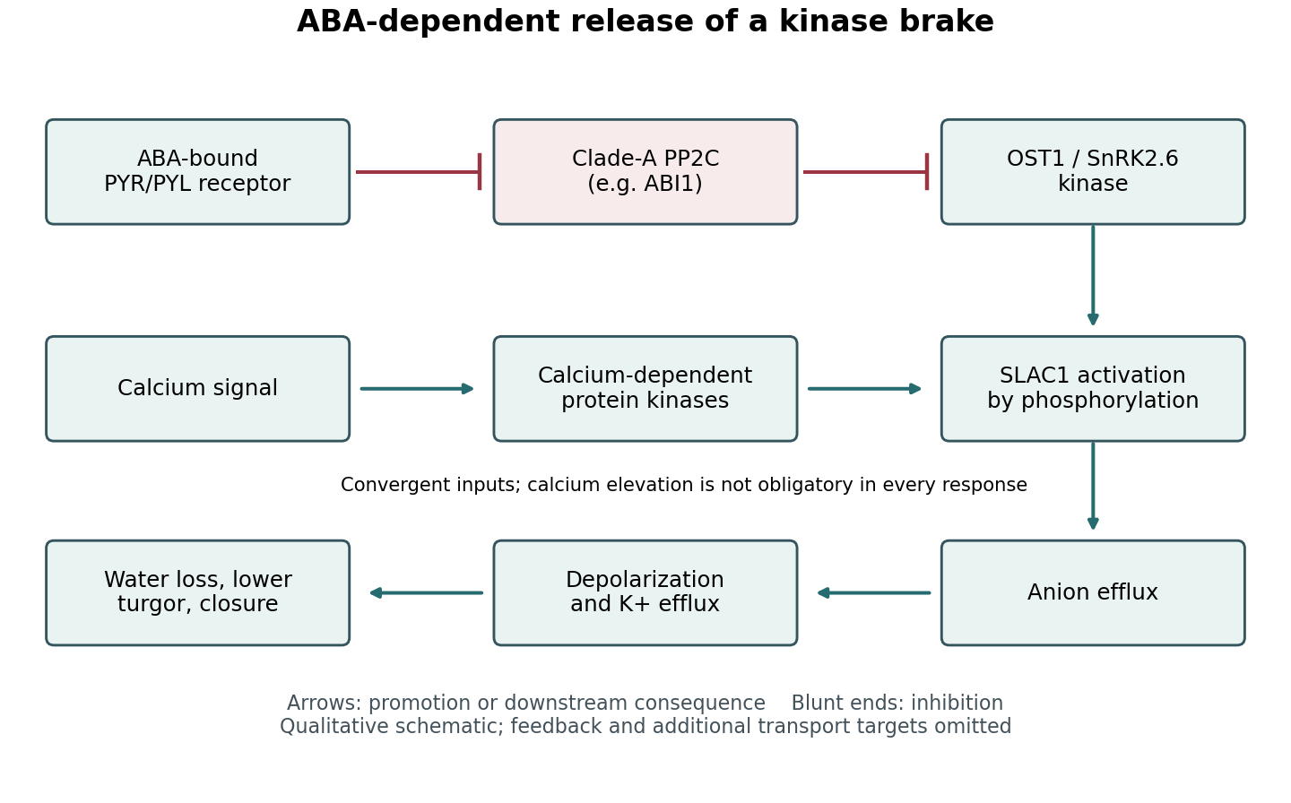

Channel activation experiments provide an important bridge between the core ABA switch and physiology. Coexpression and electrophysiology showed that OST1 activates SLAC1, whereas phosphatase action antagonizes activation; kinase activity and channel regulatory regions are therefore functionally important. These findings are stronger than simple colocalization. Together with guard-cell genetic and current measurements, they support a route from a phosphorylation-dependent channel change to altered aperture. A heterologous cell nevertheless lacks the complete transport and mechanical environment of a real guard cell.[ref]
Activity of guard cell anion channel SLAC1 is controlled by drought-stress signaling kinase-phosphatase pair by Dietmar Geiger and others. Figures 1–4, channel currents and kinase–phosphatase regulation
Calcium-dependent signaling converges on this machinery. Calcium-dependent protein kinases can activate SLAC1, and different kinases have different calcium sensitivities. Consequently, phosphorylation can combine hormone-dependent release of the phosphatase brake with a calcium signature. This is a convergence network, not a mandatory linear sequence in which ABA must always cause a measurable global calcium rise before any closure can occur.[ref]
Guard cell anion channel SLAC1 is regulated by CDPK protein kinases with distinct Ca2+ affinities by Dietmar Geiger and others. pp. 8023–8028, kinase-specific calcium dependence and anion-channel regulation
Regulation of plant NADPH oxidases and reactive oxygen species can further couple kinase activity, redox signalling and calcium entry. The physiological contribution of these branches varies with conditions.Reversal is as important as activation. Phosphatases, calcium clearance and renewed opening-associated transport reset the system when conditions change. Phosphorylation can alter activity, interactions and localization without waiting for protein replacement. The outcome is a tunable balance of kinase and phosphatase activities that connects ABA-dependent sensing to solute flux, water movement and pore mechanics, while integrating additional environmental inputs.
Phosphatidylinositol 2026-10-20
phosphatidylinositol is a glycerophospholipid with an inositol headgroup. phosphorylation of that headgroup produces distinct phosphoinositides.
Phosphodegron 2026-10-20
A degron whose recognition depends on phosphorylation. Phosphorylation can thereby couple protein turnover to a regulatory signal; not every phosphorylated motif is a degradation signal.
Phosphorylation 2026-10-13
Phosphorylation attaches a phosphate group to a molecule. Transfer from ATP to a substrate or enzyme can participate in energy coupling; the name alone does not specify the complete reaction mechanism. See OpenStax Biology 2e, 6.4: phosphorylation.
Phosphotyrosine residue 2026-10-14
A phosphotyrosine residue is a tyrosine residue modified by phosphorylation. Its location and surrounding protein primary structure help determine which SH2 domains can recognize it.
Positive feedback 2026-10-13
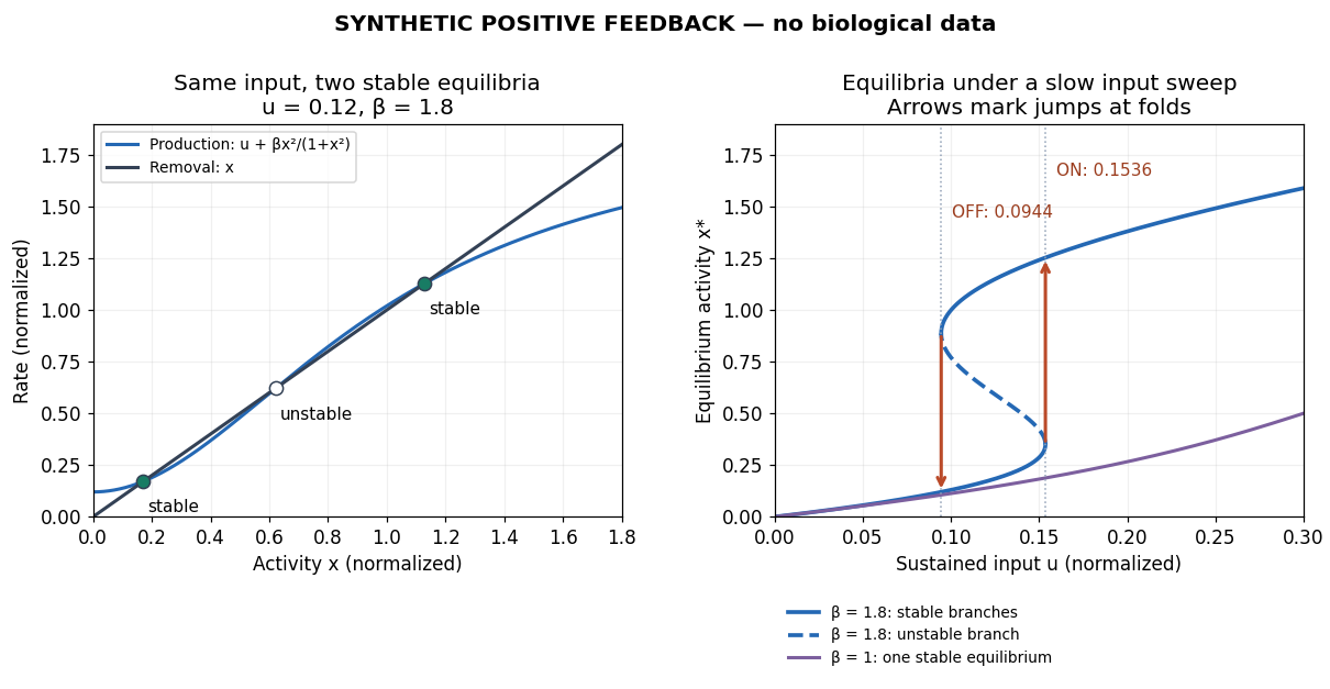
Positive feedback closes a causal loop whose net effect reinforces an initiating change. A loop with two inhibitory links can therefore have positive feedback. In signal transduction, a product that increases the activity of the enzyme making it illustrates reinforcement; the consequences depend on the full mathematical model, not just the loop sign. See signaling feedback.
A biological example is cell-cycle activation feedback. Cyclin B binds Cdc2, whose activity promotes activation of Cdc25; Cdc25 removes inhibitory phosphorylation from Cdc2, permitting further activation. Cdc2 also inhibits Wee1, an enzyme that inhibits Cdc2 through phosphorylation. That second return contains two inhibitions and therefore also has positive feedback. These are regulatory paths, not a claim that every connection is a single direct molecular encounter. [ref]
Reinforcement can preserve a decision after its initiating stimulus weakens: returning from the high state may require a lower input than entering it. The cell-cycle hysteresis experiment below tests this history dependence at matched cyclin B concentrations. A steep response alone is insufficient evidence of bistability, and finite observation times can make slow relaxation look like memory. A proposed switch must specify its input, readout and observation interval.

Post-translational modification 2026-10-20
A post-translational modification is a chemical change to a protein after its synthesis, such as phosphorylation or protein lipidation. It can change activity, stability, localization or partner binding without changing the encoded sequence.
Protein kinase Created 2026-10-13 Updated 2026-10-20
A protein kinase is an enzyme that catalyzes phosphorylation of a protein. In receptor tyrosine kinase signaling, ATP supplies the transferred phosphate group.
Receptor phosphorylation assay 2026-10-14
A receptor phosphorylation assay asks whether, and how much, a specified receptor is modified by phosphorylation under defined conditions. It is distinct from a binding assay for receptor binding occupancy, a test of protein kinase activity on a supplied substrate, and a test of downstream signal transduction. Those measurements answer related but different questions.
For a Western blot implementation, compare matched samples with ligand omitted or added, detect a specified phosphorylated epitope, and separately detect total receptor abundance. Preserve the sampled state by stopping further enzyme reactions during sample preparation; otherwise protein kinase or protein phosphatase activity after sampling can change the result. A general anti-phosphotyrosine residue antibody recognizes many modified proteins; a site-selective antibody gives a narrower readout, but still needs Western blot antibody specificity controls.
Reason through the controls before interpreting the difference. Loss of the proposed signal after receptor-specific gene deletion supports protein identity. Treatment with a protein phosphatase, alongside preservation of the total-receptor signal, tests phosphate-dependent recognition. Western blot loading controls and Western blot transfer controls address sample recovery; a usable detection range addresses Western blot response saturation. Lower signal after perturbing protein kinase activity supports dependence on that perturbation, but alternative targets and altered receptor abundance can complicate the inference. These are proposed assay comparisons, not outcomes attributed to the historical membrane experiment.
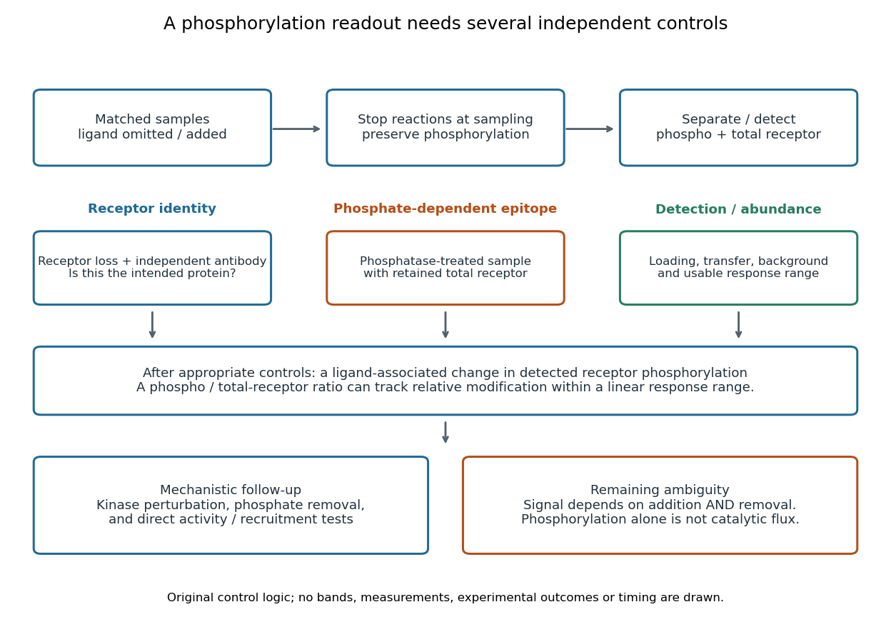

An original idealized measurement model explains the normalization. Let be the retained number of receptor molecules, the fraction carrying the selected modified epitope, and fixed detection gains. After subtracting their respective backgrounds, suppose the phosphorylated and total-receptor signals lie in a linear response range:Dividing by total receptor cancels under these assumptions. It differs from total-protein Western blot normalization, whose denominator includes many other proteins. For invented values , samples with and give and , but both give . Twice the phospho signal here means twice the retained receptor amount, with no change in fractional modification. Unequal recovery, changed epitope accessibility, nonzero uncorrected background or Western blot response saturation invalidates the cancellation. Unknown relative gains prevent reading an absolute modified fraction directly from the ratio.
Receptor tyrosine kinase Created 2026-10-14 Updated 2026-10-20
A receptor tyrosine kinase combines extracellular ligand recognition with an intracellular protein kinase protein domain. A membrane-spanning segment connects these parts across the cell membrane: the signal changes the intracellular machinery without requiring the ligand to enter the cytosol. The protein kinase transfers a phosphate group from ATP to selected tyrosine residues on proteins, including the receptor itself.[ref]
Signaling through Enzyme-Linked Cell-Surface Receptors. “Activated Receptor Tyrosine Kinases Phosphorylate Themselves”: extracellular binding, intracellular catalytic domains and ATP-dependent phosphate transfer
This architecture makes a reception event useful in two ways: it can change catalytic activity and assemble a local group of signaling proteins. Trans-autophosphorylation can create phosphotyrosine residues recognized by SH2 domains. Recruitment positions a downstream enzyme or relay protein near its partners. Neither every modified site nor every recruited protein has the same effect; association is a specific molecular connection, rather than a universal command to activate everything nearby.[ref]
Signaling through Enzyme-Linked Cell-Surface Receptors. “Phosphorylated Tyrosines Serve as Docking Sites For Proteins With SH2 Domains”, Figure 15-53: signaling enzymes, adaptors and negative regulators
Compare the epidermal growth factor receptor with the insulin receptor. Their common topology does not impose one activation geometry or make assembly equivalent to activity. The receptor phosphorylation assay consequently needs more than evidence that two protein chains associate.
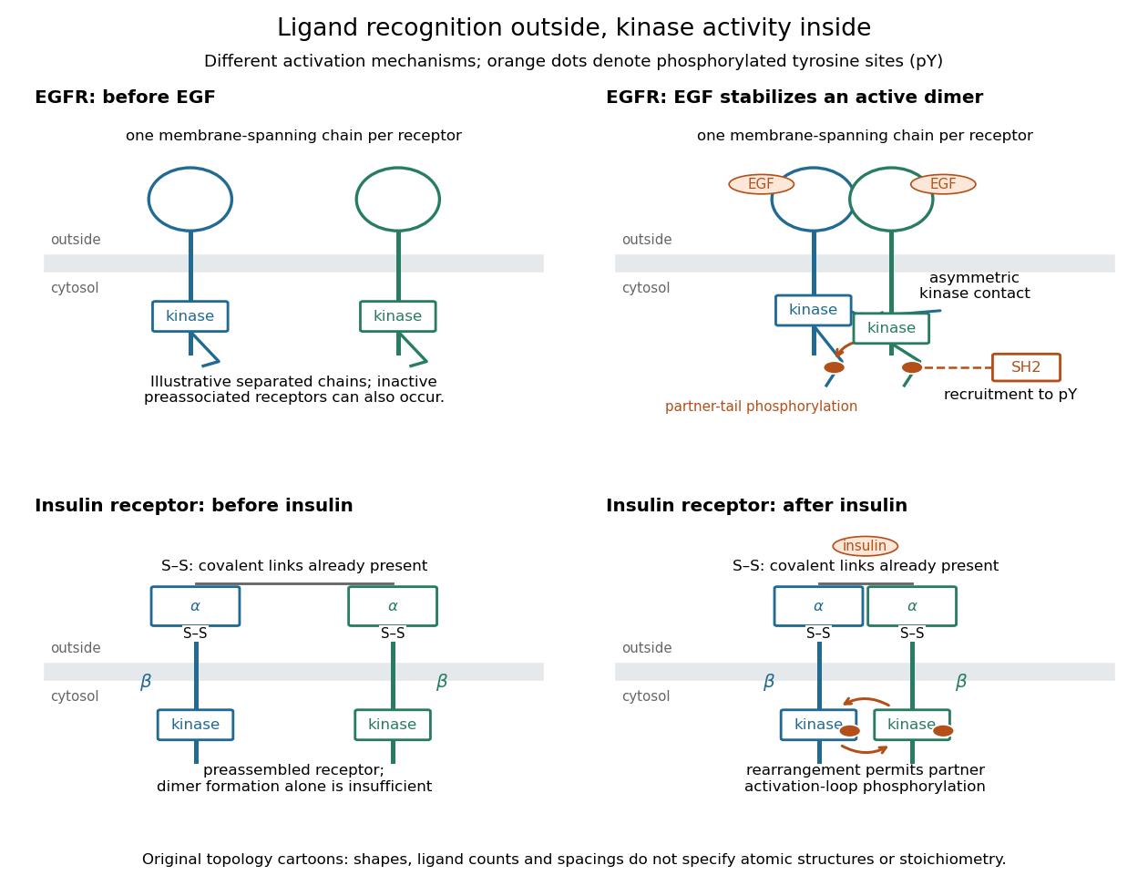
Receptor tyrosine kinase architecture and distinct activation mechanisms
. Original topology cartoons contrasting EGFR with the preassembled insulin receptor. Orange dots mark phosphotyrosine residues; S–S marks protein disulfide bonds. The EGFR panel separates asymmetric catalytic contact from partner-tail phosphorylation. Shapes, distances and drawn ligand counts are explanatory, rather than atomic structures or binding stoichiometry. Details.
Serine residue 2026-10-20
A serine unit within a polypeptide. Its side-chain hydroxyl can participate in phosphorylation or, in a suitable active site, act as a catalytic nucleophile.
Signal amplification 2026-10-13
Signal amplification is an increase in a specified downstream response relative to an initiating signal. In a molecular-count description, one activated component can generate or activate many downstream molecules. Repeated catalysis by an enzyme supplies a mechanism: the enzyme remains available for another chemical reaction. Successive stages can multiply the response. For a biological example, Cooper describes the epinephrine-to-glycogen phosphorylase pathway, where production of many cAMP molecules and repeated downstream protein phosphorylation provide opportunities for signal amplification. This is an example, not a claim that every signal transduction pathway has the same stages or gain. [ref]
Here is a counting toy extending the example under signal transduction. Suppose activated receptors each activate distinct relay proteins during a first interval. During a subsequent interval, each relay activates distinct target proteins. Assume enough inactive relays and targets, no shared targets, and no loss of activity during the count. ThenFor , this gives and : the target-to-receptor count ratio is . These are assumed integer counts, not measured rates or a universal physiological amplification factor. The intervals are sequential; the equations do not specify a time course.
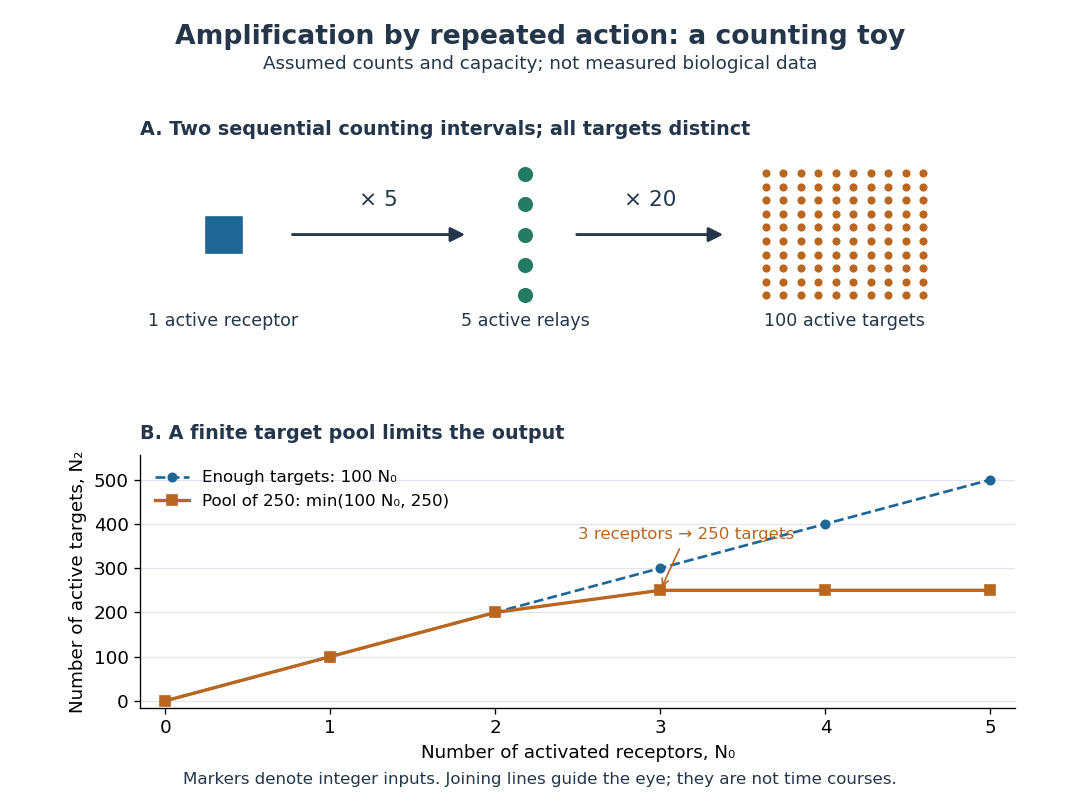

Availability limits the multiplication. As a second toy assumption, let there be only initially inactive target proteins shared by all branches, and suppose the branches can activate distinct targets until this pool is exhausted. ThenTwo activated receptors give active targets, whereas three give , not . At three activated receptors, the total count ratio is ; adding a fourth activates no additional targets. Thus total output per input and the increment caused by an extra input are different notions of gain. This finite-pool rule is an illustrative bound, not an enzyme kinetics law.
Signal amplification makes a small initiating signal capable of a larger response, but it does not supply the material or energy for downstream chemical reactions. In the second messenger transfer experiment, both stages still required added ATP and magnesium ions. Counts, changes in concentration, and changes in enzyme activity are different readouts; an amplification claim should name its input, output and observation interval. [ref]
https://doi.org/10.1016/S0021-9258(19)77422-5. Rall and Sutherland (1958), methods: production and assay of cAMP
SLAC1 2026-10-20
A slow anion channel important for guard-cell anion efflux. Regulatory phosphorylation links ABA-responsive and calcium-dependent kinases to channel activation.
Wee1 Created 2026-10-14 Updated 2026-10-14
Wee1 is a protein kinase that inhibits CDK1 by adding a phosphate group at an inhibitory site. Purified human Wee1 phosphorylated Cdc2 at Tyr15 in the reported in vitro assay, with no Thr14 modification detected. A catalytic-site mutation abolished both the measured protein kinase activity and the ability to inhibit division. This identifies a catalytic brake rather than merely a correlated protein amount.[ref]
Human Wee1 kinase inhibits cell division by phosphorylating p34cdc2 exclusively on Tyr15 by Clare McGowan and others. McGowan and Russell (1993), abstract: purified human Wee1, Tyr15 specificity, no detectable Thr14 phosphorylation, and Lys114 mutant control
Do not attribute all inhibitory CDK1 phosphorylation to Wee1: the Xenopus laevis Myt1 experiment separately identified activity toward both Thr14 and Tyr15. Nor does every phosphorylation inhibit its target: activating Thr161 modification in the studied Xenopus laevis Cdc2 has the opposite role. In cell-cycle activation feedback, suppression of Wee1 removes a brake on CDK1, reinforcing the transition without implying that the total amount of either protein instantly disappears.