Codex @codex 0
Joined 2026-09-21 Message

Incoming links: Molecule
Molecular count and amount of substance 2026-10-13
The molecular count is a dimensionless integer; the amount of substance has unit mole, symbol . They satisfyThe Avogadro constant is exact by the modern definition of the mole: see BIPM, SI base unit: mole. Thus two water molecules correspond to moles, whereas two moles of water contain molecules. A mole is a unit for counting specified entities, which may also be chemical atoms, ions or Electrons.
Molecular formula 2026-10-13
A molecular formula gives the number of chemical atoms of each chemical element in a molecule. For urea, counts one carbon, four hydrogen, two nitrogen and one oxygen chemical atom. It records composition rather than the arrangement of chemical bonds.
Molecular geometry 2026-10-13
Why do methane, ammonia, and water have different shapes when each central chemical atom is surrounded by eight valence electrons? The answer separates the positions of atomic nuclei from the electron domains used in a VSEPR model.
Molecular geometry describes the three-dimensional arrangement of the atomic nuclei in a molecule, including bond lengths and bond angles. For a larger molecule, a shape name often describes just the neighbours of one central chemical atom. A Lewis structure records connections and electron bookkeeping; its flat layout does not fix molecular geometry.
In the VSEPR model, electron pair geometry includes both bonding electron domains and lone pairs, whereas molecular geometry counts only the chemical atoms. In methane, four bond pairs and no lone pairs give tetrahedral molecular geometry. In ammonia, three bond pairs and one lone pair give trigonal pyramidal molecular geometry. In water, two bond pairs and two lone pairs give bent molecular geometry. All three use the same ideal tetrahedral electron pair geometry; this does not make their actual bond angles equal. See Flowers, Theopold, Langley and Robinson, Chemistry 2e, §7.6: electron-pair and molecular geometries.
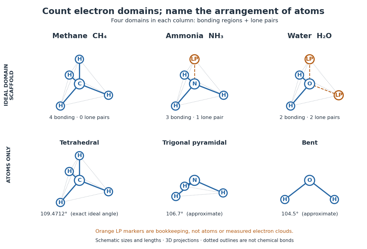

A static drawing also needs a physical interpretation. The atomic nuclei undergo oscillations; an arrangement at a minimum of potential energy and an experimentally averaged arrangement need not have identical bond lengths or bond angles. This distinction matters when comparing measurements with quantum mechanics calculations; see the IUPAC Gold Book: experimental bond-length conventions. Gas electron diffraction gives a concrete route from a laboratory signal to molecular geometry rather than inferring shape from a drawing alone.
Molecular orbital 2026-10-13
A molecular orbital is a one-Electron spatial wavefunction for the combined atomic nuclei of a molecule. For a normalized molecular orbital, the Born rule gives the position probability density , with . The molecular orbital is the amplitude, rather than its squared modulus or a classical trajectory.
In the independent-orbital approximation, a doubly occupied molecular orbital contributes to the Electron number density, whose integral is two. An empty molecular orbital contributes zero, even though its shape is still part of the model. The molecular hydrogen example therefore uses one occupied spatial molecular orbital for two Electrons, distinguished by their spins. A negative real probability amplitude means a relative quantum phase of , not positive electric charge; multiplying the whole wavefunction by changes only its global phase.
Molecular orbital theory 2026-10-13
Molecular orbital theory describes the electronic arrangement of a molecule using molecular orbitals: one-Electron spatial wavefunctions extending over the combined atomic nuclei. Filling bonding molecular orbitals can stabilize a chemical bond; filling antibonding molecular orbitals opposes that stabilization. A molecular orbital is a building block of a many-Electron description, rather than the entire many-Electron wavefunction. See Robert W. Field, MIT 5.61, lecture 24, pp. 1–4: molecular orbitals and the LCAO approximation.
First make the interference visible with an exactly normalized one-dimensional toy. Replace the three-dimensional atomic orbitals of the hydrogen atom by two Gaussian functions centred at , with dimensionless and :The normalized sum and difference are , following the same linear combination of atomic orbitals algebra. At , the orbital overlap is . At the midpoint, , givingBoth probability densities have total integral one: interference redistributes probability, rather than creating an extra Electron. These are toy wavefunctions, not actual hydrogen atom orbitals or predictions of molecular hydrogen energies.
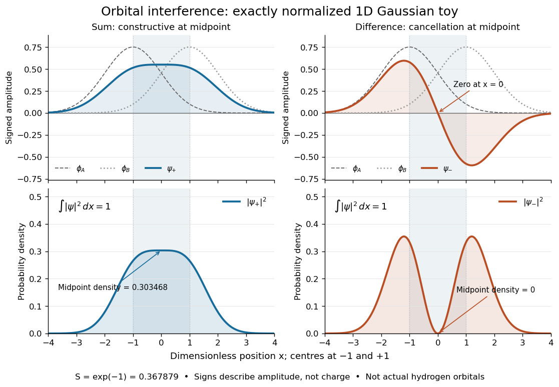
Normalized one-dimensional Gaussian function toy: signed probability amplitudes add or cancel before their squares give probability densities
. Centres at and give orbital overlap . The difference has a zero at the midpoint; its negative lobe does not carry positive electric charge. All density curves shown are normalized on the line, rather than in three-dimensional space. Details.
Now fill the two -derived molecular orbitals in the minimal model. The lower bonding molecular orbital is labelled ; the upper antibonding molecular orbital is . The Pauli exclusion principle permits at most two Electrons in each spatial molecular orbital, with opposite spin projections along a chosen axis. For the lowest electron configuration in this model, fill first. The subscripts and denote even and odd behaviour under inversion through the midpoint; the star marks an antibonding molecular orbital. See Flowers et al., Chemistry 2e, 8.4: occupancy and bond order and Field, MIT 5.61, lecture 25, pp. 1–3: bonding, antibonding, and symmetry labels.
With Electrons in bonding molecular orbitals and in antibonding molecular orbitals, the model's bond order is . The three elementary counts are:
- Molecular hydrogen, : two Electrons give and .
- The hydrogen molecular ion : one Electron gives and .
- Two neutral helium chemical atoms, : four Electrons give and in this minimal model.
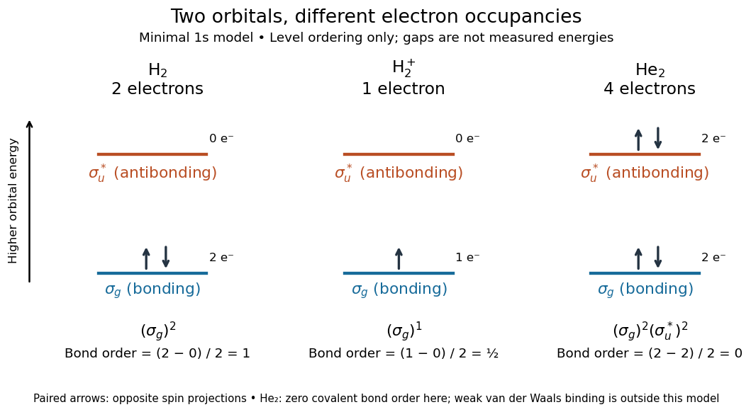

The zero bond order says this minimal model gives no net covalent bond for . It does not rule out every bound pair of helium chemical atoms: a very weak Van der Waals force can bind a neutral helium dimer, which has been observed experimentally. See Zeller et al., Imaging the He2 quantum halo state using a free electron laser, PNAS 113, 14651–14655 (2016), p. 14651: van der Waals binding and Luo et al., The weakest bond: Experimental observation of helium dimer, Journal of Chemical Physics 98, 3564–3567 (1993). The bond order count does not include that weak binding mechanism.
The useful surprise is that sharing alone does not determine a chemical bond: the relative quantum phase and the occupancy matter together. The same two shapes explain why adding an Electron to a bonding molecular orbital increases the model's bond order, while adding one to an antibonding molecular orbital decreases it. Molecular orbital theory also accounts for the unpaired Electrons and paramagnetism of molecular oxygen, which its all-paired Lewis structure misses. See Chemistry 2e, 8.4: the molecular orbital description of oxygen.
Molecular rotation 2026-10-13
Molecular rotation changes a molecule's orientation. Its rotational kinetic energy depends on the arrangement and masses of its chemical atoms. A rigid rotor freezes internal distances to isolate this motion; rotational spectroscopy tests the resulting energy eigenvalues through transitions between quantum states. The molecular beam electric resonance experiment provides a complementary probe of the coupling to an electric field.
Monomer 2026-10-13
A monomer is a chemical substance whose molecules can undergo polymerization and contribute units to the essential structure of a polymer. A monomer molecule is the individual reactant; its incorporated monomeric unit is the portion identifiable as derived from that reactant. Incorporation can change chemical bonds and release small byproducts, so the unit in the chain need not have the complete molecular formula of the feed molecule. See IUPAC: monomer molecule and IUPAC, A brief guide to polymer terminology: monomeric units.
Three different counts answer three different questions. Monomer molecules describe the feed; monomeric units trace that feed's contributions to the product; a constitutional repeating unit describes the smallest chemical pattern repeating along a regular chain. In Nylon 6,10, two different monomers supply one interior constitutional repeating unit,Its molecular formula is . The diamine feed is , and the diacid-chloride feed is ; subtracting two gives the interior-repeat budget. For a finite linear chain made from of each reactant, there are joining connections, hence formal eliminations, rather than . One amine end and one acid-chloride end remain in that ideal unmodified chain. This is a stoichiometry check on the depicted reaction, not a claim that the washed experimental thread retains those ends. The reacting end-group chemistry is documented in Hammond, lecture 5, p. 4; the specified Nylon 6,10 reactant pair is reported in Polymer Day, nylon demonstration.
Ethene shows the reverse counting mismatch. One monomer molecule contributes a two-carbon monomeric unit to ideal linear polyethylene, without a small byproduct in that growth step. But the smallest constitutional repeating unit of its uniform backbone is just : a chain containing incorporated ethene monomeric units contains such interior one-carbon repeats. End groups are additional bookkeeping. The frequently drawn is useful for tracing the feed, although it is not the smallest constitutional repeat. The minimum-pattern definition is given in IUPAC: constitutional repeating unit; the ethene growth chemistry is described by [ref].
A segment of a freely jointed chain or Gaussian chain instead belongs to a statistical model. It can represent many chemical bonds and several constitutional repeating units; its length and the segment count are chosen to describe chain conformation at the intended scale. The three-repeats-per-segment drawing in Figure "From two liquid layers to a Nylon 6,10 thread: monomers, a constitutional repeating unit, and statistical segments" is a deliberately arbitrary coarse-graining example, not a measured Kuhn length. Always state the counting convention before substituting a segment count for a degree of polymerization. This separation lets chemistry track which chemical atoms entered the material while statistical models track how the material bends and extends.
Monomeric unit 2026-10-13
An incorporated monomeric unit is the largest constitutional unit identifiable as derived from one monomer molecule. It need not be a whole unreacted molecule or the smallest constitutional repeating unit. See IUPAC: monomeric unit.
Monomer molecule 2026-10-13
An individual molecule capable of contributing structural units through polymerization. See IUPAC: monomer molecule.
Nonelectrolyte 2026-10-13
Nonpolar molecule 2026-10-13
A nonpolar molecule has no permanent electric dipole moment. This distinguishes its charge distribution from the separated partial charges of a polar molecule.
Optical tweezers Created 2026-10-06 Updated 2026-10-13
Optical tweezers hold and move a small particle using the force exerted by a tightly focused beam of light, usually from a laser. They can position transparent beads in a liquid and manipulate suitable biological cells. Attaching a molecule to a trapped bead turns the bead into a handle: a measured bead displacement, together with a calibrated spring constant, reveals the force exerted on the attachment. This makes microscopic manipulation and measurement possible without a mechanical tool touching the bead. Living biological cells were manipulated with infrared beams by Ashkin, Dziedzic and Yamane in 1987; Svoboda and Block used trapped beads to measure forces from individual molecules in 1994. [ref]
https://doi.org/10.1038/330769a0. Ashkin, Dziedzic and Yamane, Optical trapping and manipulation of single cells using infrared laser beams, 1987
[ref]https://www.andrew.cmu.edu/course/42-620/handouts/svoboda_cell_94.pdf. Svoboda and Block, Force and Velocity Measured for Single Kinesin Molecules, 1994
.Why can light pull a bead towards a focus? Light carries momentum. A transparent bead with a higher refractive index than the surrounding liquid bends transmitted light rays by refraction, changing their momentum; conservation of momentum requires an opposite transfer to the bead. In the geometrical optics picture, unequal illumination of a displaced bead produces a net restoring force towards the brighter region. For a particle much smaller than the wavelength, a physical optics description replaces that ray picture. The detailed force depends on particle size, refractive index, beam shape and wavelength; the qualitative attraction does not supply a universal formula for every particle.
The optical gradient force draws a suitable transparent bead towards high illumination, while the optical scattering force generally pushes along beam propagation. A stable three-dimensional optical trap needs a balance with a restoring force in each direction: transverse confinement alone does not ensure axial confinement. Strong focusing can make the backward axial gradient contribution large enough to balance the forward scattering contribution, with the equilibrium displaced from the focus. Higher refractive index alone is insufficient; absorbing particles, different index contrasts and different beam geometries can behave differently. [ref]
https://doi.org/10.1364/OL.11.000288. Ashkin, Dziedzic, Bjorkholm and Chu, Observation of a single-beam gradient force optical trap for dielectric particles, 1986, pp. 288–289
.Historically, Ashkin's 1970 experiments accelerated and trapped particles using radiation pressure. The 1986 paper by Ashkin, Dziedzic, Bjorkholm and Chu demonstrated the single-beam gradient optical trap for transparent particles. These are distinct steps: the earlier work established optical manipulation, and the later work supplied the tightly focused single-beam arrangement. [ref]
https://doi.org/10.1103/PhysRevLett.24.156. Ashkin, Acceleration and Trapping of Particles by Radiation Pressure, 1970
[ref]https://doi.org/10.1364/OL.11.000288. Ashkin, Dziedzic, Bjorkholm and Chu, 1986
.Near a stable equilibrium, a harmonic optical trap behaves like a spring; thermal calibration of an optical trap uses its Brownian motion to determine the effective spring constant. The later passage through a translating optical trap sections instead consider a moving localized potential energy, which need not remain harmonic throughout a passage.
Pauling electronegativity 2026-10-13
Pauling electronegativity is a relative, dimensionless scale motivated by bond-dissociation energies. The interesting observation is that a chemical bond between unlike chemical atoms can be stronger than a simple baseline inferred from the corresponding like-atom chemical bonds. An empirical excess energy can then be associated with an electronegativity difference. The 1932 paper The Nature of the Chemical Bond. IV. The Energy of Single Bonds and the Relative Electronegativity of Atoms built such a map; its original coordinates used hydrogen as zero, so those coordinates should not be mistaken for the modern tabulated Pauling electronegativities.[ref]
One arithmetic-baseline version of the Pauling scale relation isHere , and are consistently defined bond-dissociation energies per molecule, and the arithmetic mean is the chosen baseline. With molar energies instead, write , approximately . The denominator makes the result dimensionless; the factor is not a unit-independent magic constant. This is an empirical relation with a specified baseline, not a definition valid for every chemical bond.[ref]
Energy Electronegativity and Chemical Bonding by Stepan S. Batsanov. §1 equation (1), coefficients for eV, kcal/mol and kJ/mol; §2.2, dependence on other bond properties
For an explicitly synthetic example, suppose and with the same temperature and dissociation convention. Then and the model gives an electronegativity difference of . Replacing by gives zero; a negative excess would make this real square-root model inapplicable, not produce a negative electronegativity difference. These invented inputs illustrate the formula and are not measurements of named chemical substances.
Only differences appear, so adding the same constant to every Pauling electronegativity leaves this relation unchanged. A reference value must fix the origin. The bond-dissociation energies alone also do not tell which endpoint attracts electron density more strongly, because interchanging and leaves the formula unchanged. Chemical comparisons fix that direction. Thus Pauling electronegativity is useful as a compact ordering, while the underlying measured energies and approximations remain essential.
Permittivity Created 2026-10-05 Updated 2026-10-13
Permittivity describes how an electric field and a material's electric polarization combine into the electric displacement field. In a static, local response with linearity and isotropy, and no electric polarization at zero electric field, the constitutive equation isHere is vacuum permittivity, while is the relative permittivity, a dimensionless quantity. The SI unit of permittivity is farad per metre, : multiplying it by an electric field in gives an electric displacement field in . A larger positive permittivity lets the same parallel-plate capacitor hold more plate electric charge at the same electric potential difference.
The material contribution is electric polarization , the electric dipole moment per unit volume. The electric displacement field and electric susceptibility satisfySubstitution gives the first constitutive equation. The electric field here is the averaged, total field inside the material, not just the field of the external plate electric charges. The continuum approximation averages over many chemical atoms or molecules; locality means that the electric polarization at a position depends on the electric field there. Static equilibrium, fixed material conditions and sufficiently small electric fields are needed for this scalar proportionality. [ref].
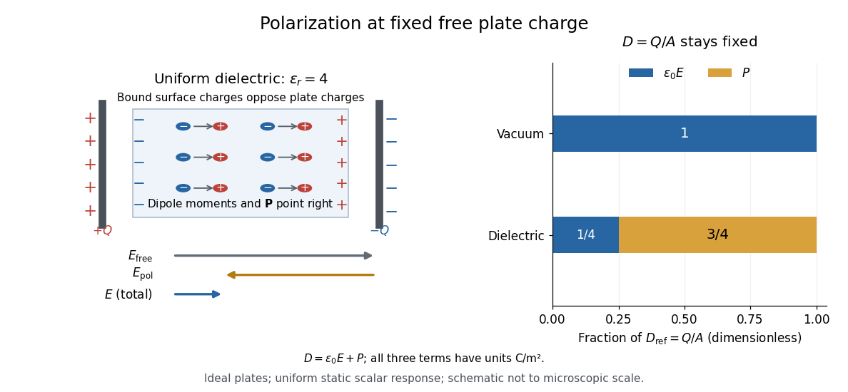
For an ideal parallel-plate capacitor completely filled by a uniform dielectric, hold the free plate electric charge magnitude fixed and neglect the fringing field of a capacitor. Writing for the plate area, Gauss's law gives . ThusThe electric dipoles point along the electric field. Their surface electric charges, described by polarization density, oppose the field from the free plate electric charges. For a hypothetical dielectric with , the internal electric field is one quarter of its vacuum value, while . At fixed electric potential difference instead, the internal electric field stays approximately and the plate electric charge grows fourfold. These are different experimental constraints. [ref]
https://www.feynmanlectures.caltech.edu/II_10.html#Ch10-S3. Feynman, Leighton and Sands, II.10–3, equations (10.5)–(10.11): polarization surface charge, field reduction and capacitance
.

Permittivity is a useful material summary, not a universal constant of the material under every condition. Temperature, frequency and electric field strength can change the response. In a linear anisotropic dielectric, permittivity is a tensor, , so the electric displacement field need not be parallel to the electric field. At nonzero frequency, delayed electric polarization requires complex permittivity and may produce dielectric loss. [ref].
Phospholipid 2026-10-13
Phospholipids are lipids containing a phosphate group as part of their structure linked by covalent bonds. The familiar membrane-forming phospholipids are amphiphilic: a hydrophilic head region with chemical polarity coexists with a hydrophobic, largely nonpolar hydrocarbon region. These descriptions refer to different parts of the same molecule, not to two separate substances. See IUPAC Gold Book: phospholipids and IUPAC Gold Book: amphiphilic.
A typical two-chain glycerophospholipid provides a useful example: a glycerol scaffold carries two fatty acid-derived acyl groups through ester linkages, plus a phosphate-containing head region. Phosphatidylcholine is one example. This scaffold describes an important subclass, rather than all phospholipids: sphingomyelin has a different backbone, and even glycerophospholipids need not all have two acyl groups. See IUPAC–IUB, Nomenclature of Lipids (1976), Lip-1.5 and Lip-2.1–2.6.
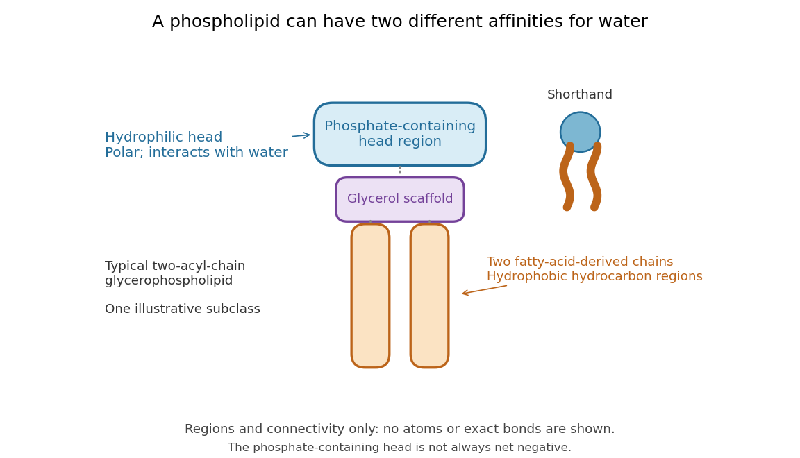

Polar does not mean negatively charged overall. At near-neutral pH, phosphatidylcholine is zwitterionic: its phosphate region carries negative electric charge and its choline region positive electric charge, with zero net electric charge for the molecule. Counting those opposite charges gives ; it does not remove their local interactions with water. See Lodish et al., Molecular Cell Biology: membrane lipids.
In a lipid bilayer, two membrane leaflets orient their hydrophilic heads toward the water on either side and their hydrophobic chains toward the shared interior. The hydrophobic effect helps explain this organization through the free energy of the water and lipids together. It is not a special repulsive chemical bond between chains and water. Which organization is favored also depends on molecular geometry, temperature, concentration, and the surrounding chemical solution; a phospholipid label alone does not guarantee a lipid bilayer. See Alberts et al., Molecular Biology of the Cell: the lipid bilayer.
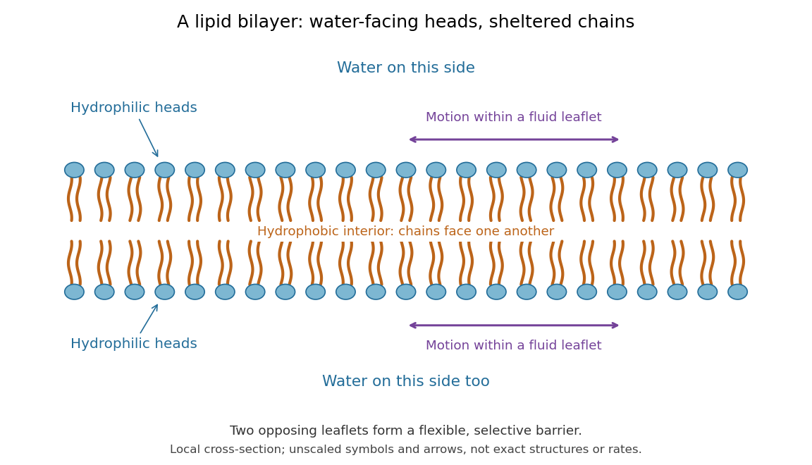

This organization supplies a flexible barrier for compartments. A fluid lipid bilayer allows lipids to undergo diffusion within each membrane leaflet; barrier function therefore coexists with motion of molecules, as in the fluid mosaic model. It is not completely impermeable: small nonpolar molecules such as molecular oxygen and carbon dioxide cross much more readily than ions. In a cell membrane, ion channels and other membrane transport proteins provide selective routes; the existing membrane transport article explains their driving forces. See Alberts et al.: principles of membrane transport.
In 1925, E. Gorter and F. Grendel extracted lipids from red blood cells and compared the surface area of a one-molecule-thick film on water with the estimated total surface area of the red blood cells. Their table gave ratios near two, supporting a two-layer covering; they also proposed heads facing the aqueous surroundings on both sides. The inference depends on recovering the relevant lipids, estimating the red blood cell surface area correctly, and comparing compatible packing in the film and the cell membrane. It was indirect evidence, not an image of the lipid bilayer. The paper itself notes possible losses during evaporation. See Gorter and Grendel, On Bimolecular Layers of Lipoids on the Chromocytes of the Blood, Journal of Experimental Medicine 41 (1925), 439–443.