Raman scattering is inelastic scattering of light in which the outgoing photon has a different frequency because matter gains or loses internal energy. Molecular vibration is a particularly useful recipient: a visible photon can reveal an internal energy spacing normally associated with infrared frequencies. Rayleigh scattering supplies the much stronger, approximately unchanged-frequency reference. The distinction concerns the transferred energy, rather than merely a change of direction or the colour of the illuminating beam.
The elementary bookkeeping is conservation of energy:Here is the Planck constant, and are the incident and scattered photon frequencies, and are initial and final internal molecular energies. This expression neglects translational recoil and Doppler broadening; those can matter in sufficiently precise gas measurements. A positive internal energy change gives Stokes Raman scattering, and a negative one gives Anti-Stokes Raman scattering. The energy difference determines a possible line position, but it does not by itself establish that a transition is optically allowed or predict its strength.
The spontaneous molecular process developed here has a connection to the Raman transition used in atomic measurement methods: two photon interactions transfer an internal energy difference. Coherently driving that process with two applied light fields is a different experimental regime from collecting weak spontaneous Raman scattering beside an illuminating beam.
Chandrasekhara Venkata Raman and Kariamanikkam Srinivasa Krishnan jointly reported the effect in 1928. Their letter describes light from the Sun concentrated by an -aperture, -focal length telescope objective and a second optical lens onto a liquid purified by repeated vacuum distillation. With blue-violet and yellow-green optical filters together in the incident beam, the visible track disappeared; moving the yellow-green optical filter between liquid and observer restored it. The complementary arrangement rejected unchanged-colour scattering of light while admitting newly generated light. Persistence after purification and strong wave polarization in many liquids argued against ordinary fluorescent contamination.[ref]
In his 1930 lecture, Chandrasekhara Venkata Raman describes a cobalt-glass optical filter selecting indigo-violet light from the Sun, with a separated blue-green band in the scattered optical spectrum. Substituting a quartz mercury arc and condenser gave sharp new spectral lines. Small Hilger quartz spectrographs recorded them photographically; Kariamanikkam Srinivasa Krishnan obtained more precise records with a larger instrument and established violet-displaced lines. The optical prism disperses wavelengths for spatial readout; it does not create the Raman shift. The diagram gives functional optical paths rather than a reconstruction of a specific Hilger instrument.[ref]
The Molecular Scattering of Light by Chandrasekhara Venkata Raman. Nobel Lectures, Physics 1922–1941; Raman, The molecular scattering of light, Nobel lecture, 11 December 1930, printed pp. 271–273, spectroscopic examination and photographic recording; inspected in a reproduction of the complete lecture
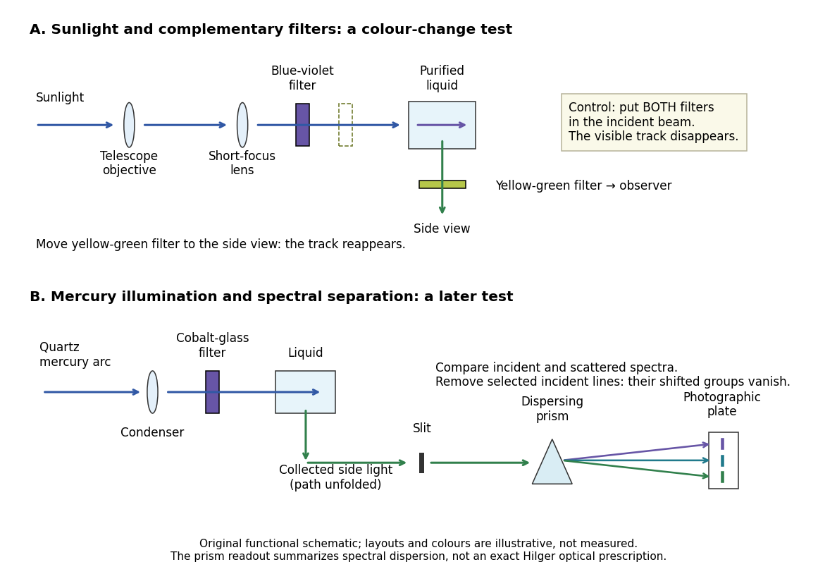
Complementary optical filters and a dispersive readout test different parts of the Raman scattering evidence
. Original functional schematic of the filter test and later spectral test. Side collection is unfolded for readability. The optical prism, spectrograph slit and photographic plate indicate dispersion and recording; the sketch supplies no historical distances, exact optical prescription or measured colour intensities. Source. Details.Their May 1928 report strengthens the comparison: filtered mercury illumination of toluene produced extra spectral lines. Removing the incident line removed its associated longer-wavelength group; a quinine filter removing the , and group removed a different shifted group while preserving the one associated with . These are actual selective-excitation controls, rather than inferred colours.[ref]
The Optical Analogue of the Compton Effect by Chandrasekhara Venkata Raman and Kariamanikkam Srinivasa Krishnan. Raman and Krishnan, The Optical Analogue of the Compton Effect, Nature 121, p. 711, Fig. 1 description and removal of individual incident-line groups
The inference has limits. A complementary-filter test alone establishes newly admitted wavelengths, not their precise spacing or a unique microscopic mechanism. Fluorescence can also survive purification when intrinsic to the liquid, and fluorescence can be polarized. Useful distinctions therefore combine resolved spectral lines, excitation dependence, wave polarization and sample preparation. A proposed modern extension is to change a single incident frequency: a molecular Raman shift should follow the same internal energy gap, whereas ordinary relaxed molecular fluorescence usually retains approximately its emission-band positions. This is a diagnostic under stated conditions, not a universal theorem about every fluorescent material or resonant scattering process. Raman scattering need not involve a populated, relaxed electronic excited state as ordinary molecular fluorescence does; neither a redder photon nor the word Stokes identifies the mechanism by itself.
The 1930 Nobel Prize in Physics went to Chandrasekhara Venkata Raman alone for his work on scattering of light and the discovery of the effect. That award allocation should be distinguished from the joint experimental report with Kariamanikkam Srinivasa Krishnan.[ref]
The Nobel Prize in Physics 1930 by Nobel Foundation. Nobel Foundation prize record: sole laureate and award motivation, Physics 1930
The signed Raman shift subtracts scattered from incident spectroscopic wavenumber:Here is the speed of light, and both wavelengths are in vacuum. Positive shifts denote Stokes Raman scattering; negative shifts denote Anti-Stokes Raman scattering. Some plots reverse the horizontal direction or report separate unsigned magnitudes, so check the convention. Spectroscopic wavenumber has no factor, unlike the common angular wavenumber . A shift of means , not a frequency of .
For a one-quantum change of molecular vibration with ordinary frequency , the magnitude is . This makes Raman spectroscopy attractive: the same molecular energy spacing can be read using different visible excitation wavelengths. Equal and opposite Raman shifts are not equal and opposite changes measured in nanometres, because wavelength is the reciprocal of spectroscopic wavenumber.
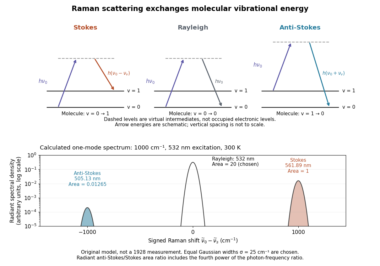
In Stokes Raman scattering, matter gains internal energy and the photon loses the same amount. For a quantum harmonic oscillator representing one molecular vibration, with . The transition gives , requiring for an outgoing photon. Even a molecule initially in its vibrational ground state can undergo this process. The shared name does not turn it into the absorption/emission-band Stokes shift used for fluorescence.
In Anti-Stokes Raman scattering, matter loses internal energy and the outgoing photon gains it. The harmonic transition requires , so an initially excited vibrational population must be available. At low temperature, high-frequency vibrational excitations are scarce and the corresponding anti-Stokes line is weak. The extra photon energy comes from the molecule; there is no conflict with conservation of energy.
The simplest off-resonant explanation is a vibrating polarizability. Let be a small normal mode coordinate of molecular vibration, and use one scalar component of polarizability, . For incident electric field , the induced electric dipole moment isThe product-to-sum formula produces the two displaced frequencies. A nonzero derivative gives vibrational Raman scattering in this linear model. Real polarizability is generally a tensor, and the selected components depend on incident and observed wave polarization. This condition differs from vibrational infrared activity, which requires a changing molecular electric dipole moment without an applied optical electric field. A vibration can therefore appear in one measurement and be absent in the other.
This classical modulation argument explains line locations but supplies equal sideband wave amplitudes; it does not explain their thermal asymmetry or the nonzero Stokes Raman scattering from a vibrational ground state. Those require the quantum harmonic oscillator population and transition strengths. The far-off-resonance drawing uses dashed virtual intermediate levels: they represent terms in a scattering probability amplitude, not a long-lived occupied electronic energy eigenstate. Near an electronic resonance, a more detailed treatment of the intermediate response is needed.
Assume one harmonic normal mode at thermal equilibrium, weak spontaneous excitation far from electronic resonances, the same collection solid angle and wave polarization, and matched response after instrumental correction. The Boltzmann distribution givesHere is the Boltzmann constant, is temperature, and is the probability of vibrational quantum number . Thus . Linear coupling to has squared oscillator matrix elements proportional to for Stokes Raman scattering and for Anti-Stokes Raman scattering. Summing over initial levels gives strengths proportional to and respectively; their ratio is , even when levels above are occupied.
Let and denote line-integrated radiant intensities, meaning power per collection solid angle. The off-resonant electric dipole radiation law adds the fourth power of emitted frequency:The photon rate divides each line's power by its own . Consequently, the ideal response-corrected anti-Stokes/Stokes count-rate ratio has the third power of the same frequency ratio. Peak heights alone do not give line-integrated radiant intensities if linewidths differ. Uncorrected detector counts can also include unequal detector response and unequal optical filter transmission. Resonant enhancement, overlapping spectral lines, nonthermal populations and local optical heating can invalidate a temperature inferred from this simple formula.
Choose a vibrational Raman shift magnitude , excitation vacuum wavelength and temperature . These are original illustrative inputs, not a measured 1928 optical spectrum or a claim about a particular liquid. They giveTake reciprocal lengths in consistent units before converting to nanometres. At , and . The predicted line-integrated radiant intensity ratio is , whereas the ideal photon count-rate ratio is . Thus the radiometric ratio is about percent, rather than the percent obtained by using the Boltzmann factor alone.
Raman spectroscopy measures shifted scattering of light to infer internal energy spacings and the molecular response that makes them visible. In a modern illustrative arrangement, narrowband laser illumination replaces the historical arc, an optical filter rejects the strong excitation line, and a spectrograph resolves the weak shifted spectral lines. The illumination, rejection and spectral dispersion are separate jobs. Increasing rejection can conceal small Raman shifts close to the excitation line.
A proposed measurement should record the excitation wavelength, a dark signal, a blank container and the liquid, then compare calibrated line positions and integrated signals under the same collection conditions. Measure optical filter transmission and detector response before using the Thermal Raman line ratio for temperature. Repeat at lower incident power to look for heating or altered sample response. These are suggested checks, not an assertion that the 1928 apparatus implemented modern calibration procedures.
Its appeal is the conversion of internal molecular energy differences into visible spectral lines: changing the excitation can move the whole pattern while preserving characteristic Raman shifts. Combined with vibrational infrared activity, Raman vibrational activity supplies complementary information about molecular vibration. A line pattern can constrain a material's identity or environment, but overlapping bands, fluorescence backgrounds and the finite spectral resolution element prevent an unqualified identification from one peak alone.
Articles by others on the same topic



Raman scattering is an inelastic scattering process that occurs when light interacts with molecular vibrations, phonons, or other low-frequency excitations in a material. This phenomenon is named after the Indian physicist C.V. Raman, who, along with his colleague, discovered it in 1928. In simple terms, when a monochromatic light source, typically a laser, shines on a sample, most of the light is elastically scattered, meaning it retains its original energy (or wavelength).