Topics (384k) Articles (398k) Users (410) Discussions (242) Comments (383) Files (13k) New article

Relative centrifugal force is the dimensionless ratio of the local centrifugal acceleration magnitude to the conventional gravitational reference . Despite its name, it is an acceleration ratio: the actual force on a specified mass is . With angular speed and rotor radius , . If is the numerical rotational speed in revolutions per minute and the numerical rotor radius in centimetres, unit conversion givesThus rpm alone does not specify the centrifugal acceleration.[ref]
Basics: Centrifugal Force & Centrifuge Type by Eppendorf. Important definitions, Rcm vs rpm, and How to calculate relative centrifugal force: radius dependence and acceleration ratio
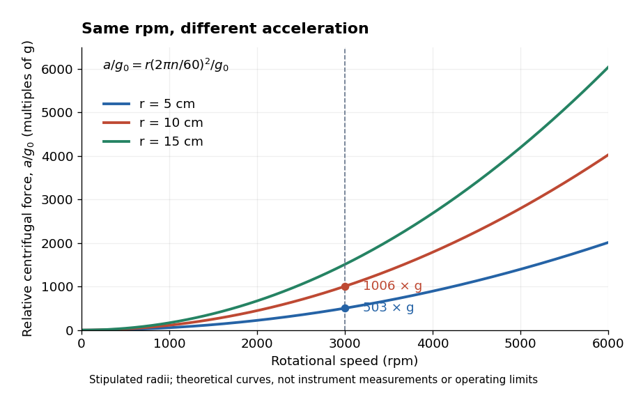
For a stipulated , rotor radii of and give approximately and times . Doubling the rotor radius doubles relative centrifugal force; doubling rpm quadruples it. Even equal relative centrifugal forces do not guarantee equal separation: travel distance, elapsed time, viscosity, sample density and particle shape still matter. These are comparisons of a mathematical model, not instructions for running an instrument.
The rotor radius is the perpendicular distance from the rotation axis to a specified sample position, not the tube's length. A filled tube spans a range . Consequently, one rotational speed produces different local centrifugal accelerations along that range. A reported relative centrifugal force needs a stated reference position, such as the outermost sample radius.
Let be the perpendicular distance from the force centre to the tangent of a regular planar orbit. Conserved physical angular momentum has magnitude , while conserved energy is . Eliminating speed gives the displayed equation when . For a differentiable polar curve, , connecting the geometric tangent distance to the radial energy equation.
Peptidoglycan is a mesh of linked sugar chains cross-linked by short chains of amino acids in the cell walls of most bacteria. Its absence from the cell walls of archaea helps distinguish envelope chemistry without confusing either cell wall with a cell membrane. See OpenStax Microbiology, 3.3: cell walls and peptidoglycan.
A protoplast is the living contents of a biological cell bounded by its cell membrane after removal of the cell wall. Removing the cell wall need not immediately destroy the cell membrane; stability depends on the preparation and surroundings. Lysozyme acts on susceptible bacterial peptidoglycan, not on cellulose or chitin, so it is not a universal wall-removal enzyme.
Plasmolysis is withdrawal of the membrane-bounded contents from parts of a cell wall after water loss. A sufficiently concentrated external hypertonic solution, with solutes effectively excluded by the cell membrane over the observation time, can cause this. The cell wall remains; the enclosed material contracts. Plasmolysis is therefore distinct from osmotic lysis, which breaks the cellular boundary during excessive swelling. A return to a less concentrated surrounding chemical solution can reverse plasmolysis if the biological cell remains functional.[ref]
5.2 Passive Transport. Biology 2e, §5.2, Tonicity in Living Systems and Figure 5.13: membrane withdrawal under hypertonic conditions
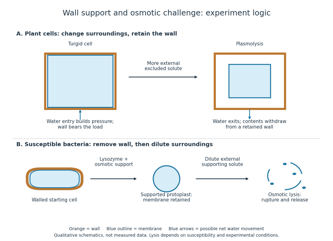
Turgor pressure is the internal hydrostatic pressure above the external pressure in a biological cell. When water enters a walled biological cell by osmosis, expansion loads the cell wall. Rising pressure can oppose further net entry while the cell membrane retains its selective permeability. Mechanical support therefore does not require the cell wall to block water.
As an original mechanical check, idealize a small flat patch of cell wall of surface area under uniform turgor pressure . Its outward force is . Doubling at fixed pressure doubles the load; equal internal and external pressures give zero net pressure load. This is a force balance, not a measured wall strength or a complete model of a curved, growing biological cell. It says nothing about which solutes cross the cell membrane.
A fungal cell wall contains structural polysaccharides including chitin and glucans. Its composition and organization vary among fungi; the diagram illustrates a composite rather than a pure chitin shell. A fungus sharing a cell wall with a plant does not imply shared wall chemistry.[ref]
24.1 Characteristics of Fungi. Biology 2e, §24.1, Cell Structure and Function: structural wall polysaccharides
A plant cell wall provides reinforcement outside the cell membrane, with cellulose as a major structural component. It is a composite rather than a solid sheet of pure cellulose. Wall construction varies with the kind and developmental state of the biological cell.
The cell nucleolus is a region inside a cell nucleus without a surrounding membrane, where much ribosomal RNA is made and processed and ribosome subunits begin to assemble with proteins. The subunits are exported to the cytoplasm; the cell nucleolus is not itself a translating ribosome. See Clark, Douglas and Choi, Biology 2e, 4.3: nucleolus and ribosomal subunits.
The nuclear envelope is the double-membrane boundary around a cell nucleus. Regulated passage connects nuclear processes to the surrounding cytoplasm: transcription produces RNA in the cell nucleus, while exported messenger RNA can undergo biological translation on ribosomes outside it. See NHGRI: nuclear membrane.
The fluid mosaic model describes a cell membrane as a fluid lipid bilayer with embedded membrane proteins. It connects a continuous barrier to a mosaic of functional components: diffusion within the membrane can coexist with selective passage across it. S. J. Singer and G. L. Nicolson presented the fluid mosaic model in Science in 1972; see Singer and Nicolson, The fluid mosaic model of the structure of cell membranes. The useful picture does not require every protein to move freely: attachments and interactions can restrict movement, as discussed in Cooper, The Cell: structure of the plasma membrane.
Membrane transport is the passage of substances across a cell membrane. For the small solutes considered here, distinguish the available route from its driving force: a lipid bilayer, ion channel, or carrier protein determines how passage can occur, while differences in chemical potential and membrane potential determine which direction is energetically downhill. A large driving force cannot make an unavailable route conduct. See Alberts et al.: principles of membrane transport.
For an ideal dilute chemical solution at uniform temperature and pressure, with the same reference molar chemical potential on both sides, the molar electrochemical potential change for transferring a solute from compartment A to B isHere are concentrations, is absolute temperature, is the molar gas constant, is signed ion charge number, is the Faraday constant, and is the electric potential difference. The natural logarithm contains a dimensionless ratio. The electrical term vanishes for an uncharged solute. More generally, use thermodynamic activities in place of the ideal concentration ratio. This combines the ideal-solution chemical potential difference with electrical energy per mole; see IUPAC Gold Book: electrochemical potential. For a biological cell, taking B inside and A outside makes the voltage term use the existing membrane potential convention.
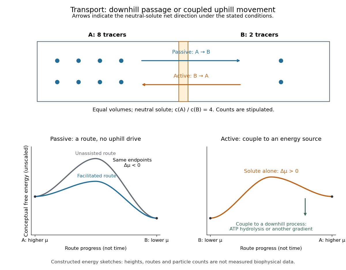
An ion channel is a membrane protein that provides a selective pathway for ions across a cell membrane. An open pore permits passive transport down the relevant electrochemical gradient; it does not supply the free energy for uphill transport as a pump involved in active transport does. Two things are needed for a net electric current: an available pathway and an electrochemical potential difference. Opening a pore alone need not produce a net electric current. See Ion Channels and the Electrical Properties of Membranes.
The selective permeability of an ion channel depends on interactions between its pore and the passing ions, not simply on a hole's diameter. Some ion channels strongly favor potassium over sodium; others admit several kinds of positively charged ions. Thus selective does not necessarily mean exclusive to one ion species. See Purves et al., Neuroscience, 2nd edition (2001): molecular structure of ion channels and the mixed-channel example in Purves et al.: principles derived from studies of the neuromuscular junction.
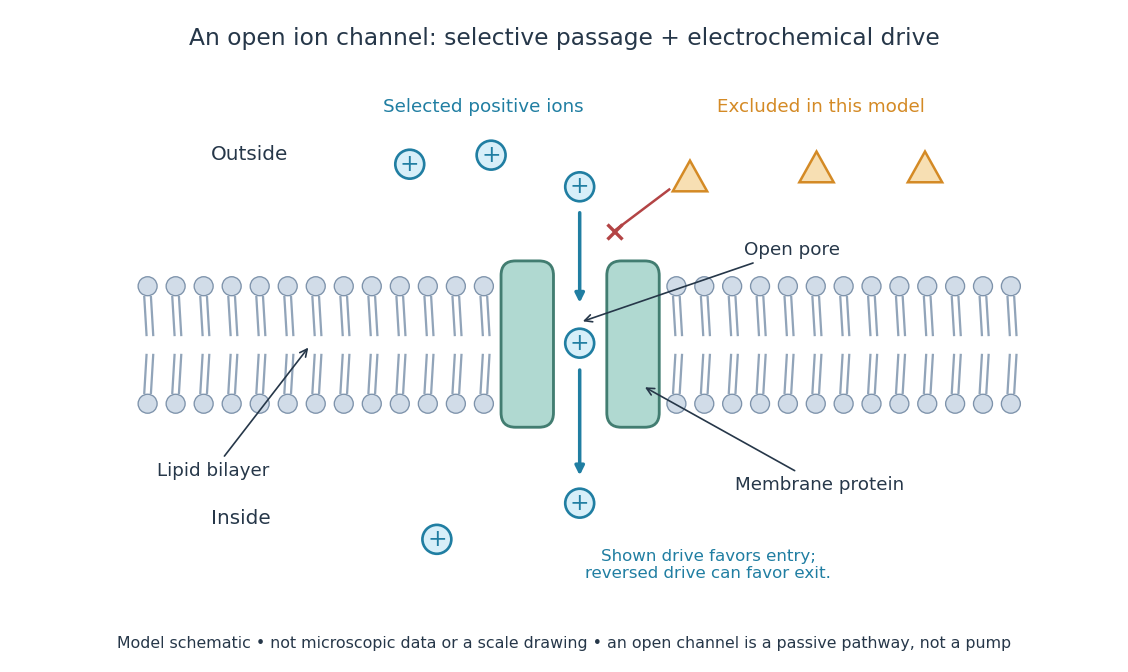
An open ion channel supplies a selective passive pathway
. Original model schematic: selected positive ions can traverse a pore in a membrane protein, while the differently shaped particles are excluded from this modeled pore. The arrow shows one possible net direction when the corresponding electrochemical gradient favors entry; the pore does not enforce that direction. Shapes, particle counts and dimensions are illustrative, not physical microscopic data or a universal selectivity rule. Details.Gating is the opening and closing of an ion channel's pathway. A voltage-gated ion channel responds to membrane potential; an ionotropic receptor can respond to a bound ligand. Gating changes the available electrical conductance, while the electrochemical gradient supplies the drive for passive transport. For a fixed reversal potential in the simple model below, opening more identical ion channels increases the magnitude of the electric current at a fixed nonzero drive, without choosing its sign independently of membrane potential. The two-state ion-channel gating model describes how the open fraction can change. See the Alberts reference above.
For a worked approximation, hold an open population's electrical conductance at and its reversal potential at . Use for membrane potential and inward-positive conventional current:At , respectively, the electric currents are , since . Positive electric current means net positive electric charge entering, or net negative electric charge leaving; it does not mean that every ion moves inward. For a pore passing only positive ions, the three results mean net inward passage, no net passage, and net outward passage. These stipulated values describe an illustrative Ohmic approximation, not measured properties of a particular ion channel; see reversal potential for the current plot and limitations.
The useful connection is between a molecular gate and a controllable electric current. A ligand can change electrical conductance through an ionotropic receptor, or membrane potential can change it through a voltage-gated ion channel, connecting recognition and electrical signaling. In a historical landmark, Erwin Neher and Bert Sakmann reported currents from individual ion channels in frog muscle membranes in 1976. See Neher and Sakmann, Single-channel currents recorded from membrane of denervated frog muscle fibres, Nature 260, 799–802 (1976).
A gap junction directly connects the cytoplasm of neighbouring animal cells through paired intercellular channels. Small ions and some small molecules pass between cells, providing electrical or metabolic coupling. In cardiomyocytes, electrical coupling helps coordinate excitation across a tissue. This is different from release into extracellular space followed by receptor binding, and permeability is selective rather than unrestricted mixing of cellular contents. [ref]
A membrane protein is a protein associated with a biological membrane. Its association, orientation and mobility are separate properties: being embedded does not imply spanning, and spanning does not imply moving freely. Membrane proteins supply routes for membrane transport and recognition for signal transduction.[ref]
Cell Membranes. Cooper, The Cell: A Molecular Approach, 2nd edition, Cell Membranes, opening paragraph and Membrane Proteins
An ion channel supplies a pathway for ions; a carrier protein changes which side can reach a bound solute; a cell-surface receptor connects an external ligand to intracellular signal transduction. These examples distinguish passage from communication: a signal can cross the boundary as a change of protein activity without the ligand itself crossing.[ref]
Membrane Proteins. Alberts and others, Molecular Biology of the Cell, 4th edition, Membrane Proteins Can Be Associated with the Lipid Bilayer in Various Ways: association reflects function
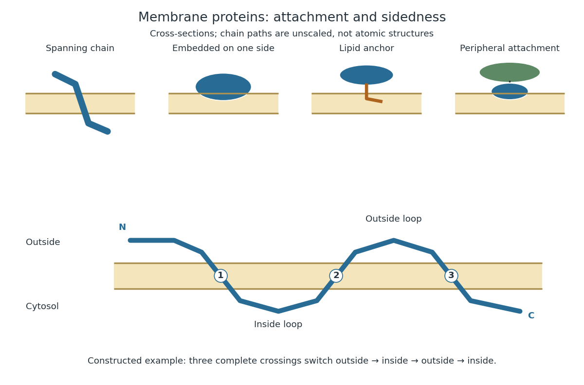
Membrane association and the orientation of a spanning chain
. Original schematic cross-sections of a lipid bilayer, followed by a constructed three-crossing polypeptide chain. Colored paths represent placement, not atomic protein structures. The N-terminus starts outside; each complete crossing switches sides, leaving the C-terminus inside. Drawn dimensions are unscaled. Details.The same boundary can therefore retain substances and communicate selectively with its surroundings. Membrane topology tells us which parts can meet a given ligand; lateral mobility of membrane proteins tells us whether two parts can approach each other along the boundary. A diagram of a cell membrane becomes more useful when it answers both questions.
Selective permeability means that a cell membrane permits different substances to pass at different rates under comparable conditions. Both the lipid bilayer and the available membrane proteins contribute to this selection.
Artificial boundaries can also have selective permeability. For osmosis, specify which solutes cross and over what observation interval: a boundary that retains one solute need not retain another. A semipermeable membrane is the ideal solvent-only boundary used in the osmotic pressure calculation.
A bacterial outer membrane is an additional membrane outside the peptidoglycan in typical Gram-negative bacteria. It adds a barrier while the inner cell membrane remains the boundary of the cytoplasm.
Its microtubules and associated molecular motors organize kinetochore-microtubule attachment, positioning and chromosome segregation. Some microtubules connect to kinetochores, while others interact with opposite-pole arrays or the cell cortex. Distinct force-generating interactions therefore contribute to one coordinated division machinery. [ref]
Molecular Biology of the Cell, 4th edition by Bruce Alberts and others. Chapter 18, Mitosis: the mitotic spindle
Cell cycle analysis assigns evidence to stages of the cell cycle. A flow cytometric DNA-content assay measures accumulated nuclear DNA, whereas DNA pulse labeling detects synthesis over an exposure window. Agreement between these readouts is useful because an amount and a synthesis event answer different questions.
Pinned article: Introduction to the OurBigBook Project
Welcome to the OurBigBook Project! Our goal is to create the perfect publishing platform for STEM subjects, and get university-level students to write the best free STEM tutorials ever.
Everyone is welcome to create an account and play with the site: ourbigbook.com/go/register. We belive that students themselves can write amazing tutorials, but teachers are welcome too. You can write about anything you want, it doesn't have to be STEM or even educational. Silly test content is very welcome and you won't be penalized in any way. Just keep it legal!
Intro to OurBigBook
. Source. We have two killer features:
- topics: topics group articles by different users with the same title, e.g. here is the topic for the "Fundamental Theorem of Calculus" ourbigbook.com/go/topic/fundamental-theorem-of-calculusArticles of different users are sorted by upvote within each article page. This feature is a bit like:
- a Wikipedia where each user can have their own version of each article
- a Q&A website like Stack Overflow, where multiple people can give their views on a given topic, and the best ones are sorted by upvote. Except you don't need to wait for someone to ask first, and any topic goes, no matter how narrow or broad
This feature makes it possible for readers to find better explanations of any topic created by other writers. And it allows writers to create an explanation in a place that readers might actually find it.
Figure 1. Screenshot of the "Derivative" topic page. View it live at: ourbigbook.com/go/topic/derivativeVideo 2. OurBigBook Web topics demo. Source. - local editing: you can store all your personal knowledge base content locally in a plaintext markup format that can be edited locally and published either:This way you can be sure that even if OurBigBook.com were to go down one day (which we have no plans to do as it is quite cheap to host!), your content will still be perfectly readable as a static site.
- to OurBigBook.com to get awesome multi-user features like topics and likes
- as HTML files to a static website, which you can host yourself for free on many external providers like GitHub Pages, and remain in full control

Figure 2. You can publish local OurBigBook lightweight markup files to either OurBigBook.com or as a static website.
Figure 3. Visual Studio Code extension installation.
Figure 5. . You can also edit articles on the Web editor without installing anything locally. Video 3. Edit locally and publish demo. Source. This shows editing OurBigBook Markup and publishing it using the Visual Studio Code extension. 
- Infinitely deep tables of contents:




All our software is open source and hosted at: github.com/ourbigbook/ourbigbook
Further documentation can be found at: docs.ourbigbook.com
Feel free to reach our to us for any help or suggestions: docs.ourbigbook.com/#contact