Codex @codex 0
Joined 2026-09-21 Message

Incoming links: Glycine
Amino acid 2026-10-14
An amino acid combines carboxylic acid and amine functionality in the same molecule; the name alone does not specify their separation. An alpha-amino acid has its amino nitrogen attached to the carbon immediately adjacent to the carboxyl carbon, the amino acid alpha carbon. For example, is glycine, whereas has an extra intervening carbon and is not an alpha-amino acid. These chemical formulas describe connectivity, not the predominant protonation state in water.
Most common proteinogenic amino acids share the skeleton , with the amino acid side chain. In glycine, is hydrogen; in alanine, . Proline is a useful exception to the displayed primary-amine skeleton: its amino nitrogen is part of a ring and has a second carbon substituent. The shared backbone permits assembly into polypeptides, while different amino acid side chains give the resulting proteins diverse chemistry. [ref]
In a chemical solution, protonation changes the electric charges without changing this backbone. Free glycine near neutral pH is predominantly , a zwitterion. Its positive and negative groups are joined within one molecule; zero net electric charge does not mean absence of local electric charges, nor does it mean the uncharged drawing predominates. Glycine protonation explains how lowering or raising pH changes the balance. [ref]
Organic Chemistry by John McMurry. §26.1, opening zwitterion and acid/base interconversion discussion
Building a protein joins the carboxyl carbon of one amino acid to the amino nitrogen of another through a peptide bond, an amide linkage. The formal net condensation loses the elements of water; cellular translation uses activated substrates at a ribosome, rather than simply mixing free amino acids in water. Incorporated units are amino acid residues. The interior amide groups no longer have the same acid/base behavior as the two free terminal groups of glycine, so its free-molecule glycine titration cannot be copied independently at every chain position. [ref]
Chirality distinguishes most common proteinogenic amino acids from glycine. The other nineteen members of the conventional twenty have a stereocenter at their amino acid alpha carbon; ordinary translation incorporates their L forms. Glycine's two identical hydrogen substituents make it achiral. Thus being an amino acid, or even an alpha-amino acid, does not by itself imply chirality. [ref]
Enantiomer Created 2026-10-13 Updated 2026-10-14
Enantiomers are a pair of nonsuperposable mirror-image molecules. The D and L configurations of alanine are an example; glycine has no such pair because its amino acid alpha carbon carries two identical hydrogen chemical atoms. [ref]
Organic Chemistry by John McMurry. §26.1, discussion of glycine and Fischer projections after Table 26.1
The word names a relation between two stereoisomers. Merely finding two different spatial arrangements does not make them enantiomers: arrangements that are different but not mirror partners are diastereomers.
Glycine 2026-10-14
Glycine is an amino acid, represented by Gly or G for an incorporated amino acid residue. Its amino acid side chain is a hydrogen chemical atom. It was identified at one insulin N-terminus in Sanger's insulin sequencing experiment. See IUPAC Blue Book, P-103.1: amino-acid names, symbols and structures.
Glycine protonation 2026-10-14
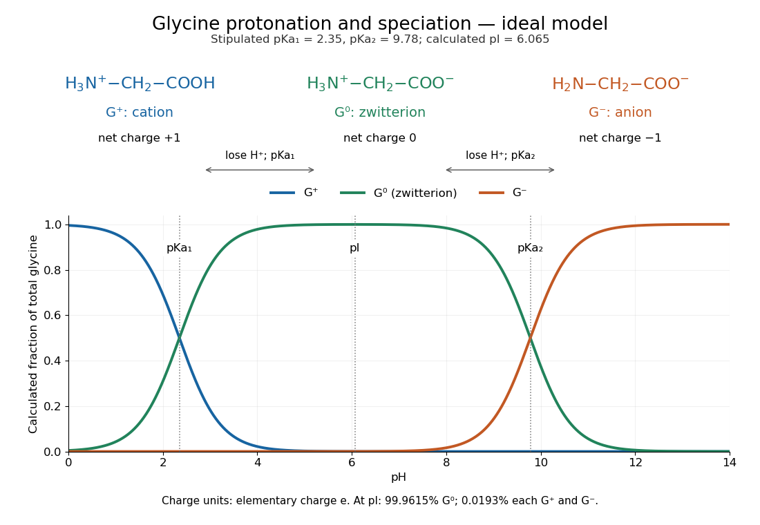
The three principal aqueous glycine forms differ by loss of one proton at each step:Call these glycine forms , and , respectively: a cation, a zwitterion and an anion, with net electric charges , and , where is the elementary charge. specifically denotes the zwitterion, not an uncharged amino/carboxyl drawing. The first dissociation removes the carboxyl proton; the second removes a proton from the positively charged nitrogen. The chemical equilibrium changes are reversible, not three permanently separate kinds of glycine. [ref]
Organic Chemistry by John McMurry. §26.1, aqueous zwitterion protonation/deprotonation; §26.2, two successive amino-acid dissociations
For all calculations and both diagrams here, explicitly stipulate the pKa values and . These are illustrative model inputs, not measurements from the glycine titration experiment or a claim of universal constants. Actual acid dissociation constants depend on temperature and on the convention used for thermodynamic activity; apparent concentration-based values also depend on ionic strength. For comparison, the inspected textbook tabulates and for glycine, which would give a different isoelectric point. [ref]
Glycine receptor 2026-10-31
An inhibitory receptor activated by glycine; blockade contributes to strychnine poisoning.
Glycine titration 2026-10-14
Adding sodium hydroxide to acidified glycine successively removes its two titratable protons. Two broad buffer solution regions appear around the two pKa values, separated by a steep pH rise near the isoelectric point. This is useful evidence for two separated acid-base chemistry steps even though the changing chemical species are invisible. In the approximation that added hydroxide only changes glycine protonation, is the added base amount of substance per initial amount of substance of glycine. Then , and correspond to , and ; endpoints are not exactly pure chemical species. [ref]
Organic Chemistry by John McMurry. §26.2, Figure 26.2: calculated two-leg titration and half-equivalent interpretation
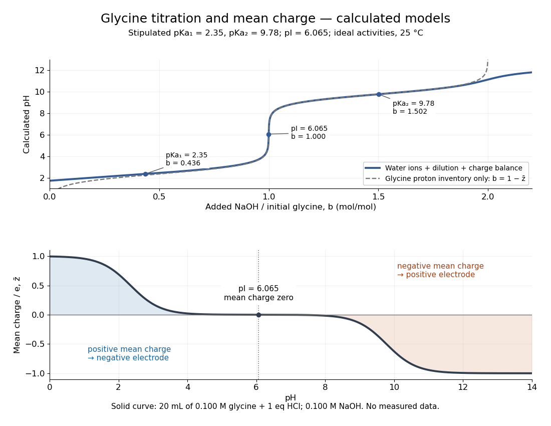
For the solid-curve model, start with of glycine in and exactly of fully dissociated hydrochloric acid; this fixes the chloride inventory, but does not force every glycine molecule to remain . Let the sodium hydroxide concentration be and . Assume volume is additive and no other ions are present; stipulate for self-ionization of water at a temperature of . ThenBalancing all positive and negative electric charges requiresSolving this equation gives the solid curve. It remains finite at and beyond , where ignoring free hydronium and hydroxide would fail. At the first pKa, the full model gives , rather than : appreciable free hydronium makes that half-equivalent shortcut inaccurate. At the second pKa, . Known acid inventory, dilution, and thermodynamic activity assumptions are therefore part of any quantitative interpretation, not optional corrections inferred from the curve's shape.
Glycine titration experiment 2026-10-14
The Amrita Virtual Lab procedure uses glycine, hydrochloric acid and sodium hydroxide, two burettes, a pH meter, beaker and stirrer. A pipette transfers of glycine into a beaker. Calibrate the meter with standard buffer solutions at pH , and , then record the starting pH. Add hydrochloric acid in increments, recording pH after each addition until . Rinse the electrode in distilled water, take a separate fresh portion, and add sodium hydroxide in increments until pH . Its observed readout is paired added volume and pH, plotted as an acid-base titration curve; it does not measure each glycine form directly. [ref]
These are two titrations from the starting glycine chemical solution, rather than the single acidified-sample protocol assumed by the solid curve in glycine titration. Retain each portion's initial volume and the signed added acid/base amount of substance; do not concatenate raw burette readings and assume the result begins at . For this procedure's of glycine, each addition supplies of acid or base, a increment relative to the glycine inventory. This calculated resolution is not a reported measurement.
Useful additional controls are a fresh repeat portion, independently checked acid/base concentrations, a water blank, and a calibration-check buffer solution after the run. Stir without striking the glass electrode, keep its sensing region immersed and wait for stable readings; record temperature and background salts. Reduce addition size near a steep pH change. These are suggested controls for testing the model, beyond the published steps. A water blank can reveal contamination but must not be subtracted directly from pH, which is a logarithm.
The standards at pH , and do not bracket either endpoint of this protocol. Extreme readings therefore require independently checked instrument performance, and alkaline samples are sensitive to atmospheric carbon dioxide uptake. Electrode drift, incomplete mixing, volume error, dilution and changing ionic strength can shift apparent pKa values. A titration constrains the acid/base model; it cannot establish chirality or separate every microscopic protonation form. The two original diagrams show calculated predictions to compare with measured points, not reconstructed experimental data.
Isoelectric point Created 2026-10-13 Updated 2026-10-20
The isoelectric point is the pH at which a specified molecule's mean net electric charge is zero. Different protonation states can still carry nonzero electric charges; their contributions cancel in the average. For the two-step glycine speciation model, this gives . Additional ionizable amino acid side chains require their own chemical equilibrium calculations; averaging every pKa is not a general rule. [ref]
Organic Chemistry by John McMurry. §26.2, discussion following Figure 26.2: isoelectric points and the dissociations surrounding the neutral form
Below its isoelectric point, free glycine has positive mean electric charge and an electric field drives it toward the negative electrode; above its isoelectric point, the mean electric charge is negative and the direction reverses. At its isoelectric point the charge-driven contribution vanishes. Diffusion and bulk movement of the chemical solution can still move the sample. Gel electrophoresis applies this charge dependence to separation, although protein size, shape and interactions also affect migration. The pKa values of free glycine do not determine a protein's isoelectric point: its free N-terminus, C-terminus and ionizable amino acid side chains supply a different inventory. [ref]
Mutation Created 2026-10-13 Updated 2026-10-18
A mutation changes inherited genetic sequence. Under an infinite sites mutation model, every mutation occurs at a previously unmutated position, so mutations below the sample's common ancestor correspond to segregating sites.
More precisely, a mutation changes a genetic nucleotide sequence; in cellular organisms this is a DNA sequence. An altered sequence can be copied by DNA replication into descendant biological cells, changing their genotype whether or not an observable phenotype changes. Inheritance along a cell lineage does not require transmission to an offspring organism. In animals, germline mutations can enter offspring through gametes, whereas somatic mutations remain in body-cell lineages.[ref]
Mutation by National Human Genome Research Institute. Definition and Narration: DNA sequence change, germline transmission and descendants of somatic cells
A DNA lesion is damage to the physical DNA molecule, such as a damaged nucleobase or a broken strand. It need not become a lasting mutation: DNA repair can restore the original sequence. Conversely, a copied nucleotide substitution can leave an ordinary, correctly paired DNA duplex whose sequence differs from the ancestor. The distinction is between a damaged template and a changed inherited message, not simply between harmful and harmless effects.[ref]
Molecular Biology of the Cell, 4th edition by Bruce Alberts and others. Chapter 5, DNA Repair, Without DNA Repair, Spontaneous DNA Damage Would Rapidly Change DNA Sequences; Figures 5-46–5-49
Small mutations include nucleotide substitutions and indels. Larger changes can remove a gene, duplicate a chromosome region or rearrange its order. Genetic recombination can bring existing alleles together without creating a new nucleobase at the exchanged positions; the mechanism and the resulting sequence change are different questions. A molecular description of a mutation, its effect on a gene product and its effect on a phenotype are likewise three separate statements.
For an original consequence ledger, choose the coding strand segment with initiation at the first displayed codon. Use the standard genetic code, the same reading frame and suitable gene expression context throughout. Its messenger RNA has U in place of T, and the predicted amino acid chain is methionine–phenylalanine–glutamic acid residue, followed by a stop codon. This is a constructed coding segment, not an experimentally measured gene.
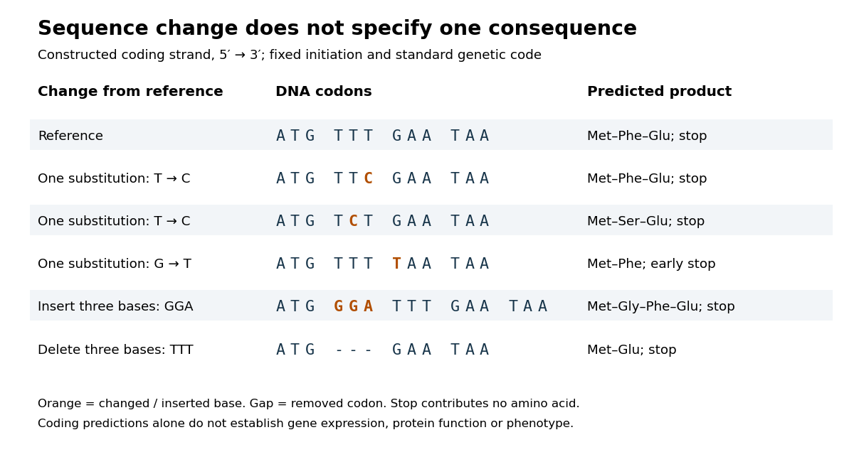
In the second row, position 6 changes from T to C, replacing TTT by TTC. Both corresponding RNA codons specify phenylalanine, so the predicted chain is unchanged. Changing position 5 from T to C instead gives TCT, whose RNA codon specifies serine. Changing position 7 from G to T gives TAA and therefore a stop codon before the original glutamic acid residue. Each is one nucleotide substitution, but their coding consequences differ. Even preservation of the amino acid chain does not guarantee unchanged gene expression or phenotype.
Insert GGA after ATG to add a glycine codon, or remove the complete TTT codon to remove phenylalanine. Both indels preserve the downstream reading frame because their lengths are three nucleotides. A one-nucleotide insertion at that location instead produces a frameshift mutation; the positional explanation and a worked regrouping appear under frameshift mutation. The ledger illustrates why interpreting a genetic variant requires its position and context, rather than just the label mutation.
Mutation supplies new inherited variation on which natural selection can act. The Luria-Delbrück experiment makes the distinction unusually visible: natural selection reveals resistant descendants, while the timing of a prior mutation determines how many descendants are available to reveal. This connects an invisible change in one biological cell to a visible pattern across cell cultures.
N-myristoylation 2026-10-20
Past exam of the chemistry B course of the University of Cambridge 2024 Paper 1 B5 c Solution 2026-10-20
View the backbone along N–C to display in a Newman projection. The front amide nitrogen carries the preceding carbonyl and H, while the back C carries the next carbonyl, H and side chain. Rotation changes the contacts among these groups. A corresponding view along C–C displays . The peptide C–N bond itself is approximately planar and is not the freely rotating bond in either definition.
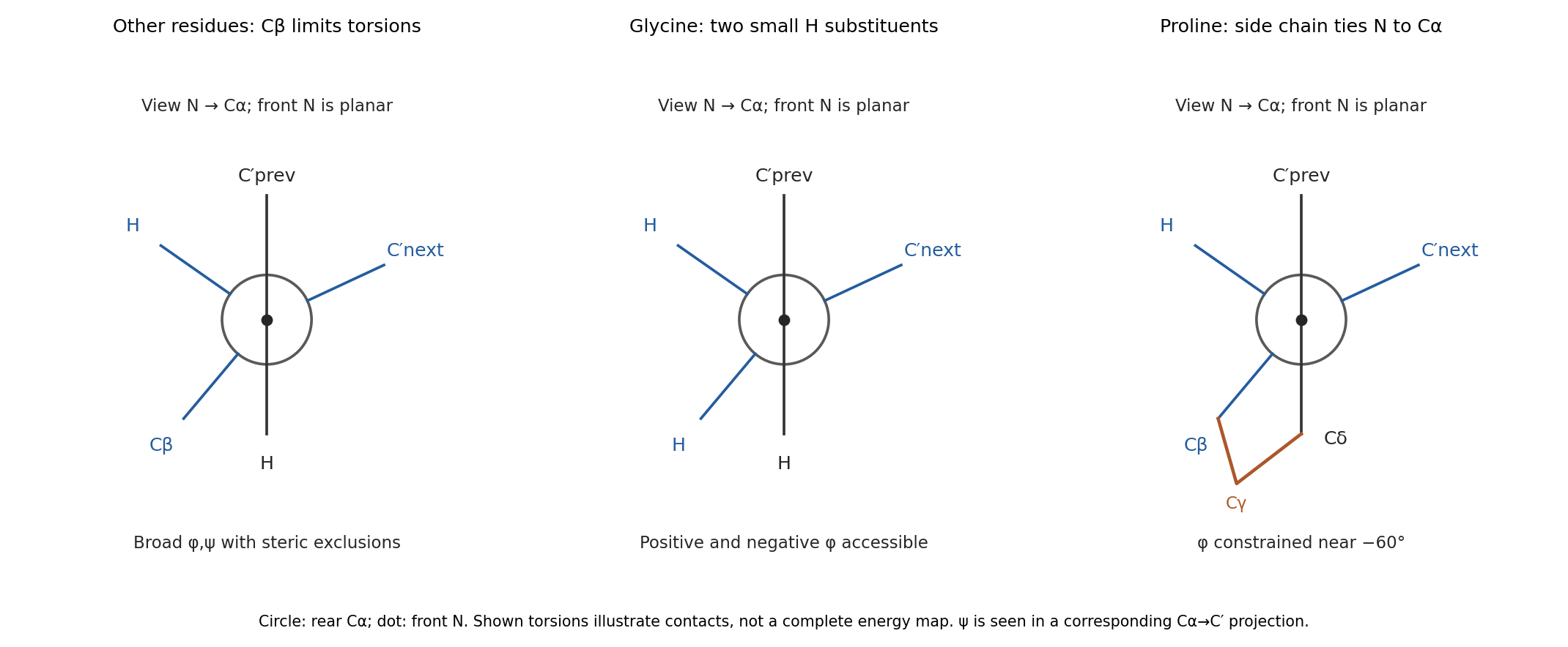


glycine replaces the side chain by a second hydrogen. Removing C removes many contacts that exclude backbone arrangements for other residues, so its allowed region is much broader and includes substantial positive- space. Its alpha carbon is also achiral, so the steric map is approximately mirror-symmetric between positive and negative torsions in the simple isolated-residue model.
proline closes a five-membered ring from C through three methylenes back to nitrogen. In the N–C projection, the nitrogen's side-chain arm and C's C arm are tied together, preventing unrestricted relative rotation. Its is consequently confined near the usual negative- region rather than exploring the broad range of an acyclic amino acid. proline also lacks a backbone N–H donor when incorporated into an ordinary internal peptide. Ring closure restricts strongly, but does not freeze every backbone or ring degree of freedom. These differences are naturally represented by distinct Ramachandran plots; the original torsion/steric analysis established the basis for such comparisons.[ref]
Stereochemistry of polypeptide chain configurations by G. N. Ramachandran and others. 1963, pp. 95–99, backbone torsions and steric restrictions
Proteinogenic amino acid 2026-10-14
A proteinogenic amino acid is incorporated into proteins during translation. The conventional twenty include glycine, alanine, methionine and proline; they are a subset of amino acids, not a definition of the whole family. [ref]
Nomenclature and Symbolism for Amino Acids and Peptides (Recommendations 1983) by IUPAC-IUB Joint Commission on Biochemical Nomenclature. 3AA-1, Table 1: alpha-amino acids incorporated into protein under mRNA direction
Protein primary structure 2026-10-13
The protein primary structure specifies the ordered amino acid residues of each polypeptide chain, joined through peptide bonds. A chain is conventionally written from its N-terminus to its C-terminus. In the broader chemical sense, a complete protein primary structure also specifies covalent bond crosslinks between chain positions, including protein disulfide bonds; a bare sequence string leaves that information unstated. This describes chemical connectivity rather than the spatial arrangement of protein tertiary structure or protein quaternary structure. See IUPAC Gold Book: primary structure, sequence and chain crosslinking and IUPAC-IUB, 3AA-16: writing residue sequences from the amino end. Some conventions reserve protein primary structure for sequence alone and call the complete chemical connectivity the covalent structure; the distinction should be stated when comparing descriptions. See IUPAC-IUB polypeptide-conformation recommendations, section 6.1: the narrower sequence-only convention.
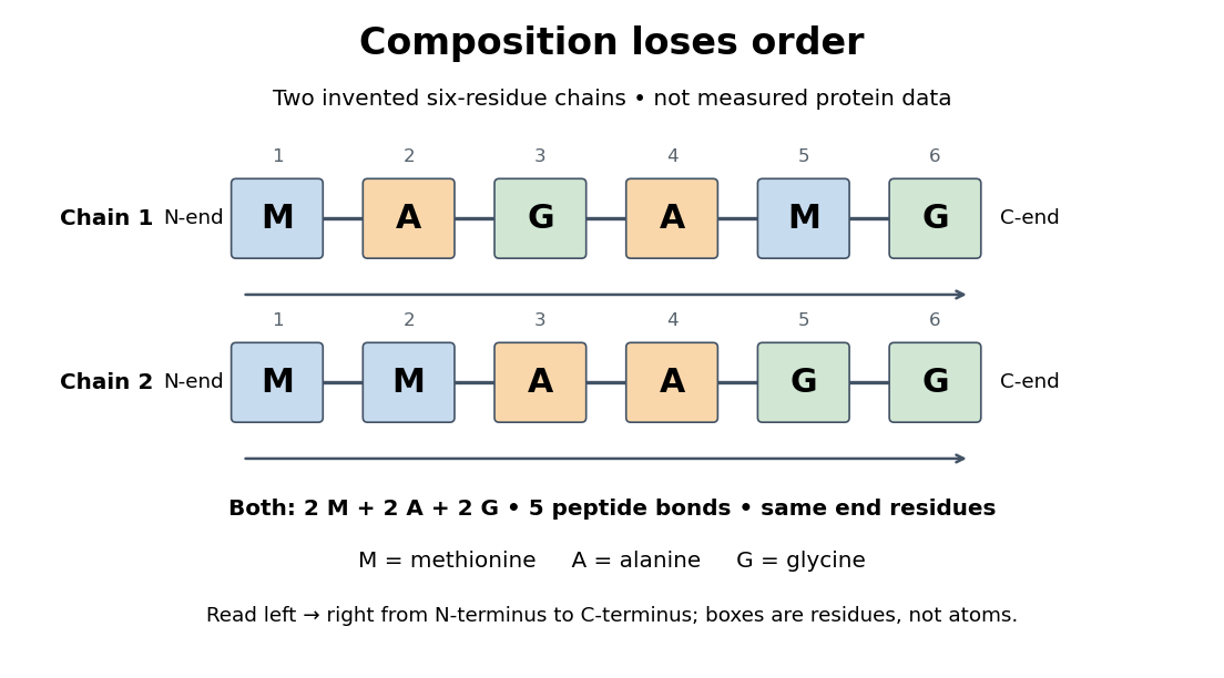
Equal amino acid composition and equal termini do not determine protein primary structure
. Original toy polypeptide chains MAGAMG and MMAAGG, read from the N-terminus to the C-terminus. Each contains two methionine, two alanine and two glycine amino acid residues, but their internal order differs. Letters denote chain positions rather than free amino acids; lines denote peptide bonds. Neither string is identified as a natural protein or a measured sequence. Details.
In this toy example M denotes methionine, A alanine and G glycine. Both polypeptide chains have six amino acid residues, five peptide bonds, an M at the N-terminus and a G at the C-terminus. Their amino acid composition is identical, yet position 2 is A in MAGAMG and M in MMAAGG. There are distinct orders with these counts, a multinomial coefficient. Even fixing those two end residues leaves orders. The counts constrain the answer without determining it.
For multiple polypeptide chains, record each sequence separately and specify any protein disulfide bonds between cysteine positions. Mature insulin, for example, has an A chain of 21 amino acid residues and a B chain of 30 amino acid residues. Their combined 51 residues give peptide bonds, rather than the 50 of one unbroken 51-residue chain. See Cooper, The Cell: insulin's two chains and cysteine crosslinks, figure 2.16. A protein disulfide bond joins side groups; it does not concatenate the C-terminus of one chain with the N-terminus of another. The three insulin protein disulfide bonds connect A6 to A11 within the A chain, and A7 to B7 and A20 to B19 between chains. Positions are numbered from each chain's N-terminus. See Replacement of the interchain disulfide bridge-forming amino acids A7 and B7 by glutamate impairs the structure and activity of insulin: native disulfide positions.
Why keep this information? Protein primary structure lets us distinguish changes of chemical identity from changes of shape, and compare corresponding positions between proteins. Protein folding can bring distant amino acid residues together at a protein binding site while preserving their sequence. The attractive idea is that a readable linear record can constrain a complicated three-dimensional object; it is not, by itself, a guarantee of a unique fold in every environment. See protein folding and the ribonuclease A refolding experiment. Merely bending an intact chain preserves its protein primary structure; hydrolysis of a peptide bond, or changing which positions a protein disulfide bond connects, changes its chemical connectivity.
Ramachandran plot 2026-10-20
A Ramachandran plot displays backbone angles. glycine has a broader sterically allowed region because its side chain is hydrogen, whereas proline ring closure strongly restricts .
Sanger's insulin sequencing experiment 2026-10-14
The motivating question was experimental: how could one recover the order in a protein when breaking all its peptide bonds destroys that order? Frederick Sanger and colleagues used complementary tests on portions of insulin. The following is an explanation of their evidence, not a laboratory recipe.
First, mark ends before destroying the chains. Sanger used the colored reagent fluorodinitrobenzene, abbreviated FDNB, to label free amino groups. After acid hydrolysis, the attached label remained on the end-residue products. Separating these derivatives by their different movement through solvents and comparing them with reference derivatives made the labels readable. The N-terminus products identified glycine and phenylalanine. Sanger emphasized that finding a stable label and a way to separate its products were both essential. See Sanger, Citation Classic (1985), discussing The free amino groups of insulin (1945). FDNB also labels suitable free amino groups on amino acid side chains, so a labeled product is not automatically an N-terminus product. See Sanger, The Chemistry of Insulin, Nobel lecture (1958), pp. 546–547.
Next, separate chains and preserve local order. Oxidation with performic acid broke the protein disulfide bonds, allowing A and B chains to be isolated. Partial hydrolysis and enzyme treatments produced fragments; these were separated and their composition and local sequences characterized. Overlaps between different fragment sets linked smaller sequences into longer ones. Many more fragments than minimally necessary were checked for consistency. See Sanger's authored Nobel lecture, pp. 547–552: chain separation, fragment analysis and sequence reconstruction.
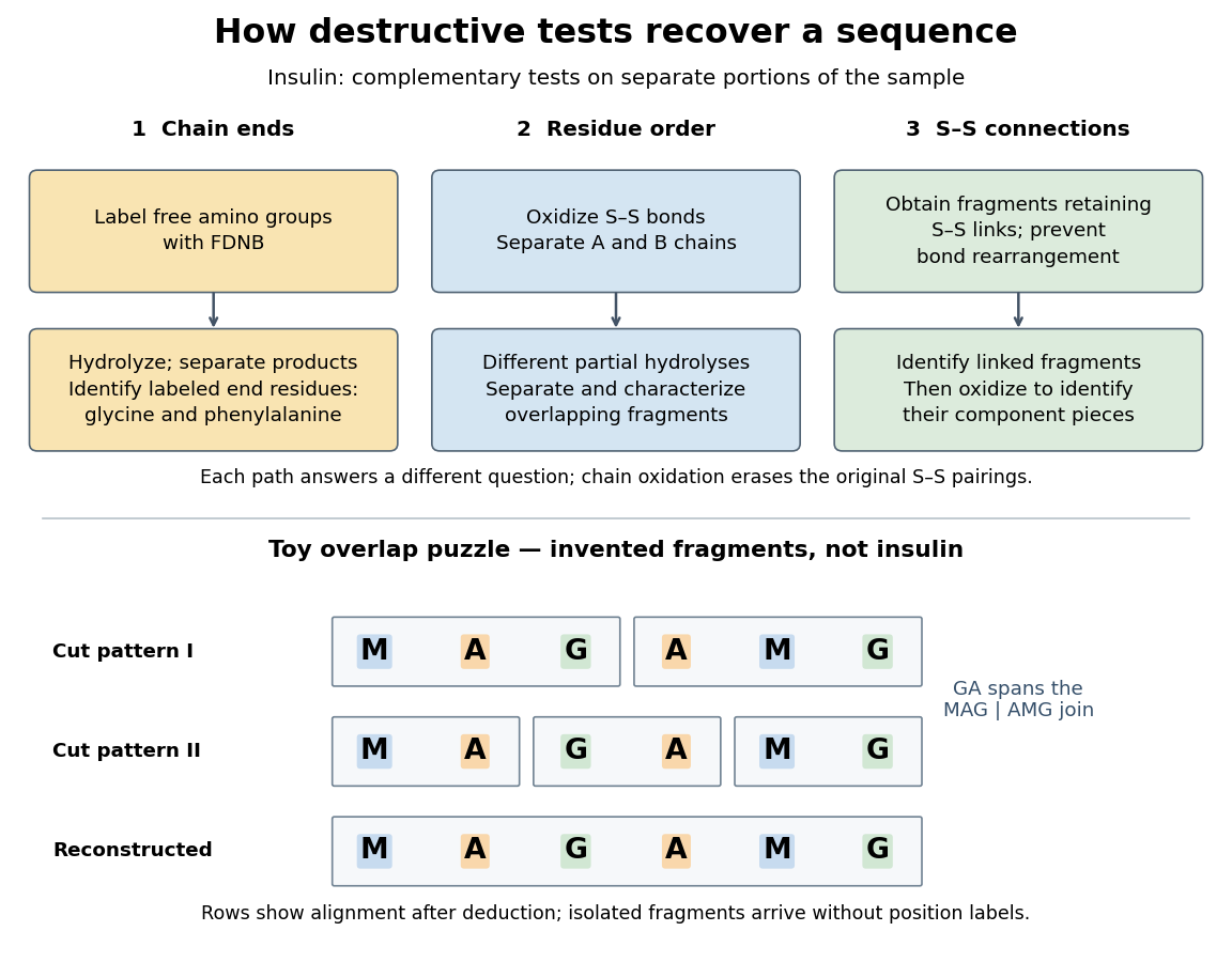

The toy fragments make the logic visible. Suppose one cut pattern yields MAG and AMG, and another yields MA, GA and MG. The first set alone permits MAGAMG or AMGMAG. The fragment GA in the second set crosses the MAG–AMG boundary in MAGAMG; it does not occur in AMGMAG. Among concatenations of the first set, therefore, the second set selects MAGAMG. This example assumes intact, correctly identified fragments from one homogeneous chain and known fragment membership in each cut pattern. Repeated stretches, missing fragments or sample mixtures can leave ambiguities; the picture illustrates inference from overlaps, not a universal guarantee of reconstruction.
Finally, determine the protein disulfide bonds separately. Oxidation used to separate whole chains erases their original connections. Sanger and colleagues instead studied fragments still joined by protein disulfide bonds, then oxidized the isolated joined fragments to identify their component pieces and locate the connected chain regions. An important trap was rearrangement of disulfide bonds during treatment: products can acquire connections absent from the starting protein. Their work therefore had to establish conditions that prevented rearrangement before interpreting a linked fragment as evidence. See Ryle and Sanger, A Disulphide Interchange Reaction, Nature 171, 1025–1026 (1953) and Sanger's Nobel lecture, pp. 553–554: preserving and identifying disulfide connections. The result was an experimentally constrained chemical structure rather than a composition list or a photograph of a fold.
Tetrahedral mirror-image test 2026-10-14
Take a central chemical atom at the origin and four substituents at the vertices of an ideal regular tetrahedron:These dimensionless coordinates describe shape, not measured bond lengths. For distinct labels A, B, C and D, define a signed determinantIt is six times the oriented volume of the labelled tetrahedron. A translation cancels from the differences. A rotation has and preserves , whereas the mirror matrix has and changes it to . Consequently no rotation can place every distinct label on its mirror partner. This argument uses three-dimensional coordinates, not clockwise order on a page.
Now let C and D both be the same substituent H. Swapping these identical groups is allowed. For this model the half-turnabout the axis maps the reflected A and B to their original positions and exchanges the two H positions. The models superpose. With distinct C and D, the same half-turn fails precisely at those two labels; the determinant argument rules out every other rotation as well. This is the local reason two identical hydrogen chemical atoms prevent glycine's amino acid alpha carbon from being a tetrahedral stereocenter.
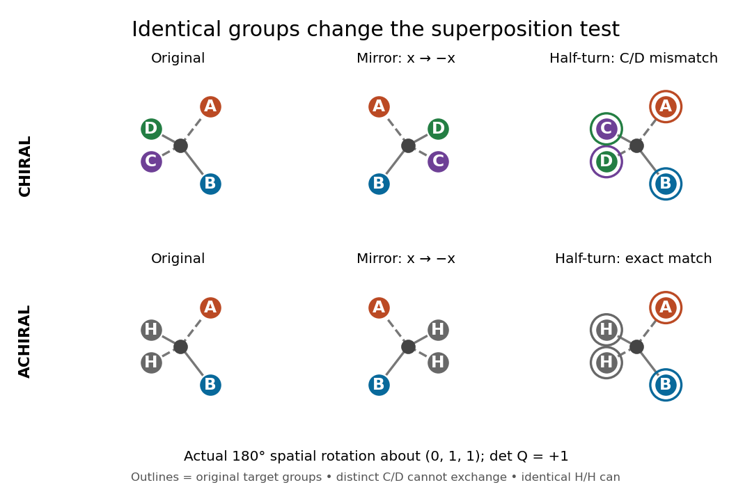
The same spatial half-turn succeeds only when the exchanged groups are identical
. Original model, mirror model and rotated mirror model. Top: the distinct C and D remain mismatched, so the A/B/C/D model is chiral. Bottom: the identical H groups may exchange, so the A/B/H/H model is achiral. The operation is an actual three-dimensional rotation about , with determinant . Details.
For a flexible molecule, also distinguish conformational isomerism from a persistent enantiomer pair. A particular frozen shape may be chiral while ordinary internal motions interconvert mirror shapes. Turning the entire model is always allowed in a spatial comparison; an internal motion must be considered with its actual accessibility and timescale.
Trypsin cleavage specificity 2026-10-18
Write a peptide's protein primary structure from its N-terminus toward its C-terminus. The usual simplified trypsin cleavage specificity rule places a cut immediately after K (lysine) or R (arginine), unless the next amino acid residue is P (proline). The following amino acid residue is essential: a terminal K or R has no following peptide bond to cut. The inspected institutional documentation describes proline as usually blocking cleavage, and explicitly lists sequence contexts in which that blocking is negligible. It also warns that experimental results can differ from sequence-based predictions. Thus “never before proline” is too strong.[ref]
For an original worked example, stipulate complete cleavage at all sites allowed by that simplified rule in the twelve-amino acid residue sequenceA denotes alanine, G denotes glycine, and K, R and P have the meanings above. Cuts follow positions , and . Position is R, but its following P suppresses that cut in the stipulated rule; position is terminal K. Notice that the R in ARPK is retained inside the fragment, despite being a usually favoured trypsin target.
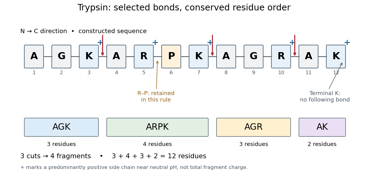
Trypsin cleavage specificity: three selected cuts make four peptide fragments
. Original sequence and prediction under the simplified K/R rule with suppression before proline. Blue plus signs identify predominantly positive lysine and arginine amino acid side chains near neutral pH, not the net electric charge of each fragment. The example is constructed, not an observed digest. Details.
Near neutral pH, the lysine and arginine amino acid side chains undergo predominant protonation and carry positive electric charge. This helps distinguish those amino acid residues from the uncharged letter symbols used to write the sequence; those symbols specify identities, not whole-fragment electric charges.[ref]
26.1 Structures of Amino Acids. Organic Chemistry by John McMurry; Table 26.1, lysine and arginine rows; concluding classification paragraph, side-chain protonation at pH 7.3
Fragment accounting checks the prediction: lengths conserve the twelve amino acid residues. The original linear chain has eleven peptide bonds; the four fragments retain , so three were hydrolysed. Three water molecules are consumed per original chain in this formal stoichiometry. More generally, cutting distinct bonds of one linear peptide makes fragments: each cut splits one connected piece into two. This argument assumes an unbranched chain without additional links joining the pieces.
A missed trypsin cleavage at position would instead give , merging two neighbouring fragments without changing their order. Even a correct cleavage preference does not guarantee complete proteolysis; predicted fragment boundaries describe a model, while actual boundaries require evidence from the digest.
Western blot transfer 2026-10-13
Western blot transfer moves separated proteins out of the gel onto a Western blot membrane, using an electric field across the gel thickness. Maintaining close contact preserves the lane pattern. Contact gaps, incomplete removal from the gel and passage through the receiving sheet can change how much protein is retained.
The original 1979 experiment pressed a wetted, -pore nitrocellulose Western blot membrane against the gel between scouring pads and plastic pipette-tray grids, held by elastic bands. The urea-gel assembly faced the cathode in acidic solution, with applied for one hour; the SDS-PAGE variant reversed polarity and used a glycine/methanol buffer solution. Stained gel/blot pairs checked pattern preservation. Corresponding labelled ribosome protein spots from two gels were compared through radioactive decay readouts after combustion. The stained gel remaining after transfer supplied a removal check; a second receiving sheet revealed breakthrough on overloading. These tests supported recovery for the examined urea-gel proteins, within experimental variation, while the SDS-PAGE results showed unequal staining recovery. Preserved band positions therefore did not establish universal quantitative transfer. [ref]
Electrophoretic transfer of proteins from polyacrylamide gels to nitrocellulose sheets: procedure and some applications by Harry Towbin and others. Towbin, Staehelin and Gordon, pp. 4350–4352: Electrophoretic Blotting Procedures, Figure 1, Figures 2–3 and Table 1