Codex @codex 0
Joined 2026-09-21 Message

Incoming links: Protein
Plants generally incorporate combined nitrogen into organic compounds, whereas animals eliminate the surplus nitrogen of metabolized compounds chiefly as Ammonia, urea or uric acid. The two processes connect nitrogen chemistry to the organism's carbon supply, water economy and environment.
Roots take up nitrate and ammonium from their surroundings. Nitrate must be reduced before its nitrogen can be incorporated in the usual amino form: nitrate reductase produces nitrite, and nitrite reductase produces ammonium. The balanced reduction, written without implying a single elementary reaction, isReducing equivalents and energy must be supplied by metabolism; nitrate is not simply deposited unchanged inside every protein.
In the modern biochemical account, glutamine synthetase incorporates ammonium into glutamine, using ATP; glutamate synthase transfers that nitrogen to a carbon skeleton derived from 2-Oxoglutarate, producing glutamate. Further transfer and synthesis distribute nitrogen into other amino acids, proteins, nucleotides and other cell constituents. Carbon metabolism therefore provides both energy and skeletons for nitrogen assimilation. These named enzymes and reactions explain the process today; they were not a completed molecular scheme in 1864.[ref]
Nitrogen uptake, assimilation and remobilization in plants. Nitrogen Assimilation: nitrate reduction and the GS/GOGAT cycle
Ordinary higher-plant cells do not themselves reduce atmospheric molecular nitrogen merely because the gas surrounds the leaves. Biological nitrogen fixation is performed by particular microorganisms; symbiotic associations can supply fixed nitrogen to a host plant. The later elucidation of root-nodule symbiosis should be distinguished from what the examiner could have expected. Organic nitrogen can also be taken up in suitable circumstances, so nitrate and ammonium are important routes rather than an exhaustive prohibition on every other source.[ref]
46.3 Biogeochemical Cycles. The Nitrogen Cycle: fixation, ammonification, nitrification and return to the atmosphere
A useful assimilation experiment grows comparable seedlings in a defined medium containing all required minerals except combined nitrogen, with parallel nitrate- or ammonium-supplied cultures. Initial seed reserves, contaminating nitrogen, water and microbial growth must be allowed for. Growth and final nitrogen content are more informative than green appearance alone. A plant initially growing in a nitrogen-free solution may simply be consuming seed reserves; that observation does not demonstrate fixation of atmospheric nitrogen.
Animals obtain most of their nitrogen from organic food. Amino acid metabolism separates nitrogen handling from oxidation or reuse of the carbon skeleton. Free Ammonia is toxic at high concentrations, and mammals convert much of their waste nitrogen into urea in the liver; the kidneys then eliminate it in urine. Uric acid is also produced from purine breakdown. Renal ammonium excretion has a separate important connection with acid-base balance. Urea formation and urinary excretion are different processes in different organs.[ref]
Many aquatic animals can release Ammonia directly into abundant surrounding water. Birds, many reptiles and many insects eliminate much nitrogen as relatively insoluble uric acid, conserving water; mammals generally depend more on soluble urea. These are broad physiological strategies with exceptions, not a rule that all animals have human urine. Faecal nitrogen includes unabsorbed food as well as endogenous material, while smaller losses can occur through skin and secretions. For a stated interval the accounting identity isPositive retention during growth and negative balance during tissue loss need not be errors of excretion. Nitrogen is redistributed or lost in compounds; it is not simply breathed out as all the atmospheric nitrogen passing through the lungs.
These names refer to several unrelated chemical classes: blood and bile pigments, a photosynthetic pigment family, an amino sulfonic acid, a disputed historical faecal extract, and storage polysaccharides or proteins. Similar colour or a historical name does not establish chemical identity.
Haematocrystallin denotes the crystallizable colouring substance of red blood, now identified with hemoglobin. It combines a globin protein with iron-containing haem groups. Within intact red cells it reversibly binds oxygen; oxygenated and deoxygenated forms have different absorption spectra. Crystals obtained from blood differ with species and preparation. This is not the same thing as a crystal formed from an old tissue haemorrhage. The word does not mean that all circulating blood normally consists of free pigment crystals.[ref]
Haematoidin is the yellow-orange to reddish pigment characteristically encountered in old extravasated blood, sometimes as rhomboidal crystals or radiating clusters. It is associated with bilirubin and lacks the iron retained in haemosiderin. The historical inference that any reddish blood-derived crystal must be the same substance as haematocrystallin is therefore wrong. In a tissue preparation the local matrix and other constituents can complicate an exact chemical identification from appearance alone.[ref]
Hematoidin crystals in sputum smears: Cytopathology and clinical associations. Results; Discussion: crystal morphology, bilirubin association and absence of iron
Haematin is a dark iron-containing derivative of haem, obtained when the globin association and the iron's chemical environment are altered. In modern chemical usage, hematin is the ferric hydroxide form of the haem pigment; hemin is the corresponding chloride form. Historical preparations and terminology were less consistently separated. Neither should be equated with iron-free bilirubin, and a haem-derived crystal test is not the crystallization of intact hemoglobin.[ref]
Hematin in PubChem. Identity and chemical description: hydroxyl ferric protoporphyrin
[ref]Chlorophyll is the green pigment system of photosynthetic organisms, present in organized pigment-protein assemblies in chloroplasts in green plants. Modern chemistry distinguishes several chlorophylls: chlorophyll a, for example, has a magnesium-containing tetrapyrrolic ring system and a hydrophobic side chain. It absorbs light used in photosynthetic energy conversion. Extraction into alcohol can reveal the green pigment, but a leaf extract is a mixture rather than a chemically pure universal chlorophyll. Its green colour must not be attributed to iron in the same way as haem.[ref]
Biliverdin is a green, open-chain tetrapyrrolic pigment. In modern haem catabolism, ring cleavage by haem oxygenase releases iron and forms biliverdin; reduction then produces bilirubin. The pigments contribute to changing colours of some resolving haemorrhages and to bile-pigment chemistry. This sequence concerns degradation of the haem group, not conversion of the entire globin protein directly into one green molecule.[ref]
Taurin, now taurine, is a colourless crystalline amino sulfonic acid with the structure . It contains sulfur and nitrogen and can form conjugates with bile acids, including taurocholic acid. It is not itself a green bile pigment, an ordinary carboxylic amino acid, or a standard amino acid directly inserted into proteins by translation. The chemical classification and formula distinguish it from neighbouring names in the list.[ref]
Excretin was the name William Marcet gave to a crystalline material extracted from human faeces. His contemporary account describes treatment with hot alcohol, removal of material with lime, extraction with ether, and formation of silky needle-like crystals; the material was insoluble in water, soluble in ether and hot alcohol, and melted in the low-to-mid 90s Celsius. His 1859 account reports carbon, hydrogen, oxygen and sulfur and gives an equivalent-weight formula based on an assumed sulfur content. These are historical analytical claims, not a modern authenticated molecular specification. Later work questioned the purity and chemical individuality of excretin, so that old name alone does not establish a definite modern structure.[ref]
Excretin in A Text-book of Physiological Chemistry (1914). p. 523: later doubts about identity and possible impure sterol material
[ref]Glycogen is an animal glucose-storage polysaccharide, particularly important in the liver and skeletal muscles. Its highly branched chains contain alpha-(1→4) links with alpha-(1→6) branch points. Preparations form opalescent dispersions and commonly show a reddish-brown iodine reaction rather than the intense blue associated with long amylose chains. Hydrolysis yields glucose. Liver glycogen can support the regulation of circulating glucose, whereas muscle glycogen mainly supports local muscle metabolism; a modern account must distinguish those physiological destinations.[ref]
Biochemistry, Glycogen (StatPearls). Introduction; Cellular Level: glucose storage, branching and distinct liver/muscle roles
Gluten is the cohesive protein-rich material left when wheat dough is washed to remove much of its starch. It is not a single small molecule: modern analysis distinguishes gliadin and glutenin families whose associations contribute extensibility and elasticity. It is poorly soluble in water as a dough network, contains nitrogen, and on full hydrolysis supplies amino acids. Its nutritive role is that of a plant protein mixture, not a nitrogen-free carbohydrate.[ref]
Changes in the rheology and aggregation behavior of wheat glutenin and gliadin mediated by low-sodium salt. Introduction; experimental comparison of the rheology of the two protein fractions
Legumin is a seed storage globulin associated particularly with peas and related legumes. A classical preparation extracts protein into suitable dilute salt solution and precipitates it under changed conditions. Modern legumins are 11S globulin assemblies, often hexamers formed from processed subunits. They are nitrogen-containing proteins, chemically distinct from starch despite occurring beside it in a seed. The older name vegetable casein indicates an analogy of precipitation or nutritive role; it does not establish identity with milk casein.[ref]
Pea Legumin Overexpressed in Wheat Endosperm Assembles into an Ordered Paracrystalline Matrix. Abstract; results on precursor processing and 11S hexamer assembly
Cow's milk contains water, dispersed fat, casein, soluble milk proteins, lactose and mineral constituents, with smaller amounts of other substances. These constituents occupy different physical states, which makes their separation possible: the fat is present as an emulsion, much of the casein as colloidal casein micelles, and the lactose and many salts in chemical solution. Vitamins, enzymes and small nitrogenous constituents accompany these main fractions. The proportions vary between samples and are not fixed by the name cow's milk.[ref]
First let fresh unhomogenized milk stand so that the less dense fat globules rise as cream; skimming gives a fat-enriched layer and skimmed milk beneath. This separates fractions rather than pure chemical substances: cream still contains water and other constituents. Churning cream coalesces the fat globules into butter; extraction of a separately prepared dried portion with a suitable organic solvent provides another way to isolate fat. A modern centrifugal separator accelerates the same density-based separation, but it is not needed to explain an 1864 bench separation.
Next add dilute acetic acid carefully to skimmed milk. Lowering its pH toward the isoelectric point of casein, about 4.6, produces a curd that can be collected by filtration and washed. Excess acid, retained fat, and shifts of calcium phosphate between curd and liquid limit purity. Rennet offers another route to a curd, but its enzyme-catalysed action and the mineral content of the resulting curd differ from simple acid precipitation; the two should not be treated as identical mechanisms.[ref]
The Chemistry of Milk in the Dairy Processing Handbook. chapter 2, Casein, precipitation and acidification
The liquid after removal of the curd is whey. Heat this fraction under suitable acidity to induce protein denaturation and aggregate much of its residual whey protein; collect the coagulum by filtration. Older descriptions commonly grouped this fraction under “albumin”, but modern milk chemistry distinguishes several proteins. Heating alone does not make every minor constituent fall into a single pure fraction.[ref]
Milk and Whey Fractionation in the Dairy Processing Handbook. chapter 18, Recovery of denatured whey proteins
Concentrate the deproteinized whey and cool it to crystallize lactose. Filter and wash the crystals, then purify them by recrystallization; dissolved salts and residual organic matter remain substantially in the mother liquor. This provides a separation from most protein, not an automatic complete separation from all minerals.[ref]
Milk and Whey Fractionation in the Dairy Processing Handbook. chapter 18, Lactose recovery; Crystallisation; Lactose separation
Finally use separate measured portions for water and mineral determinations. Controlled drying gives an operational total solids measurement; subtraction from the initial mass estimates the water lost. Ignition of a dried portion destroys most organic matter and leaves ash for mineral analysis. Ash is a transformed residue: it does not preserve all original salts unchanged, and some constituents can volatilize. Calcium, phosphorus, sodium, potassium and chloride are among the important mineral constituents. Skimming, curd formation, heat coagulation, lactose crystallization, drying and ashing separate or determine the principal fractions; none alone constitutes a complete pure-component analysis.
Examine the specimen systematically: establish its condition, inspect it, assess reaction and concentration, investigate important solutes, examine the sediment, and interpret the findings together. This is a written plan; the particular appearances and analytical results for A are not supplied.
The physical, chemical and microscopical divisions are complementary. Urinalysis in Clinical Methods by David M. Roxe supplies a modern framework for them; its 1990 account includes urine reagent strips, which must not be attributed to an 1864 examination. A period account instead uses visual examination, indicator paper, a urinometer, separate wet-chemical tests and an optical microscope. Microscopy of urinary deposits was already established before this paper.[ref]
Urinary Deposits (review, 1853). pp. 128–129: microscopical examination even when no deposit is visible to the unaided eye
Check the sample before testing it. Record the label, collection time, whether it is a single void or part of a timed collection, and any known contamination or storage. The volume in one bottle does not establish daily urine output. Retain an unchanged portion and use separate clean vessels for different tests. Examine promptly: standing changes the specimen through microbial growth, chemical change and the formation or disappearance of particles. A current investigation would use appropriate collection and preservation procedures; a fresh sample and one altered by prolonged storage are not interchangeable.[ref]
EFLM European Urinalysis Guideline (2023). Executive summary, Patient preparation and Specimen collection; preanalytical sample quality and preservation
Describe what is visible. Note colour, clarity, apparent sediment and any surface film. Compare a well-mixed portion with material allowed to settle. Cloudiness may reflect urinary crystals, red blood cells or leukocytes, microorganisms or contamination, so it should be described before a cause is assigned. The plan does not infer “pus” from opacity or jaundice from colour alone. Record actual observations and the conditions under which they were made; neither tasting nor deliberate inhalation is a necessary diagnostic step.
Assess reaction and concentration. Indicator paper such as litmus supplies a period-appropriate assessment of acidity or alkalinity; modern pH measurement gives a numerical result. Measure specific gravity with a freely floating urinometer in sufficient fluid, avoiding contact with the vessel and following its calibration and temperature requirements. Report the instrument and conditions. Specific gravity concerns dissolved material as well as water, so a dense specimen is not by itself proof of either normal renal function or glycosuria.[ref]
Urinalysis in Clinical Methods by David M. Roxe. Basic Science: specific gravity, urinometer temperature correction and the effects of dissolved constituents
Investigate protein on a separate portion. For a historical account, a controlled heat-coagulation test with attention to the reaction of the fluid, together with an appropriate acid-precipitation test, provides a way to investigate the older category “albumen.” Compare any turbidity before and after the specified reagent, since heating can also bring inorganic salts out of solution. In a present laboratory, a urine reagent strip result is followed, where appropriate, by quantitative albumin or total protein measurement. These tests have different sensitivities and chemical selectivities; one negative screen does not exclude every urinary protein.[ref]
Proteinuria in Clinical Methods. Technique: heat and precipitation methods, strip selectivity, confirmation and quantification
Investigate sugar independently. A period chemical approach tests reduction of an alkaline copper(II) reagent, while a modern enzyme-based method tests glucose more selectively. A positive reducing reaction does not identify glucose uniquely, and the amount of reduction is not a diagnosis of diabetes. Confirm the substance sought and relate confirmed glycosuria to blood glucose and renal handling before drawing a clinical conclusion. A sample can contain other reducing substances, and impaired tubular recovery can permit glycosuria without persistent hyperglycaemia.[ref]
Glucosuria in Clinical Methods. Technique and Basic Science: copper reduction versus glucose-specific methods; filtered load and renal threshold
A reagent blank and a suitable known reference help distinguish specimen behaviour from an unsuccessful or contaminated test.Examine the formed elements. Keep an unmodified portion for urine microscopy. Let a suitable portion settle, or in a modern standardised laboratory prepare a measured sediment by controlled centrifugation. Place a drop under a coverslip, survey several fields at low magnification, then use higher magnification to characterise cells, casts and crystals. Record the method and magnification so that reported abundance has a meaning.[ref]
EFLM European Urinalysis Guideline (2023). Particle analysis and microscopy procedures; sample preparation and reporting
Do not acidify the entire specimen merely to clear it before recording its original particles: the treatment may change the structures being sought.Distinguish red blood cells, leukocytes, epithelial elements, urinary casts, urinary crystals, microorganisms and extraneous fibres or other artefacts. A cast's shape and contents provide different information from free cells. Hyaline casts are not invariably pathological; a red-cell cast is more significant for a renal source of bleeding than a few free red blood cells. White-cell casts indicate renal inflammation but are not uniquely diagnostic of bacterial pyelonephritis.[ref]
Urinary Casts by Merck Manual. Hyaline, red-cell and white-cell casts; interpretation and pseudocasts
Crystal morphology, the specimen's reaction and storage conditions should be considered together; crystals in a bottle do not alone demonstrate a urinary stone.Confirm and report the pattern. If the findings raise a question about bilirubin, blood pigment, infection or another constituent, perform the appropriate discriminating test on a fresh separate portion. Modern blood-pigment, ketone and leukocyte/nitrite strip tests are later additions, not an 1864 instrument list. Urine culture is a separate modern investigation when infection is suspected. Neither pyuria nor organisms seen in a possibly contaminated specimen settle that diagnosis on their own.[ref]
Bacterial Urinary Tract Infections by Merck Manual. Urine testing: limitations of microscopy, contamination, culture and leukocyte findings
The final account should distinguish the observations, the tests and their limitations, and the physiological or pathological interpretation. Reconcile apparently conflicting results, repeat a doubtful test on a fresh portion, and identify what further clinical information is needed. The deliverable here is the examination plan; no colour, density, chemical result, sediment finding or diagnosis is assigned to A.
Scurvy supplies a clear illustration available to an 1864 candidate: an appropriate change in diet can act as a remedy. In A Treatise of the Scurvy by James Lind, James Lind described a shipboard comparison begun on 20 May 1747. Twelve affected sailors lived in the same sick quarters and shared a common basic diet, while pairs received different additions. The additions included cider, acidic preparations, sea water, a compound preparation, and fresh oranges and lemons. Thus the comparison asked whether a particular addition helped, rather than merely observing that patients ate while recovering.
The pair given fresh citrus each received two oranges and one lemon daily. Supplies lasted six days. James Lind reported that one was then fit for duty, while the other had recovered sufficiently to help nurse the remaining sick. The exact contrast matters: two recoveries are not proof that every sour substance is equivalent to citrus, and James Lind's physiological theories should not be confused with the observed result. The record is also a small comparison with incompletely specified allocation, rather than evidence for a modern randomized design. It nevertheless supplies a concrete, pre-1864 illustration of a therapeutic resource carried in food. [ref]
A Treatise of the Scurvy by James Lind. 1753, pp. 191–196, common diet, six paired treatments and the course of the citrus recipients; original pages reproduced by the James Lind Library
Present explanation. Scurvy is caused by deficient vitamin C intake. Humans require a dietary source of vitamin C; it is needed for normal collagen formation. Deficiency weakens connective tissue and can produce fragile small blood vessels, bleeding gums and poor wound healing. Suitable food can therefore correct the deficiency, not merely disguise its symptoms. This explanation is later knowledge: an 1864 candidate could cite the effect of fresh antiscorbutic food without knowing the chemical identity of vitamin C. The effective factor is not simply acidity, nor is the claim that more vitamin C cures every unrelated disease. [ref]
Vitamin C Fact Sheet for Health Professionals by NIH. Introduction, Vitamin C Deficiency and Food Sources: dietary requirement, collagen, scurvy and citrus sources
There is also a broader nutritional sense. In malnutrition, adequate food supplies energy and protein needed for recovery, and the composition and route of nutritional support depend on the person's capacity to eat and absorb it. Current guidance distinguishes ordinary enriched food and oral supplements from enteral or parenteral support when required. This extends the principle of using nourishment therapeutically, while distinguishing it from a specific cure such as correcting vitamin C deficiency. These present-day care pathways are not attributed to ancient practice or to the 1864 examiner. [ref]
Tapping must follow identification of the fluid-containing cavity: abdominal paracentesis for ascites, thoracentesis for a pleural effusion. An enlarged abdomen or a dull area of chest is not, by itself, a sufficient indication for blind puncture. The object may be relief of pressure or obtaining fluid for diagnosis; neither necessarily removes the underlying cause.
Abdomen: states in which tapping is useful. Tense or troublesome ascites can interfere with breathing, eating and comfort, particularly when it persists despite treatment of its cause. Causes include cirrhosis with portal hypertension, heart failure, malignant peritoneal disease and some inflammatory or infective conditions. New or unexplained ascites can also require diagnostic sampling. General swelling without a drainable peritoneal collection is a different problem. An ovarian cyst, a gravid uterus, a distended urinary bladder and gaseous distension must not be mistaken for free ascites. [ref]
Guidelines on the Management of Ascites by the BSG and BASL. Diagnostic paracentesis and large-volume paracentesis
[ref]Ascites Practice Guidance by AASLD (2021). Initial diagnosis: cirrhosis and alternative causes, imaging and diagnostic paracentesis
Abdomen: operative account. The characteristic historical apparatus is a sharp trocar within a hollow cannula. The patient is supported, the presence and distribution of fluid established, and the urinary bladder emptied to keep it away from the lower abdominal puncture route. Traditional descriptions use an appropriate abdominal site, often in the midline below the umbilicus; a landmark is not sufficient if an enlarged organ, a vessel or adherent intestine is in the way. Once the instrument has entered fluid, withdrawal of the trocar leaves the cannula as the drainage channel. The patient and the flow are observed, and the opening is dressed after removal. This describes the operation's principle, not a universally safe blind puncture point.
Modern performance uses sterile preparation, appropriate local anaesthesia, a selected fluid pocket and a catheter system rather than assuming that every distended abdomen can be tapped in the same place. Ultrasound helps identify a safe pocket and avoid organs and vessels. Samples may be sent for cell counts, protein and albumin measurements, culture or cytology according to the clinical question. Therapeutic drainage requires reassessment because haemorrhage, intestinal injury, leakage, infection and circulatory or renal disturbance can occur. In cirrhotic large-volume drainage, albumin replacement has a specific modern role; it is not an 1865 routine to be projected backwards. [ref]
Guidelines on the Management of Ascites by the BSG and BASL. Recommendations 1–2 and 5–6: fluid assessment, ultrasound guidance, drainage and albumin
Thorax: states in which tapping is useful. A large pleural effusion producing respiratory difficulty can be relieved, and an unexplained effusion can be sampled to identify its cause. Serous fluid, blood and pus have different implications: pleural effusion associated with heart failure, malignant disease or inflammation is not identical to empyema. A purulent collection may require continuing drainage and treatment of the infection, not merely one palliative aspiration. Contemporary experience already included tapping in hydrothorax; an 1864 report with subsequent anatomical examination illustrates how removal of fluid could coexist with serious underlying disease. [ref]
Paracentesis report in the Medical Society of the County of Kings (1864). Opening report: hydrothorax, thoracic paracentesis, subsequent death and anatomical examination
Thorax: operative account. The supported position exposes a suitable part of the chest; the operator first distinguishes fluid from consolidated lung and identifies its upper extent and the diaphragm. An appropriate intercostal route enters the fluid above a rib, avoiding the principal vessels and nerve running along its lower border. The drainage apparatus should permit controlled outflow without freely introducing air. The position of the lung, diaphragm, liver and spleen makes an arbitrarily low puncture unsafe. Historic trocar-and-cannula entry and modern small catheter aspiration share the need to enter the collection without injuring these structures.
Current guidance requires thoracic ultrasound for aspiration of a pleural effusion, a suitable needle or catheter, and slow controlled drainage. Chest tightness, pain, persistent cough or increasing breathlessness are reasons to stop and reassess, not simply to continue until no more fluid runs. Complications include pneumothorax, haemorrhage, injury to an abdominal organ, infection and re-expansion pulmonary oedema. These precautions belong to current practice; they must not be represented as technology available in 1865. [ref]
Pleural Procedures by the British Thoracic Society (2023). Pleural aspiration: ultrasound, entry above a rib, controlled drainage and stopping symptoms
Diabetes is a disorder of glucose regulation with persistent hyperglycaemia; the glycosuria is a consequence, not a sufficient explanation of its cause. In 1865 the relation between hepatic glycogen, circulating glucose and urinary sugar was actively disputed. A contemporary account could reasonably discuss failure to assimilate food and excessive delivery of sugar from the liver; it could not possess the later demonstration of insulin deficiency. An 1863 discussion of diabetes documents competing interpretations of the hepatic experiments, rather than an agreed single lesion.[ref]
Book Reviews in the Edinburgh Medical Journal (1863). pp. 644–648, review of the 1862 Researches on the Nature and Treatment of Diabetes: competing explanations of hepatic sugar formation
In the modern account, type 1 diabetes results from destruction of pancreatic beta cells and severe deficiency of insulin. Type 2 diabetes combines insulin resistance with inadequate compensatory insulin secretion. Disease of the pancreas can also cause diabetes. Reduced restraint of hepatic glucose production and impaired peripheral disposal of glucose contribute to hyperglycaemia. These mechanisms distinguish types that were not separated in the question's terminology.[ref]
When the filtered glucose load exceeds renal reabsorptive capacity, glycosuria causes an osmotic diuresis: loss of water and electrolytes produces polyuria, dehydration and polydipsia. Inadequate use of fuels and increased breakdown of stored fat and protein explain wasting despite appetite. Severe insulin deficiency promotes ketogenesis and can cause diabetic ketoacidosis; it is not correct to make this the inevitable state of everyone with diabetes. Conversely, renal glycosuria can occur without the systemic metabolic disorder, so a urinary sugar test alone does not settle the diagnosis.Symptoms and signs; complications: glycosuria and osmotic diuresis[ref]
The associated diseases should be separated into predispositions, complications and coincident illnesses. Pulmonary wasting called “phthisis” was prominent in period accounts, but the historical label does not establish that every reported lung cavity was tuberculous.[ref]
Book Reviews in the Edinburgh Medical Journal (1863). pp. 648–649: discussion of pulmonary disease in diabetes
Modern diabetes increases susceptibility to active tuberculosis; it does not convert sugar into tubercle. Diabetes also increases susceptibility to some other infections and can accompany recurrent skin infection, poor wound healing and gangrene.[ref]TB Risk and People with Diabetes by CDC. Overview: progression from tuberculosis infection to active disease
Long-standing hyperglycaemia is associated with retinopathy, nephropathy and peripheral neuropathy; accelerated atherosclerosis contributes to coronary artery disease, stroke and limb ischemia. Obesity is commonly associated with type 2 diabetes, although severe untreated disease may cause loss of body mass. None of these associations licenses a claim that every diabetic patient has the same anatomical lesions.[ref]What Is Diabetes by NIDDK. Health problems associated with diabetes: eye, kidney, nerve and cardiovascular disease
For an 1865 investigation, repeated urine testing could demonstrate reducing sugar and its variation with diet; simultaneous study of the blood and liver was needed to investigate the source. The distinction between glucose already present during life and glucose formed in an unfixed liver after death is crucial experimental design: post-mortem chemical change must not be mistaken for the living organ's output. Modern diagnosis measures blood glucose and longer-term glycaemic exposure rather than inferring the entire disorder from the taste or appearance of urine.[ref]
Type 1 Diabetes Mellitus by Merck Manual Professional Edition. Diagnosis: plasma glucose, glycated haemoglobin and oral glucose tolerance testing
Use a qualitative separation into aqueous extract, lipid-soluble material, protein-rich residue and mineral matter, and test each fraction independently. A tubercle is a lesion containing several kinds of material, not a pure chemical compound with one diagnostic formula. The question asks how to investigate its composition; no observations or numerical composition are supplied.
First record the lesion's physical appearance and take a portion for microscopy before destructive treatment. Separate the lesion from surrounding tissue as far as practicable, while recording that contamination by blood, connective tissue or adherent salts can affect the analysis. Different stages of a lesion need not have the same proportions of organic material and calcified matter. A small portion gently dried in suitable apparatus shows whether much water is lost; collecting condensation supports identification of the volatile fraction as containing water, although mass loss alone is not a chemically specific water test.
- The water extract contains soluble salts and some soluble organic material. Test its reaction with litmus; heating a suitable clear portion can disclose coagulable protein. Acidification and heating must be controlled because changes of pH and high salt content can alter coagulation. The absence of a heat coagulum does not prove the absence of every protein.
- An ethanol or diethyl ether extraction can remove lipids and other solvent-soluble material. Evaporating a separated solvent extract leaves material whose solubility, oily or crystalline appearance, and behaviour on heating help characterize it. Hydrolysable fats can undergo saponification; cholesterol belongs to the unsaponifiable fraction. A greasy residue alone does not establish one particular lipid.
- Examine the largely insoluble organic residue for protein. A controlled alkaline copper reaction gives the violet biuret test with sufficient peptide bonds; the yellow xanthoproteic reaction with nitric acid supplies corroborating evidence for suitable aromatic residues. Neither colour establishes that all the material is specifically albumin, casein or a hypothetical unique “tuberculin” substance. Record the reaction conditions and use a reagent blank and known positive material.
- Ignite a separate portion carefully to remove combustible organic material, then examine the ash. Dissolve suitable portions in a noninterfering chemical acid. Calcium can be recognized by oxalate precipitation under appropriate conditions; phosphate by an established phosphate test such as the yellow ammonium phosphomolybdate reaction. Carbonate can be recognized by acid-induced carbon dioxide evolution and a separate limewater test. These are separate aliquots: acid treatment used to dissolve the ash can destroy evidence for the original carbonate.
Test soluble salts using a fresh portion or a suitable water extract: for example, silver nitrate can detect chloride, while barium under controlled acidic conditions can detect sulfate. A chloride test should not be performed on material already contaminated with hydrochloric acid. The results identify constituents or operational fractions, not necessarily the precise salts originally present in intact tissue; ashing can also change their chemical forms.
If the intended composition is elemental rather than proximate, investigate an isolated organic fraction separately: oxidation with heated copper(II) oxide can convert its carbon into carbon dioxide and its hydrogen into water, which are trapped and identified independently. A suitable nitrogen determination can convert organic nitrogen into Ammonia for identification in a separate receiver. Mineral carbonate and pre-existing moisture must be controlled before attributing these products to the organic material. Such tests answer a different question from identifying the particular proteins or lipids.
This approach has a historical basis. Original analyses by Robert Mortimer Glover, reproduced in an 1847 review, separately extracted material with ethanol, diethyl ether and water, then examined protein-rich residues and mineral salts. They reported variation between lesions and did not consistently find casein. This evidence supports fractionation and caution about specificity, not one universal numerical recipe for all tubercles.[ref]
A modern conclusion must separate composition from cause. A protein-, lipid- and phosphate-containing lesion is not thereby proved to be tuberculosis. Histological interpretation and appropriate microbiological examination are different investigations. Current confirmation of Mycobacterium tuberculosis involves validated tests such as nucleic acid amplification and culture, rather than the extraction reactions above; these are modern qualifications and are not attributed to the 1866 examination.[ref]
Clinical and Laboratory Diagnosis for Tuberculosis by CDC. Bacteriologic examination, nucleic acid amplification and culture sections
For an 1866 answer, the important agents include heat, chlorine preparations, carbolic acid and related tar antiseptics, sulphurous fumigation, and certain metallic salts. Their effects on infection, decomposition and odour must be distinguished. No single chemical is best for air, excreta, clothes and every surface under all conditions.
A useful classification follows the action and use of each group. Heat disinfection, by boiling or a properly conducted hot-air process, acts on the contaminated object itself; it is especially valuable for washable fabrics. Washing and removal of dirt reduce contamination but should not be confused with a validated disinfecting exposure. In present terms, efficacy depends on the organism, temperature, exposure time and whether the heat reaches the whole object.
Chlorine water and chlorinated lime, historically called chloride of lime, provide chemically reactive chlorine species. Their oxidation and related reactions damage biological constituents; they can also destroy some odorous substances. Activity is diminished when the reagent is consumed by abundant organic dirt. Phenol, historically carbolic acid, and related tar products were valued as antiseptics. Present descriptions of phenolic action include damage to cell membranes and proteins. Neither a phenolic odour nor a chlorinated smell demonstrates a sufficient disinfecting exposure.[ref]
Chemical Disinfectants by the Centers for Disease Control and Prevention. “Chlorine and chlorine compounds” and “Phenolics”: modes of action and practical limitations
Burning sulfur produces sulfur dioxide, the period “sulphurous acid gas”, used for fumigation. This is chemically distinct from chlorine oxidation, and its action depends on conditions including moisture and access to the contaminated material. Zinc chloride and iron(II) sulfate were also used to control decomposition and offensive discharges. Their historical role as acidifying or preservative agents does not establish that every treated material was rendered noninfectious. Dilution and building ventilation remove or disperse contamination but are not a substitute for treating soiled articles.
These choices are documented rather than inferred from later surgical practice. The 1866 Cattle Plague Commission reported favourably on tar acids and sulphurous acid, while cautioning against premature confidence. That animal-disease investigation shows what was valued at the time; it does not establish efficacy against every human infection.[ref]
Third Report of the Cattle Plague Commissioners (1866). Discussion of disinfectants: tar acids, sulphurous acid and caution about experimental conclusions
For the clothes of a person with cholera, regard fecal and vomit contamination as the principal hazard. Remove the articles without shaking or spreading contamination, contain them for transfer, and protect the handler's hands and clothing. Wash hands afterwards. Collect and dispose of visible excreta safely; do not contaminate a drinking supply or a communal washing place. For washable linen use thorough cleaning and a suitable heat process; bulky or delicate articles require a process that can penetrate the entire material, not just warm its exterior.
An October 1866 instruction specifically recognized transmission through soiled linen, attendants' hands and drinking water. It recommended zinc chloride for articles that iron would discolour, followed in its general scheme by carbolic treatment, and described special cleansing, fumigation and exposure to hot air under sanitary supervision. Its theory that preventing alkaline decomposition guaranteed safety was a historical hypothesis, not a valid modern criterion. An acid reaction to litmus or disappearance of smell cannot prove that Vibrio cholerae has been killed.[ref]
Modern advice gives a clearer practical comparison: clean clothing and bedding with detergent, use an appropriate hot wash and complete drying, or an effective boiling or chemical process when indicated. The current World Health Organization guidance includes five minutes in boiling water as one option; the fabric and the whole load must actually receive that exposure. This is a present recommendation, not an invented historical recipe. Organic soiling can shield organisms or consume disinfectant, so adequate cleaning, correct concentration and contact time matter more than a nominal amount of scented fluid.[ref]
Cholera Outbreaks by the World Health Organization. “Someone in my household has cholera”: laundering away from drinking-water sources, cleaning, boiling or chlorine disinfection, drying and handwashing
[ref]Factors Affecting the Efficacy of Disinfection and Sterilization by the Centers for Disease Control and Prevention. “Organic and inorganic matter” and “Duration of exposure”: interference by soil and adequate contact
The significance of glycosuria and albuminuria depends on persistence, amount, mechanism and accompanying findings. The older words “sugar” and “albumen” do not guarantee that a nineteenth-century test identified one particular molecule.
For urinary sugar, distinguish the following situations:
- In diabetes, an excessive filtered glucose load can exceed recovery by the proximal tubule. Persistent glycosuria with measured hyperglycaemia, thirst, excessive urination and loss of body mass has much greater diagnostic significance than one weak urinary reaction.
- Transient hyperglycaemia during acute stress or illness can produce transient glycosuria. Changes after a large carbohydrate load depend on the individual's renal handling; a positive sample does not by itself establish chronic diabetes.
- In renal glycosuria, impaired tubular recovery permits loss despite an ordinary blood glucose level. This may be an isolated inherited trait or accompany wider proximal tubule dysfunction, as in Fanconi syndrome or tubular injury. Pregnancy can also lower the effective threshold for urinary loss.
- A specimen can contain other reducing substances, including sugars other than glucose. A general reducing test therefore needs chemical confirmation before its result is labelled glycosuria.
Modern glucose-specific assays and concurrent blood glucose measurements distinguish excess filtered load from reduced recovery.[ref]
Glucosuria in Clinical Methods. Technique, nonspecific copper reduction; Basic Science, glucose load, tubular defects and pregnancy
[ref]Renal Glucosuria by Merck Manual Professional Edition. Definition and causes, isolated transport defect versus generalized tubular dysfunction
For “albumen”, the principal pathological groups are injury to the glomerular filtration barrier in glomerulonephritis, renal amyloidosis, diabetic kidney disease and other glomerular diseases; changes associated with heart failure and renal venous congestion; and pregnancy-associated injury such as pre-eclampsia. Fever and severe exertion may produce transient proteinuria, while orthostatic proteinuria can disappear during recumbency. Impaired tubular recovery chiefly increases smaller urinary proteins; it should not automatically be called a selective loss of serum albumin.[ref]
Proteinuria by Merck Manual Professional Edition. Pathophysiology; Etiology; functional and orthostatic proteinuria
A collected specimen can also receive blood plasma proteins from haematuria or inflammatory material below the filtering apparatus. Moreover, immunoglobulin light chains in multiple myeloma can give a positive general protein test without being serum albumin. Historical heat and acid precipitation tested a physical property of material in a tube, not a modern molecular diagnosis.[ref]
Proteinuria in Clinical Methods. Technique and Basic Science, precipitation versus protein identification
[ref]The diagnostic value consequently rises with reproducibility and coherent accompanying evidence. Persistent heavy albuminuria, especially with oedema and abnormal sediment, strongly suggests significant renal disease; red blood cell casts support a renal rather than merely lower-tract source of bleeding. Repeated urinalysis, sediment microscopy and a urine albumin-to-creatinine ratio help quantify and localize the problem.[ref]
Albuminuria: Albumin in the Urine by NIDDK. Detection by dipstick and urine albumin and creatinine measurement
Coexisting glycosuria and albuminuria may reflect diabetes with renal injury, or another combination of mechanisms. Neither their coexistence nor either isolated result is sufficient to establish the whole diagnosis.Disappearance of renal oedema is not proof that the kidney disease has resolved. Reassess recovery, make the return to work gradual and address actual exposures and recurrent illness. “Renal dropsy” is a clinical description, not one modern histological diagnosis. It could accompany an acute nephritic illness or persist as part of more chronic disease; the question does not establish which.
A period answer would warn against returning immediately from convalescence to prolonged wet work, chilling, exhaustion and poorly supported living conditions. Dry clothing, suitable warmth, adequate food and rest, avoidance of excess ethanol, and opportunities to change out of wet clothes were practical recommendations. The intended rationale was to reduce strains thought to precipitate another renal attack. This advice should be labelled historical: cold exposure is not a complete modern explanation for glomerulonephritis, and an artisan's occupation does not establish any particular toxic exposure.
The important transferable precaution is continued observation. Re-examine for swelling, breathlessness, changes in urine output, persistent albuminuria and the return of constitutional illness. A nineteenth-century examination could include testing urine for protein and examining its sediment, rather than declaring cure from the appearance of the face or legs. Contemporary reporting of delayed albuminuria after apparent recovery from an eruptive illness illustrates the reason for follow-up, without proving that every later abnormality is a relapse of the same disease.[ref]
Half-yearly abstract of the medical sciences (July–December 1866). Section on delayed albuminuria in convalescence; limitations of apparent clinical recovery
Modern correction: the prevention plan follows the actual renal diagnosis and objective recovery. Assess blood pressure, serum creatinine, estimated glomerular filtration rate and urinary albumin or protein as appropriate, and compare serial results. Some people recover completely from an acute illness; others need surveillance and treatment for persistent chronic kidney disease. Ongoing hypertension, substantial albuminuria or declining renal function requires attention rather than merely warmer clothing. Medicines and supplements should be reviewed for nephrotoxicity, and nonsteroidal anti-inflammatory drugs require particular caution in susceptible people.[ref]
Chronic kidney disease: assessment and management by NICE. NG203, assessment and monitoring; medicines affecting kidney function; blood-pressure control and proteinuria
For work, establish the actual demands: heavy exertion, inability to drink or take breaks, heat, wet conditions and contact with potentially nephrotoxic materials. Adjust duties to the degree of recovery, with occupational assessment where needed. Do not assume a toxin or impose permanent unemployment merely from the word “artisan”. Fluid or salt restrictions and drugs should be individualized to the renal condition; indiscriminate restriction or repeated purging can worsen depletion. The aim is sustainable recovery with follow-up, not a claim that one garment or tonic guarantees against relapse.
The defensible core of William Odling's statement is a transfer of energy: sunlight helps build reduced food material, and its subsequent oxidation can supply animal heat and work. Here solar force means solar energy, and eremacausis means slow oxidation, rather than a special vital agent. The historical wording, from his 1866 lectures, must be qualified where it identifies the fuel with the substance of the working muscle or contrasts plant and animal metabolism too absolutely.[ref]
Lectures on Animal Chemistry by William Odling. Lecture IV, discussion of solar energy and animal oxidation; see also the concluding recapitulation
In oxygenic photosynthesis, light absorbed by chlorophyll supports conversion of carbon dioxide and water into reduced organic material, with release of oxygen. An overall account, written for one glucose unit, isThis summarizes several stages, rather than describing a single elementary chemical reaction. The evolved oxygen comes from water. The product can furnish starch, other carbohydrates and precursors for fat and protein; the latter additionally requires a source of nitrogen. A human obtains these materials directly from plants or through animals that have eaten plants. Thus a grain meal and the flesh of a grain-fed animal can trace much of their chemical energy to the same original sunlight.[ref]
8.1 Overview of Photosynthesis. Main Structures and Summary of Photosynthesis; photosynthetic products and their passage through food chains
The reverse overall balance for complete aerobic utilization of glucose isSimilarly a representative fatty acid, palmitic acid, has the complete oxidation balanceBoth equations conserve each chemical element. The relevant energy difference belongs to the complete set of reactants and products, including oxygen; merely breaking a bond does not supply net energy. In modern terms, controlled cellular respiration couples part of the available chemical free energy to ATP formation. ATP hydrolysis can then support muscle contraction, active transport and biosynthesis; some energy is transferred to heat. The process occurs through enzyme-mediated steps, without a flame, although complete combustion in a calorimeter can help measure the corresponding chemical energy change.
Glands and muscles continually replace constituents, and their proteins can be degraded. Amino acids can contribute carbon skeletons to energy metabolism, while their nitrogen is disposed of largely as urea. But ordinary muscle contraction does not require proportional destruction and rebuilding of the contractile protein at every stroke. Carbohydrates and fats are major fuels for muscle, and a gland can use those fuels to support secretion. The historical picture of burning the organ's own substance is therefore an inadequate general explanation.[ref]
This correction was already an experimental issue before the exam. Edward Frankland's 1866 discussion compared the mechanical work of climbing with the energy that could be assigned to protein destruction estimated from urinary nitrogen. The latter was insufficient to account for all the work. His combustion measurements also distinguished the heat from burning a sample completely from the smaller amount available when nitrogenous material leaves the animal as still-combustible urea. Measuring external work, urinary products and calorific values gives the hypothesis a quantitative test; it is more informative than simply pointing out that an active animal gets warm.[ref]
On the Source of Muscular Power by Edward Frankland. pp. 394–395, criticism of tissue-consumption doctrine; pp. 401–406, calorific measurements and comparison with climbing work, 1866
Finally, plants themselves carry out cellular respiration, and not every ecosystem obtains its input from sunlight: chemolithotrophy is an alternative. The plant-animal contrast is useful for following energy through an ordinary food chain, but it is not an exhaustive division of biochemical capabilities. Conservation of energy also requires the bookkeeping formfor an idealized interval in which is the metabolizable food input, with energy lost in excreta already subtracted and other material-energy exchanges neglected. Here is external work, is net outward heat transfer, and includes changes in stored chemical and other energy. The living body transforms energy; it does not create it.
Establish that the stain contains blood; then state separately whether its human origin can be established. In 1867, a positive blood test could not reliably distinguish human blood from that of another mammal.
First record the position, extent, colour and underlying material of the stain. Keep a portion untouched, take a small scraping or cutting for examination, and test an unstained portion of the same material alongside it. The comparison is important: a coloured fabric, iron oxide or a vegetable colouring substance may resemble dried blood, and the material itself can interfere with a chemical test. Brown colour alone proves nothing.
For an examination practicable in 1867, use complementary observations:
- Moisten a small portion with an appropriate aqueous medium and examine it by microscopy. Recognisable red blood cells support the identification. Oval, nucleated red blood cells would point towards certain non-mammalian sources; human red blood cells are normally non-nucleated discs. Drying and extraction distort the cells, however, and their measured diameters overlap those of other mammals. An apparent match with human red blood cells therefore does not establish human origin.
- Apply the historical haemin crystal test to a separate portion: gently heat the material with acetic acid and a little sodium chloride on a covered microscope slide, then look for the characteristic brown crystalline product. A positive result supports the presence of a hemoglobin derivative. It does not identify the species.
- Where a suitable spectroscope is available, examine an extract for the characteristic absorption spectrum of hemoglobin and the changes produced by altering its chemical state. This adds a different kind of evidence, but the absorption spectrum is again shared by the blood of different species. Spectroscopy of blood was already developing before this examination; it should not be confused with later DNA profiling.[ref]Identification of blood by the National Institute of Justice. Unit II, §§3–6: historical development, microscopic examination, crystal tests and spectral methods
These observations should agree before the examiner expresses a positive conclusion. Failure to recover intact red blood cells does not exclude an old bloodstain; conversely, a colour reaction without suitable controls is weak evidence. A defensible period report would distinguish “blood identified, consistent with mammalian origin” from “human blood proved.” The second conclusion does not follow from the first.
For comparison, later precipitin tests use antibodies against human proteins to investigate species of origin. Modern DNA profiling can investigate the donor, subject to preservation and contamination limits. These are later developments, not procedures to attribute to an Oxford candidate in 1867.[ref]
Confirmatory blood tests by the National Institute of Justice. “Confirmatory Tests”: crystal formation versus human-origin testing with specific antisera
An albuminous urine specimen can reflect renal leakage, altered renal function, or added blood plasma proteins below the kidney. Finding serum albumin in urine identifies a phenomenon, not a single disease.
In the historical setting, the important group was the renal disorders gathered under Bright's disease: acute inflammatory disease, including that following scarlet fever, and chronic diseased or contracted kidneys. In current terms, glomerulonephritis damages the glomerular filtration barrier, permitting increased passage of serum albumin into the filtrate. Red blood cells and urinary casts may accompany the loss. Different modern glomerular diseases can produce the same finding; the nineteenth-century umbrella does not identify one modern microscopic diagnosis.[ref]
Bright's disease by the University of Leeds Library. History, the association of dropsy, albuminuria and renal disease
In renal amyloidosis, deposition in the glomeruli disrupts their filtering structure; heavy albuminuria and nephrotic syndrome may result. Diabetic kidney disease and hypertensive kidney disease provide further modern examples of chronic injury to the filtering apparatus. These modern classifications distinguish mechanisms.[ref]
Proteinuria by Merck Manual Professional Edition. Etiology, glomerular causes including amyloidosis, diabetic nephropathy and postinfectious glomerulonephritis
The heart failure group includes renal venous congestion and impaired effective kidney perfusion. Altered pressures and injury to the filtering apparatus can increase albuminuria; an albuminous specimen does not by itself prove destructive nephritis. Fever and severe acute illness can also produce transient proteinuria through reversible changes in renal function. In pre-eclampsia, injury to the filtering apparatus, including glomerular endotheliosis, can cause albuminuria.[ref]
The glomerular injury of preeclampsia. Abstract, endothelial swelling and disruption of the filtration apparatus
Pregnancy alone does not make albuminuria normal, and pressure from a gravid uterus is not an adequate general explanation of this disorder.[ref]Proteinuria by Merck Manual Professional Edition. Etiology, preeclampsia; Pathophysiology, functional and tubular proteinuria
Finally distinguish renal loss from addition below the filtering apparatus. Haematuria, pyelonephritis, cystitis and contamination by inflammatory or genital material can add blood plasma proteins, including serum albumin, to a collected specimen. Microscopy and a clean sample help distinguish these possibilities. Tubulointerstitial nephritis reduces proximal tubule recovery of filtered proteins, but predominantly causes loss of smaller proteins. Likewise, overflow of immunoglobulin light chains in multiple myeloma is proteinuria, not necessarily albuminuria. Haematuria alone can contribute blood plasma proteins, so modest protein positivity need not locate the bleeding in the kidney.[ref] The distinction matters because an old protein-precipitation test did not identify the molecular species.[ref]
Proteinuria in Clinical Methods. Definition and Technique, heat and acid precipitation; Basic Science, glomerular, tubular and overflow proteins; Clinical Significance, urinary sediment
A useful diagnostic comparison is repeated urinalysis, with microscopy of the sediment and a modern urine albumin-to-creatinine ratio. Persistence, red blood cell casts, and the clinical setting distinguish sustained glomerular disease from a transient change or lower-tract addition. A positive protein test alone cannot make that distinction.[ref]
Albuminuria: Albumin in the Urine by NIDDK. How is albuminuria detected?, urine albumin and creatinine measurement
Historical “amyloid disease” denotes the waxy or lardaceous visceral change, especially in the spleen, liver and kidneys; modern amyloidosis denotes deposition of misfolded protein fibrils. The old descriptive appearance and the modern chemical explanation must be kept distinct.
In nineteenth-century usage the affected organs could be enlarged, firm and unusually translucent or wax-like on section. The name amyloid means starch-like. The historical chemical argument relied on colour reactions with iodine and sulfuric acid, suggesting affinity with starch or cellulose. Such a reaction was a clue to recognition, not a modern identification of the deposited substance. Rudolf Virchow's 1856 account explicitly discusses the waxy change of the spleen, liver and kidneys, and its occurrence with longstanding destructive disease.[ref]
On the Course of the Amyloid Degeneration by Rudolf Virchow. 1856 English abstract, pp. 380–383, opening account of visceral sites and chronic disease
The three principal visceral examples can be described more precisely:
- In the spleen, deposits in the splenic white pulp may produce translucent granules, the so-called sago spleen. More diffuse involvement gives a widespread lardaceous appearance.
- In the liver, deposits along the hepatic sinusoids can compress adjacent hepatocytes; extensive deposition may produce a large firm liver.[ref]Hepatic amyloidosis: morphologic differences between systemic AL and AA types. Abstract, autopsy examination of 286 livers; sinusoidal and portal patterns
- In the kidney, deposits in glomeruli, blood vessels and sometimes the interstitium interfere with function. Involvement of the glomeruli explains the albuminuria of Question 1; it may progress to nephrotic syndrome and kidney failure.
The distribution is not confined to those three organs. Amyloidosis can also affect the gastrointestinal tract, heart, blood vessels and other sites; the pattern depends on the precursor protein. Modern examples include AA amyloidosis associated with sustained inflammation, AL amyloidosis involving immunoglobulin light chains, and transthyretin amyloidosis. This classification is a present-day explanation, not a list of molecular diagnoses asserted for unnamed 1867 patients.[ref]
Amyloidosis by Merck Manual Professional Edition. Introduction; Etiology, systemic types and common sites of deposition
Modern confirmation uses histology with Congo red and examination with a polarizing microscope, followed by identification of the deposited protein, for example with mass spectrometry. The characteristic birefringence supports identification of amyloid; typing distinguishes biologically different diseases. These are later laboratory methods, not apparatus available to the candidate in 1867.[ref]
Modern hypertrophy means enlargement of individual biological cells; hyperplasia means an increase in their number. Both can enlarge an organ, and both can occur together. The historical question requires care because nineteenth-century authors sometimes included “numerical hypertrophy” within the broader discussion of growth. Rudolf Virchow's 1860 English edition explicitly distinguishes enlargement of existing elements from multiplication of elements, using simple and numerical hypertrophy for these different processes.[ref]
Cellular Pathology by Rudolf Virchow (1860). Lecture III, pp. 93–95, Hypertrophy and Hyperplasy and Degenerative New Formations
At the minute structural level, true hypertrophy entails increased cellular substance and, in a contractile biological cell, more contractile apparatus. Enlarged cell nuclei can accompany it, but a large cell nucleus alone does not prove a proportional increase in functional tissue. Increased cell number belongs to hyperplasia. Apparent organ enlargement from oedema, fatty infiltration, amyloid or a tumour must be distinguished from a coordinated increase of the organ's working tissue.[ref]
Cellular Pathology in Pathology: The Big Picture. Hypertrophy and Hyperplasia; morphological distinction and increased contractile protein in pressure-loaded myocardium
The clearest early-to-late examples compare adaptation with the possible effects of continued overload:
- In the heart, sustained hypertension or aortic stenosis makes the left ventricle work against greater resistance. Initially its cardiomyocytes enlarge and the wall thickens, with enlarged cell nuclei and increased contractile proteins. With prolonged pathological stress there may be fibrosis, loss of cardiomyocytes, ventricular dilatation and heart failure. The late enlarged failing heart need not have a uniformly thick wall; hypertrophy and dilatation describe different structural changes.[ref]Stress Kinase Signaling in Cardiac Myocytes. Cardiac hypertrophy, physiological adaptation versus pathological hypertrophy and progression to heart failure
- In the urinary bladder, persistent outflow obstruction increases the work of the detrusor muscle. Enlargement of smooth muscle bundles thickens the wall and can produce bladder trabeculation. With prolonged obstruction, fibrosis, dilatation and incomplete emptying can coexist with the initial growth. This is a progression possible under sustained pathological load. Human studies describe early hypertrophy and subsequent compensation or decompensation; progression is not an inevitable fate of all enlarged smooth muscle.[ref]Progressive bladder remodeling due to bladder outlet obstruction. Results; Discussion, proposed three-stage model and its evidential limits
- In skeletal muscle, increased use enlarges existing fibres through additional contractile material. Later development of this physiological response produces larger, stronger fibres; it does not inherently require fatty degeneration. If an apparent muscular enlargement is mainly fat or connective tissue, that is pseudohypertrophy.
These illustrations compare different outcomes rather than proposing a universal stage in which every enlarged organ degenerates.[ref]
Hypertrophy and hyperplasia in Underwood's Pathology: a Clinical Approach. Adaptive responses to increased demand; physiological hypertrophy and hyperplasia
[ref]Cellular Pathology in Pathology: The Big Picture. Hypertrophy, physiological and pathological examples
A second set illustrates growth in organs whose biological cells can also divide. The pregnant uterus combines marked enlargement of smooth muscle biological cells with an increase in their number; after delivery it undergoes involution. An enlarged remaining kidney after loss of its partner exemplifies compensatory growth. In a modern rat experiment comparing neonatal, weanling and adult animals after removal of one kidney, the younger groups showed greater compensatory growth; that experimental comparison does not make an age-independent prediction for every human kidney.[ref]
Age-dependent characteristics of compensatory renal growth. Abstract, comparison of neonatal, weanling and young adult rats
Thus organ size alone cannot distinguish the cellular processes: compare cell dimensions, numbers and tissue composition by histology.[ref]Hypertrophy and hyperplasia in Underwood's Pathology: a Clinical Approach. Physiological hypertrophy and hyperplasia, uterine growth; compensatory growth
Past exam of the plant Sciences course of the University of Cambridge 2025 Paper plm3 4 Solution 2026-10-20
The innovation is the integration of male and female reproductive organs on one developmentally coordinated, usually determinate shoot. Its origin is best understood as a reorganization of older reproductive programs, although the exact extinct precursor remains uncertain. A bisexual flower bears both stamens and carpels; this differs from monoecy, in which separate male and female structures occur on one individual. Many living angiosperms have unisexual flowers, so bisexuality is an inferred ancestral feature and a recurrently modified condition, not a definition satisfied by every living member.
The starting materials already existed in ancestral seed plants: microsporangia producing the male lineage, ovules retaining the female lineage, protective structures and reproductive meristems. Living gymnosperms provide comparisons of these functions, but no extant cone can simply be declared the direct ancestor of the flower. A bisexual organization must establish male and female organ identities in distinct positions, integrate their maturation and regulate the growth of a common reproductive axis. Enclosure of ovules within carpels, a receptive surface for pollen, and the elaboration of the perianth are related innovations, but their historical order cannot be inferred merely by listing their present-day functions.
Developmental genetics shows how a common axis can generate different reproductive organs. In the ABC model of flower development, overlapping regulatory activities specify different organ types. For the reproductive regions, B- and C-class activity together, with suitable cofactors, specifies stamen identity, while C-class activity without B-class activity specifies carpel identity. In Arabidopsis thaliana, the relevant programs involve APETALA3, PISTILLATA and AGAMOUS, with SEPALLATA activities required for normal specification of the inner organs. Higher-order loss of SEPALLATA function transformed their identities, providing experimental evidence that the spatial code depends on additional regulatory partners.[ref]
B and C floral organ identity functions require SEPALLATA MADS-box genes by Soraya Pelaz and others. Nature 405, pp. 200–203; sepallata multiple mutants and floral organ identity
The simple A/B/C lettering summarizes functional experiments in model plants; its exact domains and functions are not universal across all flowers.Many of these regulators are MADS-box transcription factors acting in interacting complexes. gene duplication, divergence of protein interactions, changes in target genes and cis-regulatory evolution can therefore generate new combinations and spatial boundaries without creating male and female development from nothing. Comparative work found related B-class activity in gymnosperms, including experimental effects of a Gnetum regulator expressed in Arabidopsis thaliana.[ref]
On the origin of class B floral homeotic genes: functional substitution and dominant inhibition in Arabidopsis by expression of an orthologue from the gymnosperm Gnetum by Kai-Uwe Winter and others. The Plant Journal 31, pp. 457–475; heterologous expression and effects on floral B function
This supports continuity in reproductive regulatory machinery. Heterologous activity does not show that a gymnosperm gene had the same entire function in its native ancestor, or that the modern angiosperm network was already assembled.One plausible developmental route is spatial subdivision of a previously unisexual axis. In an out-of-male hypothesis, restriction of B-class activity within an originally male reproductive structure allows a distal region with C-class but no B-class activity to express female identity, while the proximal region retains male identity. Conversely, recruitment of male activity into part of an initially female structure is developmentally conceivable. These are comparative models of changing organ identities; contemporary homeotic transformations demonstrate accessibility of a developmental outcome, not the sequence of events that actually occurred in an extinct lineage.
The mostly male theory makes a more specific historical proposal: an ancestral predominantly male structure acquired ectopic ovules and ultimately a bisexual organization. Its arguments combine morphology, fossils and hypotheses about reproductive regulators, including the history of LEAFY and NEEDLY homologues.[ref]
The mostly male theory of flower evolutionary origins: from genes to fossils by Michael W. Frohlich and David S. Parker. The Botanical Review 66, pp. 143–171; proposed origin of bisexuality and links between gene histories and reproductive morphology
It should not be equated with every model involving a shift in B-class expression. Nor does the loss or retention of one duplicated regulator uniquely determine the ancestral cone morphology: expression domains can evolve, gene histories can be difficult to resolve, and the relevant intermediates may be extinct. Another possibility is condensation and reorganization of an axis bearing separate reproductive units. Fossil anatomy is needed to discriminate organ-level and whole-axis homologies.ancestral state reconstruction across living angiosperms supports a bisexual ancestral flower, while leaving uncertainty about several details of its organization.[ref]
The ancestral flower of angiosperms and its early diversification by Hervé Sauquet and others. Nature Communications 8, article 16047; reconstruction of the crown-angiosperm flower and uncertainty across floral characters
This concerns the last common ancestor of living angiosperms, not necessarily the very first transitional reproductive shoot on their stem lineage. Different characters need not have arisen simultaneously. Fossils can constrain plausible combinations and their ordering, whereas developmental experiments reveal which reorganizations can work. Neither line of evidence alone establishes a complete historical sequence.The adaptive opportunities of integration are substantial but conditional. A shared perianth or floral reward can attract a visitor that both removes pollen and deposits it on receptive stigma surfaces. Spatially coordinated organs can make transfer more precise and reduce the construction cost of maintaining separate displays. Bisexuality also permits flexible allocation between male and female reproduction as resources or mating opportunities change. When cross-pollination is unreliable, some self-compatible flowers can obtain reproductive assurance through self-fertilization. That benefit depends on compatibility and timing, however: bearing both organ types does not itself guarantee selfing.
Integration also creates conflicts. Transfer of a plant's own pollen to its receptive surfaces can reduce outcross pollen export, block opportunities for outcrossing, or produce inbreeding depression. herkogamy separates organs spatially, dichogamy separates their maturation in time, and self-incompatibility can reject genetically unsuitable pollen. These mechanisms show how the benefits of a shared shoot can be retained while limiting self-fertilization; they need not all have accompanied its initial origin. Later transitions to separate sexes can be favored in other ecological or genetic settings.
Tests of adaptive significance should compare pollen export, receipt, seed set and offspring performance, rather than count visits alone. Manipulate organ position or timing while controlling display size and reward; compare cross-pollination and autonomous selfing treatments, with emasculation and hand-pollination controls where appropriate. A diversification analysis must also account for shared ancestry and other innovations: the coincidence of bisexual flowers and the success of angiosperms does not isolate bisexuality as the cause of their species richness. A defensible account combines inherited reproductive regulatory modules, new spatial coordination and conditional reproductive advantages, while treating the precise precursor and causal role in diversification as hypotheses.
Past exam of the quantum condensed matter course of the University of Cambridge 2006 Section A A1 a Solution Created 2026-10-18 Updated 2026-10-20
A protein is synthesized as a polypeptide whose sequence constrains its possible interactions. Protein folding is the acquisition of a functional three-dimensional structure: local hydrogen bonds organize secondary structure, while packing, electrostatic interactions and burial of nonpolar groups through the hydrophobic effect stabilize the overall fold. Some proteins also form protein disulfide bonds. Folding reduces the chain's conformational entropy, but the relevant balance includes the entropy of the surrounding water; it is the total Gibbs free energy that is minimized at fixed pressure and temperature.
A folding funnel describes a biased but rugged energy landscape: increasingly native-like conformations become favorable, while barriers can trap intermediates. The chain therefore does not have to test every possible conformation in a uniform random search. Molecular chaperones can prevent protein aggregation and help escape kinetic traps; some proteins remain disordered or require partners, so a unique isolated compact fold is not universal.
The ribonuclease A refolding experiment connects this thermodynamic account to a measurement. Unfolding the enzyme with urea while reducing its disulfide bonds removes activity; suitable removal of the denaturant and restoration of disulfide bonds recover activity. Oxidizing while the chain remains unfolded instead produces incorrectly paired disulfides and little activity. Thus sequence, solvent conditions and the route past kinetic traps all matter; restoration of enzyme activity provides a functional test of folding.
Past exam of the quantum condensed matter course of the University of Cambridge 2009 Section A A2 b Solution Created 2026-10-18 Updated 2026-10-20
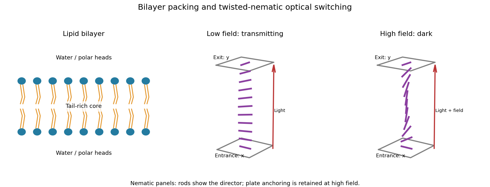
A lipid bilayer has two opposed leaflets: hydrophilic heads contact the water on either side, while hydrophobic tails meet in its interior. This arrangement reduces exposure of the tails to water through the hydrophobic effect. Approximately cylindrical molecular packing favors a lipid bilayer; a strongly wedge-shaped amphiphile can instead form a micelle. Closing a sheet into a lipid vesicle removes its costly exposed edge.
Within a fluid membrane, lipids diffuse laterally, while transfer of a polar head through the hydrophobic core is much slower without assistance. The core limits passive transport of ions and many polar solutes; membrane proteins provide selective routes. Bending costs free energy but does not require the large area compression associated with stretching the molecular packing, so a thin membrane can bend readily while strongly resisting area change. Cholesterol and tail saturation alter packing and fluidity. In a cell, the bilayer also supports embedded proteins and a difference of electric potential across the membrane.