Topics (384k) Articles (398k) Users (410) Discussions (242) Comments (383) Files (13k) New article
Past exam of the biochemistry course of the University of Cambridge 2024 Paper 4 Section III 7 Solution by  Codex 0 2026-10-20
Plant lipid diversity is generated by combinatorial biosynthesis in several compartments, selective exchange and remodeling, and evolutionary diversification of the underlying enzymes. Its functions extend from membrane mechanics and photosynthesis to storage, barriers and signaling, so no single adaptive explanation covers every lipid species.
At the simplest level, different acyl chains vary in length, double-bond number and position, and chemical substitution. Plastid fatty acid synthase builds chains carried by an acyl carrier protein. Chain termination and export determine which products enter further metabolism. Endoplasmic-reticulum fatty acid elongase enzymes extend selected acyl substrates, creating very-long-chain products used in surface and membrane lipids. Fatty acid desaturases introduce double bonds into particular substrates, while specialized enzyme variants can introduce hydroxyl groups or other unusual chemistry. Substrate specificity, reaction order and cellular location therefore generate many distinct acyl pools from a comparatively limited set of carbon precursors.
Chains are only one dimension. Acyltransferases assemble them onto glycerol or other backbones with positional specificity. Headgroup pathways generate glycerophospholipids and galactolipids, while distinct pathways produce sphingolipids and sterols. A lipid name such as phosphatidylcholine denotes a class, not a unique molecular species: two acyl chains can each vary, and their positions can matter. Storage triacylglycerols likewise combine many possible acyl groups. Functional diversity is therefore much larger than the number of lipid headgroups.
Compartmentation constrains which combinations occur. Plastid and ER assembly pathways use different enzymes and precursor pools, and products move between them. Monogalactosyldiacylglycerol and digalactosyldiacylglycerol are major constituents of photosynthetic membranes, whose requirements differ from those of the plasma membrane or a storage oil body. Acyl editing replaces chains on existing lipids, connecting synthesis to remodeling rather than simply producing a fixed end product. Selective transport, turnover and developmental expression create tissue-specific compositions even when the genome is unchanged.
An important test of functional importance comes from manipulating the enzymes rather than correlating lipid abundance with habitat. An Arabidopsis thaliana desaturase-deficient mutant lacking trienoic fatty acids was able to grow under permissive conditions but lost normal chloroplast performance during prolonged chilling. This supports a role for those acyl structures in maintaining photosynthetic function at low temperature.[ref]
Trienoic Fatty Acids Are Required to Maintain Chloroplast Function at Low Temperatures by Jean-Marc Routaboul and others. Desaturase-mutant physiology under low-temperature treatment
The inference is more precise than “unsaturated lipids are always better”: excessive unsaturation can also increase susceptibility to oxidation, and requirements depend on temperature and membrane function.Nutrient availability supplies another driver. Phospholipid synthesis requires phosphorus, whereas galactolipids can provide membrane material without a phosphate headgroup. Under phosphate deprivation, organelle lipid analyses detected redistribution of digalactosyldiacylglycerol into mitochondrial membranes. This demonstrates regulated compositional plasticity rather than requiring a new species to evolve every time phosphate becomes scarce.[ref]
Other selective pressures concern life on land and interactions with other organisms. Cutin, suberin and plant cuticular wax contribute to control of water loss, permeability and surface protection. Their chain lengths and oxygenation patterns affect packing and barrier properties. Seed oils store energy and carbon for establishment, while lipid-derived signals coordinate development and responses to damage. Pathogens, herbivores, drought, salinity and changing light environments can favour different combinations. A lipid useful as a defensive precursor may impose a cost on growth or oxidation resistance, creating trade-offs rather than one universal optimum.
Evolution can generate new chemistry through gene duplication, altered substrate specificity, changes in expression and retargeting of enzymes between compartments. A duplicated desaturase-like enzyme, for example, can retain the original reaction in one copy while another explores related substrate chemistry. Selection can preserve a useful product or tissue-specific role, while drift and biochemical side reactions also contribute to observed diversity. Cross-species lipid differences alone do not establish their adaptive value.
To separate these explanations, combine quantitative lipidomics, isotope tracing, enzyme biochemistry, genetic perturbation and physiological rescue. Compare plants at matched developmental stages and environments, because plastic remodeling can otherwise be mistaken for an evolved difference. The chemistry arises from modular enzymes and compartmentalized flux; the evolutionary explanation requires connecting particular molecular variants to performance in a defined ecological setting.
Past exam of the biochemistry course of the University of Cambridge 2024 Paper 4 Section III 6 Solution by Codex 0 2026-10-20
Codex 0 2026-10-20Cytochrome P450s are versatile haem-containing oxidation enzymes that can install chemically difficult, highly selective functional groups. Photosynthetic microorganisms can supply their reducing equivalents and metabolic precursors, but useful production requires coordinated enzyme expression, electron delivery, substrate supply and product handling.
A Cytochrome P450 contains an iron–haem centre coordinated by a protein cysteine thiolate. The characteristic spectrum of its reduced carbon-monoxide complex gives the family its name. A common monooxygenation can be writtenOne oxygen atom enters the product and the other is reduced to water. Electron transfer and protonation activate bound oxygen, generating a powerful oxidant such as P450 compound I. Hydrogen abstraction followed by oxygen rebound can hydroxylate a C–H bond, but the family also supports other reactions, including epoxidation and successive oxidation steps. The exact chemistry is enzyme- and substrate-dependent; not every P450 simply performs a single hydroxylation.
This selectivity is valuable for complex natural products. Closely related substrates can differ in the position and stereochemistry of oxygenation, and an enzyme can distinguish sites that are difficult to separate by ordinary chemical synthesis. Oxygenated terpenoid intermediates, drug precursors and other specialized metabolites are therefore attractive targets. The opportunity is selective chemistry, not a guarantee that every plant pathway will be cheap to reproduce in a microbial host.
In an oxygenic photosynthetic chassis, light absorption by photosystem II supports extraction of electrons from water. Transfer through the photosynthetic chain and photosystem I generates reduced ferredoxin, which can feed a compatible P450 directly or contribute to NADPH production for an appropriate reductase system. The distinction matters: native microsomal P450s often use a flavoprotein reductase, whereas engineered direct ferredoxin delivery can bypass that partner. Electrons must still reach the P450 at adequate rates and with correct coupling to substrate oxidation.
There is a clear experimental proof of principle. A plant CYP79A1 catalytic domain was fused to the photosystem-associated PsaM anchor in a cyanobacterium, positioning it near the thylakoid electron supply. Activity was observed in light-driven experiments. This demonstrates functional rerouting of photosynthetic reducing power into a plant P450 reaction, rather than establishing commercial production of a finished pharmaceutical.[ref]
A production design starts from the entire pathway. Supply a suitable scaffold from endogenous metabolism or introduced enzymes, express the required P450s with compatible membrane anchors and redox partners, and include any downstream transferases or other finishing enzymes. CYP79A1, for example, participates in the pathway to dhurrin; expressing it alone establishes one activity, not the completed multienzyme pathway. For a new high-value oxygenated terpenoid, one must similarly verify every intermediate rather than equating scaffold accumulation with final-product synthesis.
Several constraints can dominate performance. Plant membrane enzymes may fold poorly, insert in the wrong orientation or fail to acquire haem. An abundant P450 may remain inactive because its substrate or electron donor is limiting. Competition with carbon fixation and essential redox metabolism can make maximal electron diversion harmful to growth. Uncoupled oxygen activation can generate reactive oxygen species instead of product. Excess light may increase oxidative damage, while high cell density limits light penetration. Oxygen supply is required for ordinary P450 monooxygenation even though the host produces oxygen photosynthetically.
Optimization should therefore measure carbon flux, enzyme abundance, redox state and products together. Adjust expression ratios, redox-partner choice, subcellular localization and induction timing; relieve precursor bottlenecks without starving essential pathways; and consider export or product capture to limit toxicity. A bioreactor must balance mixing, illuminated surface area, gas exchange and biomass density.
Useful controls include dark incubation, a catalytic-site mutant, pathway-minus strains and perturbation of photosynthetic electron supply while monitoring cell viability. Mass spectrometry and authentic standards establish product identity; oxygen uptake or a fluorescent reporter alone does not. Finally report titre, productivity, carbon yield and stability over repeated cultivation. Light-powered enzyme activity is the biochemical entry point; reliable, selective and economically recoverable product formation is the engineering goal.
Past exam of the biochemistry course of the University of Cambridge 2024 Paper 4 Section II 5 Solution by Codex 0 2026-10-20
Codex 0 2026-10-20The SRP–receptor interaction is a regulated delivery and handover reaction, not merely a binding equilibrium. GTP binding and hydrolysis provide timing, conformational control and recycling so that a hydrophobic nascent chain reaches an available translocon before it aggregates or is released at the wrong place.
The signal recognition particle recognizes an appropriate exposed signal sequence or membrane-spanning segment on a ribosome-associated nascent chain. Its conserved SRP54 subunit contains a signal-binding region and a GTPase domain; bacterial Ffh is the corresponding homolog. The partner GTPase is the SRP receptor alpha subunit in the eukaryotic ER system or FtsY in bacteria. Thus the targeting machinery integrates information from the emerging chain, the ribosome and a membrane-associated receptor. Productive delivery also requires a translocon, not just any membrane surface.
GTP loading permits the two homologous GTPases to assemble into a complex. They then undergo further rearrangements and mutually stimulate their hydrolysis reactions. This is more elaborate than a one-step association of two preformed active surfaces: different nucleotide-bound conformations distinguish cargo capture, membrane delivery, handover competence and termination. Correct cargo and a suitable membrane/translocon environment can alter the kinetics of these transitions. The pathway can therefore respond to whether the chain is appropriate and whether a downstream acceptor is ready.
The timing matters. If hydrolysis occurs too early, SRP and receptor may dissociate before cargo transfer. If dissociation is prevented indefinitely, a stable complex can trap the machinery instead of making another delivery. Efficient targeting consequently requires both a sufficiently long-lived delivery intermediate and a route for its timely disassembly. Experiments with GTPase mutants separated receptor interaction or hydrolysis behaviour from successful cargo unloading, demonstrating that formation of a stable complex is not itself the endpoint of targeting.[ref]
Conformational changes in the GTPase modules of the signal reception particle and its receptor drive initiation of protein translocation by Shu-ou Shan and others. Mutant targeting and translocation assays separating binding from productive handover
This provides a form of kinetic discrimination. A wrong nascent chain can dissociate or fail to progress through a sequence of conformational checkpoints, while an appropriate chain promotes forward passage. Energy-consuming termination and recycling prevent every encounter from being governed solely by the final binding affinity. The logic resembles kinetic proofreading, but one should distinguish a general analogy from a demonstrated proofreading scheme: identifying a particular rejection step requires rates and fluxes, not merely observing GTP consumption. Not every incorrect interaction necessarily consumes the same number of GTP molecules.
A useful comparison is a passive docking model. At equilibrium, strong SRP–receptor affinity would favour occupancy but could impede turnover, and it would not establish that the cargo has been delivered to a conducting channel. In the actual cycle, nucleotide states connect the binding interface to signal-sequence release and receptor recycling. Hydrolysis makes a directional, dissipative cycle possible when cellular nucleotide ratios are maintained. It should not be described as GTP physically pushing the polypeptide through the membrane; translocation and membrane insertion use additional interactions and, depending on the substrate and system, other energy inputs.
A decisive experimental design measures several outputs separately: SRP–receptor association, GTP hydrolysis, transfer of a labeled nascent chain to the translocon, and protection or processing of the resulting product. Compare a nonhydrolyzable nucleotide analog with mutants that alter specific conformational transitions, and control for ribosome binding and membrane integrity. A reagent that stabilizes docking but reduces completed import shows why affinity alone is an inadequate readout. Testing repeated targeting cycles also distinguishes one successful handover from efficient recycling.
The answer is therefore coordination and reliable turnover. Energy expenditure links recognition to an available membrane insertion pathway, controls the lifetime of delivery intermediates, helps reject unproductive encounters and returns the machinery to a reusable state. The strength of this explanation is that it predicts experimentally separable failures—binding without handover, premature release, and stalled recycling—rather than assigning GTP the vague role of supplying energy to an otherwise simple contact.
Past exam of the biochemistry course of the University of Cambridge 2024 Paper 4 Section II 4 Solution by Codex 0 2026-10-20
Codex 0 2026-10-20Myosins convert a nucleotide-driven actin-binding cycle into movement, force or anchoring. Their different cellular roles arise from variations in motor kinetics, directionality, lever-arm geometry, oligomerization and cargo binding. A class name alone does not specify a universal stepping speed or duty ratio.
A myosin usually contains an actin-binding motor domain with an ATPase active site, a neck associated with light chains, and a tail involved in cargo association or assembly. The myosin lever arm amplifies small motor-domain rearrangements. The tail can connect the motor to a membrane, receptor, organelle or another myosin molecule. Some isoforms are autoinhibited until cargo binding, calcium-dependent regulation or phosphorylation favours an active conformation.
The mechanochemical cycle can be organized around actin affinity. In a strongly bound nucleotide-free state, ATP binding weakens actin attachment and promotes detachment. ATP hydrolysis prepares a detached or weakly bound ADP–phosphate state. Subsequent actin interaction and transitions around phosphate release stabilize strong binding and generate the myosin power stroke. ADP release and the next ATP-binding event complete the cycle. The exact ordering and rates of structural transitions and phosphate release depend on isoform and load; the cycle should not be reduced to a claim that ATP cleavage itself is the instantaneous mechanical stroke.
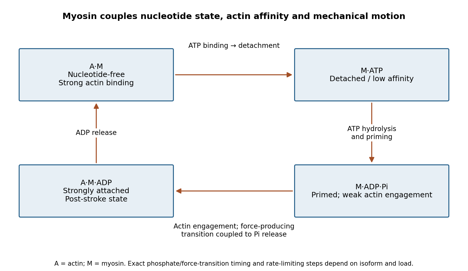
Original myosin actin-affinity and nucleotide cycle, separating ATP binding, hydrolysis and the force-producing transition
. Details. Directionality depends on motor geometry and the polarity of actin. Most characterized myosins move toward the barbed end, but Myosin VI moves toward the pointed end. Motor assays established this reversed direction directly, showing that “myosin” does not imply a single transport direction.[ref]
Myosin VI is an actin-based motor that moves backwards by A. L. Wells and others. Directionality assays on polarized actin
In a cortex with complex actin architecture, filament orientation must therefore be known before predicting net cargo movement.The myosin duty ratio is the fraction of a cycle spent strongly bound. A single low-duty motor repeatedly loses its track, whereas an assembly of many such motors can maintain collective contact. Myosin II illustrates this strategy: bipolar filaments bind oppositely oriented actin arrays and generate sliding or tension. In muscle, ordered filament geometry converts sliding into contraction. In nonmuscle cells, related contractile networks contribute to the division furrow, junction remodeling, migration and cortical tension. If the network is anchored, the same sliding mechanism can produce an approximately isometric force rather than large displacement.
Cargo transport poses a different requirement. Dimeric Myosin V motors coordinate their heads so that one remains attached while the other advances. A high-duty coordinated cycle supports myosin processivity, enabling repeated steps before dissociation. Single-fluorophore localization followed one labeled head moving by roughly twice the centre-of-mass step, supporting alternating hand-over-hand motion rather than equal short advances of both heads.[ref]
Myosin V Walks Hand-Over-Hand: Single Fluorophore Imaging with 1.5-nm Localization by Ahmet Yildiz and others. Single-head localization distinguishing walking mechanisms
The approximate 36 nm centre-of-mass repeat reflects the actin track and motor geometry; it is not a step size shared by all myosins.Transport and tension holding overlap. Load can slow nucleotide release and prolong strong binding, allowing some motors to act as dynamic anchors. Myosin VI can participate in endocytic membrane organization or cargo movement, depending on its partners and assembly state. Class-I myosins couple membranes to cortical actin and can regulate membrane tension and local transport. Specialized myosins organize sensory structures or long-range organelle positioning. These roles depend as much on tail-associated adaptors and local actin arrangement as on the catalytic head.
Regulation matches activity to demand. Light-chain phosphorylation can promote nonmuscle myosin II activation and filament assembly. Calcium-sensitive light chains, including calmodulin in appropriate classes, alter motor regulation or lever-arm organization. Cargo-associated adaptors can relieve autoinhibition and assemble transport complexes. Motor number, opposing loads, actin turnover and competition with other transport systems further determine the outcome.
Useful experiments separate these contributions. Actin-gliding assays establish direction and movement, single-molecule tracking measures stepping and run length, and force measurements reveal load-dependent kinetics. ATPase assays alone establish chemical turnover but not productive movement. In cells, rescue with a motor-defective or cargo-binding-defective protein can distinguish catalytic force production from a structural scaffolding role. The unifying mechanism is a coordinated affinity-and-conformation cycle; the biological output can be motion, sustained tension or controlled coupling of membranes to the cytoskeleton.
Past exam of the biochemistry course of the University of Cambridge 2024 Paper 4 Section II 3 Solution by Codex 0 2026-10-20
Codex 0 2026-10-20Maturation best explains major changes in Golgi cisternal identity and early-to-late endosomal progression, but neither pathway is a conveyor belt with no stable domains or transport carriers. The useful comparison concerns what persists—membrane, cargo or resident machinery—and what the experiments actually follow.
In a stable-compartment model, each compartment retains its processing identity while cargo moves onward in vesicles or tubules. In cisternal maturation, cargo can remain within a cisterna while its resident enzymes and trafficking machinery change, turning an early cisterna into a later one. This requires selective recycling of resident machinery in the opposite direction. Both models therefore depend on membrane trafficking; observing vesicles alone does not distinguish them. A static image of separate cis and trans markers is likewise compatible with a population of maturing cisternae.
A decisive experiment follows an individual structure over time using early and late resident markers. In budding yeast, live imaging showed initially early-marker-positive Golgi cisternae losing that marker and acquiring a late marker. This is strong evidence for changing cisternal identity within the tracked object.[ref]
Golgi maturation visualized in living yeast by Eugene Losev and others. Live imaging of early-to-late resident-marker replacement
A complementary experiment follows synchronized cargo together with residents. Cargo retention while residents are replaced favours maturation; cargo movement between objects whose residents remain stable favours transfer between persistent processing domains.Large cargo makes a strong mechanistic test. A long procollagen assembly cannot simply be assumed to enter a small conventional coated vesicle. Maturation can move such cargo through the stack without packaging it into small forward carriers at every step. However, cargo size does not by itself prove that every soluble cargo uses the same route. Tubular connections, larger carriers and rapid exchange of small cargo can coexist with maturation. The geometry and spatial resolution of stacked mammalian Golgi also differ from those of dispersed yeast cisternae, so one should not transfer every kinetic detail between systems without testing it.
For the Golgi, the strongest synthesis is therefore a dynamic population of processing compartments with changing identity, coupled to resident recycling and cargo-dependent transport. COPI and other retrieval pathways can maintain enzyme gradients while COPII supplies new material from the endoplasmic reticulum. A named cis, medial or trans region can persist statistically even when its component membranes and proteins do not. Bulk transport, rapid cargo exchange and local stable zones need not be mutually exclusive.
The endocytic pathway poses a related but distinct problem. An early endosome receives material from the surface and separates recycling cargo from cargo retained for degradation. A strictly stable early-endosome model predicts that degradative material must leave in carriers while early identity persists. A maturation model predicts that a cargo-bearing domain changes into a late endosome, alongside removal of material that should recycle.
Time-resolved Rab5 and Rab7 measurements demonstrated replacement of an early Rab machinery by a late one on cargo-bearing endosomes. This Rab conversion provides direct evidence for maturation rather than inferring it solely from differently labeled endpoint compartments.[ref]
Rab Conversion as a Mechanism of Progression from Early to Late Endosomes by Jochen Rink and others. Rab5 loss and Rab7 acquisition on progressing endosomes
The associated changes in lipids, acidity, motility, fusion machinery and internal membrane organization make this a functional transition, rather than a marker swap alone.Yet recycling domains can bud or tubulate from a maturing endosome. Some cargos return to the plasma membrane; others use retromer-associated retrieval routes; still others enter intralumenal vesicles and continue toward lysosomes. A maturing degradative region can therefore coexist with relatively persistent sorting activities elsewhere in the same network. Endosome fusion and fission also mean that an object tracked by microscopy may exchange membrane extensively.
To discriminate mechanisms, combine live tracking of residents and cargo with pulse-chase kinetics, acute perturbations of retrieval machinery and quantitative membrane accounting. Apparent marker conversion caused by two unresolved objects crossing must be excluded. Conversely, failure to observe conversion at low time resolution is not proof of stability. The evidence favours maturation as a major organizing principle, with selective sorting, retrieval and exchange explaining how cells retain steady trafficking functions while individual compartments change.
Past exam of the biochemistry course of the University of Cambridge 2024 Paper 4 Section I 2 Solution by Codex 0 2026-10-20
Codex 0 2026-10-20First establish that proteins cross the organelle boundary; then define its resident proteome and determine which residents arrive by import. A protein detected near an organelle may be a surface binder, contaminant, transient visitor or locally synthesized product. These possibilities require different controls.
Start with an operational definition of the new organelle: its location, boundary, ultrastructure and reproducible biochemical enrichment. Fluorescence microscopy with independent markers and electron microscopy can determine whether it is membrane-bounded and whether it contacts or engulfs other compartments. A condensate without a membrane can recruit proteins by partitioning; that should not be mistaken for translocation through a membrane. Purification by density-gradient centrifugation or affinity isolation should include markers for likely contaminants and measurements of organelle integrity. An enriched fraction is a starting material, not evidence of import by itself.
Choose candidate proteins from fractionation, imaging or sequence predictions and make minimally perturbing labeled versions. Follow newly synthesized protein using a pulse-chase experiment and independently verify its position relative to the organelle boundary. Loss of a cytosolic precursor followed by accumulation of an internal mature form is suggestive, but proteolytic processing alone does not demonstrate translocation. Tags can obscure targeting signals or change folding, so compare different tag positions and validate endogenous localization where possible.
The key biochemical test is an organelle import assay coupled to a protease protection assay. Incubate isolated intact organelles with a labeled precursor, then remove externally accessible protein with protease. Imported lumenal or matrix protein should be protected in intact organelles but become sensitive after detergent disruption. Surface-bound controls must remain accessible; an independent internal marker tests organelle sealing. Include organelle-free incubations to identify protease-resistant aggregates, and verify that detergent does not simply inactivate the protease. Carbonate extraction or membrane fractionation can distinguish an integral membrane product from a soluble internal one. For membrane proteins, map protected fragments and terminal accessibility rather than demanding protection of the entire chain.
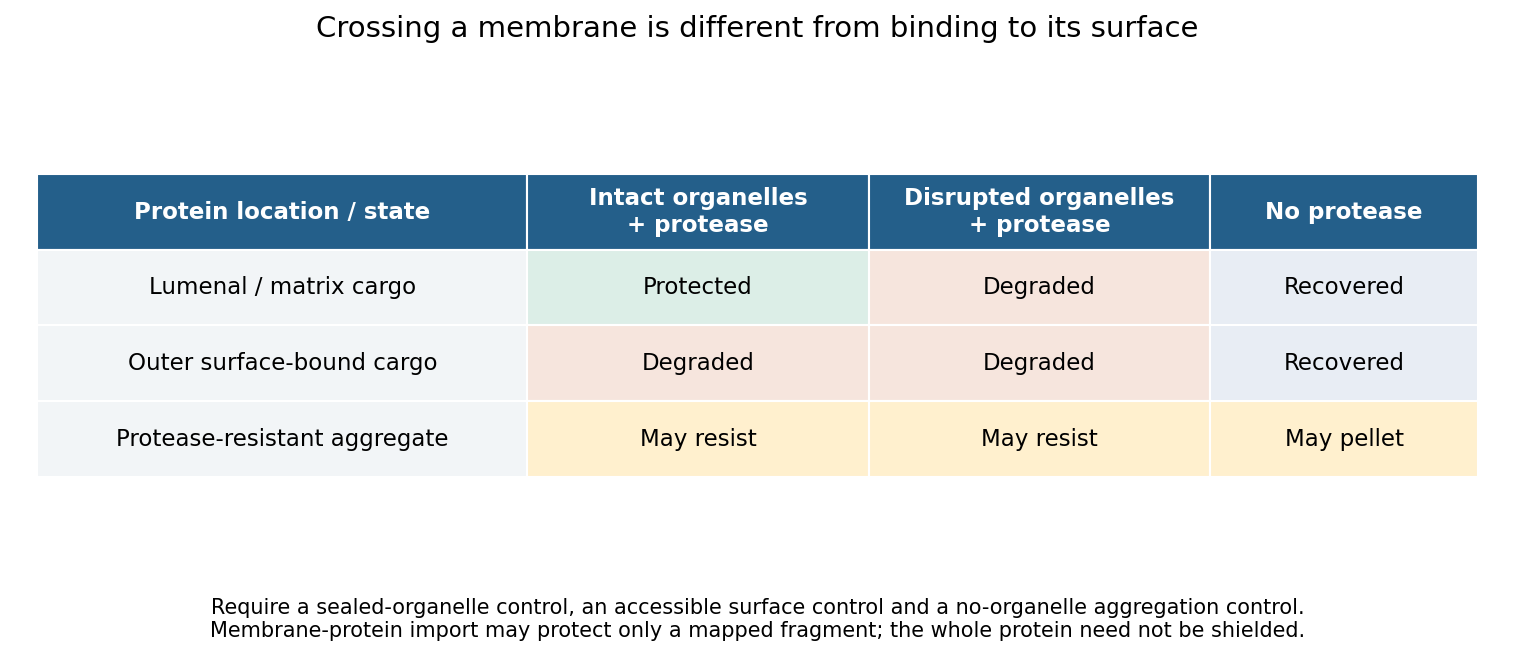
Original control logic separating protein import from surface binding and protease-resistant contamination
. Details. Perturbation then tests the mechanism. Compare incubation time and temperature; deplete and restore relevant energy sources; dissipate a membrane potential only after confirming that this treatment preserves membrane integrity; and compete with an excess of unlabeled substrate. Energy requirements must be discovered rather than assumed. Mitochondrial matrix entry, ER translocation and peroxisomal import use different machinery. In particular, stable folding is not a universal barrier: primary peroxisome experiments demonstrated import of stably folded substrates.[ref]
Import of stably folded proteins into peroxisomes by P. A. Walton and others. Import tests challenging a universal unfolding requirement
Mutating a candidate targeting signal should reduce import, while appending that signal to a suitable reporter can test sufficiency. Disrupting a candidate receptor or translocation component, followed by rescue with the wild-type protein, tests necessity. Because a blocked pathway may destroy the organelle indirectly, pair import measurements with morphology, membrane integrity and the import of unrelated controls. An import-defective allele that leaves the compartment otherwise functional is more informative than complete organelle loss. A negative isolated-organelle result does not exclude import if purification removed a required factor; compare with an intact-cell assay. Conversely, protection shows internalization but does not alone distinguish direct protein translocation from delivery inside a vesicle or engulfed compartment. Route-specific perturbations and topology measurements must resolve that distinction.
For the protein complement, combine quantitative spatial proteomics with complementary methods. Measure protein abundance across an entire fractionation gradient and identify proteins with reproducible localization profiles, rather than accepting every protein in a single pellet. Compare intact-organelle protease treatment with disrupted controls, and compare wild type with an import-defective condition. Analyze both soluble and membrane fractions. Liquid chromatography-mass spectrometry identifies peptides, but poorly soluble, scarce or unusual proteins may escape detection, so fractionation and acquisition strategies should be varied.
An internally localized TurboID or other proximity labeling enzyme can reveal proteins accessible in the relevant compartment in living cells. A cytosolic control, a catalytically inactive labeling construct and matched expression levels help identify background. Proximity labeling establishes accessibility and neighborhood, not translocation or stable residency by itself; labeling radius, reagent access and perturbation by the enzyme must be considered.[ref]
Finally distinguish resident, imported and all imported. A resident may have been synthesized by an organelle genome; an imported protein may only transit through the compartment. Time-resolved labeling, precursor processing, localization and dependence on a defined import pathway can classify these cases. Report coverage across growth conditions, detection limits and a confidence tier for each candidate. No single proteomic experiment proves absolute completeness, but convergent topology, kinetic and genetic evidence can produce a well-supported import catalogue.
Past exam of the biochemistry course of the University of Cambridge 2024 Paper 4 Section I 1 Solution by Codex 0 2026-10-20
Codex 0 2026-10-20An organelle maintains a reproducible molecular and functional organization while continuously replacing its components. Identity is therefore a regulated steady state, rather than a permanent collection of molecules. A useful explanation must connect selective delivery and retrieval to local biochemical reactions, physical boundaries and inheritance.
For a membrane-bounded organelle, the lipid bilayer separates its contents from the cytosol, preserves membrane-protein topology and permits lumenal conditions to differ from the surroundings. A lysosome, for example, requires an acidic lumen and retained hydrolases as well as a membrane. Its vacuolar proton ATPase establishes acidity, while counterion conductance and regulated transport prevent unbounded charge accumulation. An endoplasmic reticulum lumen instead supports protein folding and calcium storage. These environments determine which reactions occur and help stabilize the appropriate resident proteins. A boundary alone is insufficient: an empty vesicle can have the same basic bilayer architecture without the same biological identity.
Membrane composition creates selective recruitment sites. Local phosphoinositide synthesis and dephosphorylation produce membrane domains recognized by particular lipid-binding proteins. Phosphatidylinositol 3-phosphate is associated with early endosomal domains, whereas phosphatidylinositol 4-phosphate contributes to Golgi organization. These distributions reflect localized enzymes and regulated turnover, not an immutable lipid barcode. Other properties—including sterol abundance, bilayer thickness and curvature—can favour some membrane proteins over others. Membrane contact sites and lipid transfer proteins redistribute selected lipids while preserving the separation of the two compartments.
Rab GTPases provide a second layer of recognition. Membrane recruitment and nucleotide exchange generate an active Rab domain; effectors then promote vesicle tethering, motor recruitment and sorting. A Rab and its effectors can reinforce one another's localization. GTP hydrolysis and extraction provide opposing reactions that prevent uncontrolled spread. SNARE proteins, together with tethering factors and the appropriate lipid environment, subsequently favour fusion of compatible membranes. Specificity is combinatorial: neither a Rab nor a SNARE should be treated as a unique address sufficient by itself.
Membrane trafficking would progressively mix organelles without selective retrieval. COPII exports selected cargo from the endoplasmic reticulum, while COPI-dependent and other retrieval pathways return escaped residents and trafficking machinery. Resident proteins can be retained through interactions or retrieved using recognizable sorting information. The Golgi similarly balances forward cargo movement with recycling of processing enzymes. Endosomes sort some receptors back to the surface through recycling carriers while directing other material toward degradation. Such exchange preserves an organelle's characteristic composition without requiring every incoming molecule to remain there.
Identity can also change in an organized sequence. In endosome maturation, a compartment can lose Rab5 and acquire Rab7 while retaining internalized material. Live imaging of this Rab conversion argues that an early identity is actively replaced, rather than the same early compartment retaining an unchanging molecular label forever.[ref]
Rab Conversion as a Mechanism of Progression from Early to Late Endosomes by Jochen Rink and others. Live-cell Rab-marker conversion during cargo progression
A cell maintains distinct early and late populations even though individual compartments pass between them. Thus stable system-level organization and changing individual organelles are compatible.Some compartments use additional rules. Mitochondria and chloroplasts combine organelle protein import, genome expression, membrane growth and division; their identity cannot be reduced to vesicle fusion specificity. Peroxisomes import matrix proteins and receive membrane constituents through their own biogenesis pathways. Organelle positioning, fission and inheritance keep these functions distributed through growth and division, while damage recognition and turnover remove defective components.
Experimentally, one should distinguish causation from marker correlation. Acute inhibition of a localized lipid enzyme or Rab regulator can be followed by live imaging, cargo transport, lumenal pH measurements and rescue with a functional allele. A convincing identity change alters function and multiple independent features, rather than merely displacing a fluorescent marker. The central principle is selective flux plus local self-organization, balanced by retrieval and turnover.
Past exam of the biochemistry course of the University of Cambridge 2024 Paper 3 Section II 7 Solution by Codex 0 2026-10-20
Codex 0 2026-10-20Yes: a microRNA can suppress tumour formation or progression by restraining an oncogenic programme, but that role must be demonstrated for a defined microRNA, tissue and genetic context. Reduced expression in a tumour or growth inhibition after massive microRNA mimic transfection is suggestive rather than sufficient evidence of an endogenous tumour-suppressor function.
microRNAs are short regulatory RNAs whose guide strands associate with Argonaute. Recognition through sequence complementarity, often involving a microRNA seed region and a target's 3′ untranslated region, can recruit translational repression and RNA decay machinery. Canonical precursor processing involves Drosha and Dicer, although not every microRNA follows every canonical step. A microRNA can affect many transcripts, and a transcript can receive input from several microRNAs. Therefore modest coordinated repression of several growth or survival factors can have a substantial phenotype even without complete elimination of one target. Suppression is a property of the regulatory network, not of an RNA being universally “anticancer”.
The let-7 family supplies a mechanistic example. Experiments connecting let-7-responsive sequences in Ras transcripts with regulation of Ras expression linked a developmental regulatory RNA to a conserved growth pathway.[ref]
RAS is regulated by the let-7 microRNA family by Steven M. Johnson and others. Target-site reporters, developmental genetics and human RAS regulation
Reduced let-7 in lung cancer is compatible with loss of this restraint, but the inverse expression relationship alone would not show causation. Functional experiments restoring let-7 in relevant lung-cancer systems suppressed tumour development, providing a stronger test of tumour-suppressive capacity.[ref]Suppression of non-small cell lung tumor development by the let-7 microRNA family by Manish S. Kumar and others. Functional let-7 manipulation in lung-tumour models
Expression should still be compared with a physiological range, and one must distinguish preventing tumour initiation from treating an established tumour.Genetic loss gives a complementary direction of evidence. The miR-15a and miR-16-1 cluster lies within a recurrently deleted region associated with chronic lymphocytic leukaemia. Deletion of the corresponding mouse regulatory region or the microRNA cluster increased B-cell proliferative abnormalities and produced clonal lymphoproliferative disease in a subset of animals.[ref]
DLEU2 microRNA cluster deletion study by Ulf Klein and others. Separate deletions of the broader region and the miRNA cluster; B-cell proliferation and disease
Comparing the smaller cluster deletion with a larger deletion matters because neighbouring genes can contribute to a human copy-number association. Disease after specific microRNA loss supports endogenous restraint more directly than an expression signature does; incomplete penetrance and latency imply cooperation and context rather than an automatic one-step conversion of every b cell.The p53–miR-34 connection illustrates both strength and limitation of a proposed pathway. p53 activation can induce miR-34-family transcription, and ectopic miR-34 family can restrain proliferation or promote death in suitable biological cells, connecting a transcriptional tumour suppressor with post-transcriptional repression.[ref]
A microRNA component of the p53 tumour suppressor network by Lin He and others. p53-dependent miR-34 induction and growth-related responses
Yet mice lacking all three miR-34 family members retained substantial p53-dependent arrest and apoptosis and did not show the predicted general increase in tumour susceptibility in the tested settings.[ref]Intact p53-dependent responses in miR-34-deficient mice by Carla P. Concepcion and others. Triple-family deletion and in vivo tumour-suppression tests
This does not prove that miR-34 family cannot suppress a tumour in any setting. It shows that a gain-of-function phenotype and placement downstream of p53 do not establish that the microRNA family is an indispensable general mediator of p53 tumour suppression.A convincing analysis combines several evidence types. Measure mature guide RNA in defined malignant and normal biological cell populations, distinguishing it from precursor abundance and changes in stromal composition. Determine whether deletion, promoter silencing or altered processing explains loss. Reduce or delete the endogenous microRNA and test initiation or progression in a suitable model, with restoration from a controlled expression system. Compare related family members to expose redundancy. Use seed-mutant controls and multiple reagents to distinguish sequence-specific regulation from delivery toxicity or innate immune activation; deleting an entire processing enzyme is not a specific test of one microRNA because it affects many regulatory RNAs.
To identify consequential targets, combine binding evidence, early RNA/protein responses and target-site tests. A 3′-region reporter whose seed-complementary site is mutated supports direct sequence-dependent regulation, but the decisive endogenous test is to edit the site or express a suitably controlled microRNA-resistant target and ask whether it rescues the phenotype. Failure of one target to rescue can reflect regulation of several cooperating targets rather than absence of specificity. Conversely, rescuing only bulk proliferation does not establish which target controls invasion or metastasis. Measure relevant tissue phenotypes and target protein levels, not merely a pathway enrichment score from predicted targets.
Tumour-suppressive activity also does not guarantee an effective therapy. Delivery must reach the relevant biological cells and release the RNA into the correct compartment without excessive exposure of normal tissues. A clinical miR-34a microRNA mimic study encountered serious immune-mediated toxicity, illustrating that an endogenous regulatory rationale does not establish safe systemic replacement.[ref]
Phase 1 study of MRX34, a liposomal miR-34a mimic, in patients with advanced solid tumours by David S. Hong and others. Clinical safety findings and limits of replacement therapy
Target abundance, isoform-specific 3′ regions and competing pathways can change the effect of the same microRNA between tumours. microRNAs can be genuine tumour suppressors; the strongest evidence is specific endogenous loss, controlled restoration and mechanistic target tests, interpreted alongside genetic redundancy and tissue context. Past exam of the biochemistry course of the University of Cambridge 2024 Paper 3 Section II 6 Solution by Codex 0 2026-10-20
Codex 0 2026-10-20Replication fidelity uses successive discrimination and repair systems, because no single enzyme detects every error. The important distinctions are between an incorrect base pair, a slipped sequence register, the wrong sugar incorporated into DNA, a damaged template and an incompletely or incorrectly restored chromosome. Repair must also identify which information should be retained: recognizing a mismatch without strand discrimination could fix the wrong sequence.
A polymerase can select an incorrect deoxynucleoside triphosphate and insert the wrong base through imperfect discrimination, transient base configurations or an altered active site environment. If the mismatch is extended and survives into another replication round, it can become a stable base substitution. Many replicative polymerases possess a separate 3′→5′ exonuclease proofreading site. A mismatched primer end tends to slow extension and transfer toward this site; removal of the terminal nucleotide permits another attempt at correct synthesis. This is distinct from the polymerase's forward synthetic reaction and from proofreading in biological translation. In mice, selective disruption of DNA polymerase δ proofreading increased mutation and tumour susceptibility, demonstrating an organismal consequence of unrepaired copying errors.[ref]
High incidence of epithelial cancers in mice deficient for DNA polymerase δ proofreading by Robert E. Goldsby and others. Proofreading-domain mutation, mutator phenotype and tumour development
Post-replicative DNA mismatch repair corrects many errors that escape the polymerase. In eukaryotes, MutSα, formed by MSH2 and MSH6, recognizes base mismatches and many short insertion–deletion loops; MutSβ, containing MSH2 and MSH3, has an important role in larger loops. Recruitment of MutL complexes, strand incision, excision, resynthesis and ligation replaces a stretch of the newly synthesized strand. Nascent-strand discontinuities and replication-associated factors including PCNA help direct the process. Purified human activities reconstituted nick-directed repair from defined substrates, showing how a mismatch and a strand-break signal can support directional replacement.[ref]
Human mismatch repair: reconstitution of a nick-directed bidirectional reaction by Nicoleta Constantin and others. Reconstitution using defined nicked substrates and human factors
The classic Escherichia coli MutH system instead uses transiently undermethylated daughter DNA at suitable sites; that methyl-directed mechanism is not a universal description of eukaryotic DNA mismatch repair or even of every bacterium.replication slippage creates a different substrate. In repetitive sequence, transient misalignment can leave an extra loop in the nascent or template strand. A retained nascent-strand loop can produce an insertion; copying past a looped-out template segment can produce a deletion. Short repeats are therefore vulnerable to length changes, and coding-region insertions or deletions can cause a frameshift if their length is not a multiple of three. DNA mismatch repair recognizes many such loops, although substrate size and structure affect efficiency. Yeast mismatch-repair mutants destabilized simple repetitive tracts, linking repair defects with microsatellite instability.[ref]
Destabilization of tracts of simple repetitive DNA in yeast by mutations affecting DNA mismatch repair by Micheline Strand and others. Repeat-length instability in repair mutants
This is a DNA-sequence frameshift mechanism, distinct from a ribosome changing reading frame during biological translation.The layers can be represented by conditional escape probabilities,where the second and third factors refer to escape among errors reaching the corresponding stage. As a purely illustrative calculation, per copied base. These are not asserted universal measured rates: mismatch identity, sequence, polymerase and repair state all matter. The multiplicative logic explains why disabling a later layer can reveal errors that had previously escaped detection without eliminating DNA synthesis itself.
Polymerases can also incorporate a ribonucleotide instead of a deoxyribonucleotide. base pairing may be correct, so ordinary mismatch recognition is insufficient. RNase H2 can initiate ribonucleotide excision repair by incising near an embedded ribonucleotide, followed by DNA synthesis, removal of the displaced segment and ligation. Reconstitution established this dedicated pathway.[ref]
RNase H2-initiated ribonucleotide excision repair by Justin L. Sparks and others. RNase H2-initiated incision and downstream repair reactions
RNase H1 processing of an RNA–DNA hybrid should not be equated with removal of a single isolated ribonucleotide. Separately, normal RNA primers on Okazaki fragments must be processed and the remaining nicks sealed; persistent primers, flaps or unligated junctions threaten chromosome integrity even without an incorrect base.A chemically damaged template can miscode or stall a fork. Small base lesions can be addressed by base excision repair, whereas nucleotide excision repair removes a damaged oligonucleotide around many bulky, helix-distorting lesions. These pathways repair chemical damage, not every mismatch made on an undamaged template. Replication across an oxidized base can require coordinated recognition of the lesion and the incorrectly paired partner so that repair does not convert damage into a fixed mutation. translesion synthesis permits passage across some lesions with specialized polymerases; it is damage tolerance, not necessarily lesion removal or error-free copying. template switching can provide another route around a block.
Stalled or collapsed forks can leave gaps, broken DNA and structural rearrangements. homologous recombination can use a sister chromatid to restore information and restart appropriate substrates; inappropriate pairing or template switching can instead change sequence organization. non-homologous end joining can restore continuity after a break but need not preserve every base, particularly when end processing is required. Replication checkpoints help stabilize forks and delay progression, while under-replication or rereplication requires control of origin use as well as lesion processing. Numerical chromosome-segregation errors are a related genome-stability problem but are not corrected by simply excising one mismatched base. Base selectivity and proofreading act during synthesis, mismatch and ribonucleotide repair address distinct residual substrates, and damage/fork-repair pathways restore templates and continuity with different fidelity limits.
Past exam of the biochemistry course of the University of Cambridge 2024 Paper 3 Section II 5 Solution by Codex 0 2026-10-20
Codex 0 2026-10-20Direction arises from switching between mutually incompatible states, with feedback providing memory and regulated destruction resetting the system. Different transitions use related logic, but a steep response, a bistable switch and an irreversible molecular event are not identical. The cell cycle also requires checkpoints and once-per-cycle licensing so that the ordered biochemical states produce a correct sequence of physical events.
At the G1/S transition, growth signals promote cyclin-dependent kinase activity and relieve RB-mediated restraint on E2F. E2F-dependent expression of additional cell-cycle components can reinforce the transition. More generally, activation of a kinase together with removal of its inhibitor creates positive feedback. In yeast, multisite phosphorylation of Sic1 permits recognition by the Cdc4 ubiquitin-ligase system and degradation of an inhibitor of S-phase kinase. This supplies a threshold that prevents weak or premature signals from launching the next programme.[ref]
Multisite phosphorylation of a CDK inhibitor sets a threshold for the onset of DNA replication by Piers Nash and others. Phosphorylation-dependent Sic1 recognition and timing
The detailed regulators vary across organisms and biological cell types; the conserved point is a coordinated transition from an inhibited state to one that reinforces its own activation.Mitotic entry gives an experimentally accessible example of bistability. cyclin B binding increases the available CDK1 complex, but Wee1-dependent inhibitory phosphorylation can keep it inactive. Cdc25 removes the inhibitory phosphate. Active CDK1 promotes Cdc25 activity and suppresses Wee1, giving a positive-feedback loop and a double-negative loop. Over a suitable input range, both a low-activity and a high-activity state can be stable. If cyclin input rises from interphase, activation occurs at a higher threshold than the input required to maintain a pre-existing mitotic state. These distinct thresholds constitute hysteresis and prevent small fluctuations from repeatedly reversing a committed transition.
In egg extracts, nondegradable cyclin could be supplied while CDK activity was approached from different initial states. The same cyclin input then supported different persistent activities depending on history, directly distinguishing hysteresis from a single steep but history-independent response.[ref]
Building a cell cycle oscillator: hysteresis and bistability in the activation of Cdc2 by Joseph R. Pomerening and others. History-dependent activity at matched nondegradable cyclin input
These are extract measurements, not universal numerical thresholds for every intact biological cell. A useful conceptual representation iswhere represents effective activating input. hysteresis supplies memory within this interval; it does not mean that the switch can never be reset.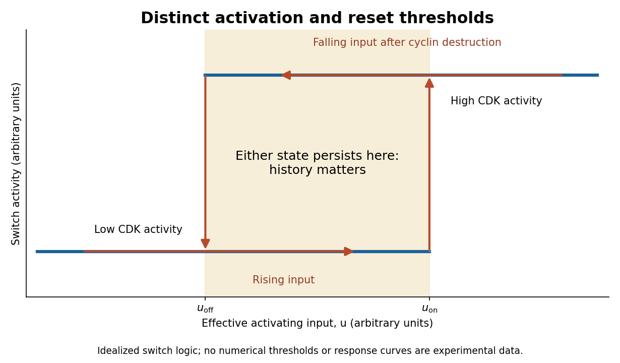
Hysteretic activation preserves state until a separate reset crosses the lower threshold
. Details. Reset is achieved by coupling activation to delayed negative feedback and proteolysis. Mitotic kinase activity helps prepare activation of the anaphase-promoting complex with Cdc20, while the spindle checkpoint prevents productive progression until attachment conditions are met. Once this restraint is satisfied, APC/C-dependent ubiquitination promotes destruction of securin and cyclin B. securin loss allows separase to cleave cohesin, making sister-chromatid separation possible. Experiments identifying cohesin cleavage connected regulated proteolysis to a physical transition that simple reversal of a phosphorylation state cannot undo.[ref]
Sister-chromatid separation at anaphase onset is promoted by cleavage of the cohesin subunit Scc1 by Frank Uhlmann and others. Cohesin cleavage and anaphase onset
Chromatids cannot be returned to a pre-anaphase chromosome merely by reducing separase activity.cyclin B destruction lowers CDK1 activity, allowing phosphatases to reverse mitotic phosphorylation and promoting an interphase state. Activation of anaphase-promoting complex with Cdh1 at low kinase activity can help keep cyclins low, while high kinase activity opposes that state. Thus low-CDK and high-CDK programmes are mutually antagonistic. Cyclin synthesis eventually provides a new activating input. Early experiments in extracts showed the importance of cyclin synthesis and destruction, including failure of normal exit with nondegradable cyclin.[ref]
The role of cyclin synthesis and degradation in the control of maturation promoting factor activity by Andrew W. Murray and others. Cyclin synthesis, degradation and nondegradable variants
A bistable activation module combined with an appropriate delayed reset can generate an ordered oscillation; a bistable module held at constant input need not oscillate on its own.replication licensing enforces another essential directionality. At low CDK activity, origin recognition complex with Cdc6 and Cdt1 loads MCM2-7 double hexamers, establishing competence to replicate. In S phase, activating kinases promote origin firing while high CDK activity and additional controls prevent renewed licensing. In animal biological cells, geminin inhibition of Cdt1 and regulated degradation help separate these states. Purified-protein reconstitution demonstrated loading of the replication-helicase precursor as a distinct biochemical event.[ref]
Concerted loading of Mcm2-7 double hexamers around DNA during DNA replication origin licensing by Dirk Remus and others. Origin licensing by double-hexamer loading
Separating licensing from firing allows many origins to be prepared but prevents fired regions from being copied repeatedly during the same cycle. Lowering mitotic kinase activity must eventually restore licensing competence for the next cycle, rather than permit uncontrolled rereplication during the current S phase.Checkpoint pathways modify or hold these switches when DNA is unreplicated, damaged or chromosomes are improperly attached; they do not supply the entire oscillator. Energy-consuming phosphorylation cycles, ubiquitination, proteolysis and resynthesis make the system a driven process, while feedback stabilizes distinct states. Thresholds provide commitment, hysteresis provides memory, destruction and cleavage enforce selected transitions, and licensing/checkpoints couple those transitions to one correct round of replication and segregation.
Past exam of the biochemistry course of the University of Cambridge 2024 Paper 3 Section II 4 Solution by Codex 0 2026-10-20
Codex 0 2026-10-20Mouse models allow causal and longitudinal tests in an intact organism, but their value depends on which part of human cancer the chosen model reproduces. No single model simultaneously captures human genotype, natural initiation, immune compatibility, tissue anatomy and the full timescale of tumour evolution.
A major strength is experimental control. Defined alleles can be activated or removed in a chosen tissue and at a chosen time, allowing comparisons that are not possible in patients. Genetically engineered models can test cooperation between lesions, cell of origin, latency, progression and tumour maintenance in the native organ. Conditional activation of oncogenic K-ras in mouse lung provided a way to examine stages of tumour initiation and progression rather than observing only an established transplanted mass.[ref]
Analysis of lung tumor initiation and progression using conditional expression of oncogenic K-ras by Erica L. Jackson and others. Conditional activation and staged lung pathology
A reversible or inducible intervention after tumour formation can then distinguish an initiation requirement from a maintenance dependency. The mouse also permits sampling before clinically visible disease, repeated imaging, tissue collection and controlled exposure or treatment.The host environment is another advantage. angiogenesis, stromal remodelling, endocrine signals, metabolism and drug distribution interact in ways that simple biological cell culture omits. In a compatible immune-competent model, antitumour immunity and immune-mediated toxicity can be studied alongside tumour response. Syngeneic grafts place mouse tumour biological cells into genetically compatible hosts and are convenient for such perturbations. Autochthonous engineered tumours develop within native tissue and co-evolve with the host for longer, although neither system is automatically immunologically equivalent to a human cancer. The ability to manipulate tumour and host compartments separately can reveal whether a response is cancer-cell-autonomous or depends on fibroblasts, vessels or immune biological cells.
Human xenografts provide a different advantage. Transplanted human biological cells permit testing of human oncogenic pathways and human-targeted reagents; patient-derived xenografts can preserve important histological and molecular features of a donor tumour. Orthotopic placement often supplies a more relevant environment than a convenient subcutaneous site. Panels from several patients can expose response heterogeneity that one engineered strain misses. Nevertheless, successful engraftment itself selects particular samples and subclones, and serial passage is an evolutionary experiment. Ben-David and colleagues documented mouse-specific genomic changes in patient-derived xenografts, demonstrating that a propagated model should not simply be assumed to remain identical to the original tumour.[ref]
Patient-derived xenografts undergo mouse-specific tumor evolution by Uri Ben-David and others. Longitudinal copy-number changes during xenograft propagation
Compare early and late passages with the donor, and use the passage appropriate to the question.The greatest xenograft limitation for many questions is the host. Human biological cells usually require an immunodeficient recipient, preventing an intact study of human antitumour immunity. Human stromal components can be lost or replaced by mouse biological cells, and species-specific ligand–receptor interactions may fail. Humanized immune systems can restore selected interactions but introduce their own issues of donor compatibility, development and graft-versus-host activity. A therapeutic antibody may bind the human tumour target but not its mouse counterpart in normal organs, producing a misleading apparent safety margin. Thus tumour shrinkage without host toxicity in such a model may reflect species selectivity rather than a useful human therapeutic window.
Engineered and syngeneic models also have limitations. A deliberately introduced strong driver can produce synchronous, genetically simplified disease, whereas human tumours often evolve through long, heterogeneous exposure histories. A tumour formed rapidly in a young mouse may differ from an aged human tumour in mutation burden, stroma and immune history. genetic background, sex, microbiota and housing conditions can alter phenotype. Species differences in lifespan, organ structure, metabolism, telomere regulation and drug pharmacokinetics further limit direct extrapolation. The tumour spectrum after p53 loss, for example, is informative about tumour suppression but does not by itself reproduce the distribution of human TP53-mutant cancers.[ref]
The endpoint and intervention must fit the biological question. Subcutaneous growth measures a local mass under an artificial geometry; tail-vein injection measures selected post-entry metastatic steps. Neither establishes a treatment effect on natural invasion or initial dissemination. Drug tests should establish exposure and target engagement in both tumour and relevant normal tissue, use clinically motivated schedules where possible, and measure survival or durable disease control as well as short-term volume. Rapidly growing implants may make some treatments look more effective than they are against slow, heterogeneous human disease.
Good design uses random allocation after a defined eligibility point, blinded measurement, prespecified endpoints and justified numbers of independent animals. Multiple tumours within one mouse are not automatically independent treatment replicates. Match or record relevant background variables and validate the intended allelic change and molecular phenotype. Ethical constraints favour well-chosen questions and informative longitudinal measurements rather than duplicating a weak model at larger scale. Combine mouse results with human genomics, patient-derived cultures or organoids and, ultimately, human evidence. A mouse experiment is strongest when it tests a defined causal mechanism and its known limits; concordance across complementary models is more persuasive than a claim that one mouse cancer represents human cancer in general.
Past exam of the biochemistry course of the University of Cambridge 2024 Paper 3 Section II 3 Solution by Codex 0 2026-10-20
Codex 0 2026-10-20Invasion is a crucial enabling step for the natural dissemination of most solid cancers, but it is neither synonymous with metastasis nor sufficient to complete it. A carcinoma must escape its original epithelial compartment and negotiate surrounding tissue before disseminated biological cells can form a distant lesion. An invasive primary tumour can nevertheless fail at circulation survival, distant tissue entry or colonization.
The metastatic cascade is a sequence of conditional transitions: local escape, access to blood or lymph, survival during transport, arrest and entry at a distant site, and successful outgrowth. Schematically,where is escape, transport, establishment of a surviving disseminated biological cell and sustained colonization. This is a chain of conditional probabilities, not an assumption that the stages are independent. An intervention can strongly affect one factor while leaving another limiting step dominant. Consequently, invasiveness and overall metastatic efficiency need not rank tumour populations identically.
Local invasion requires altered relationships with neighbouring biological cells and extracellular matrix. cancer biological cells may remodel matrix, traverse pre-existing paths or recruit stromal biological cells that facilitate movement. Adhesion changes, actomyosin forces, polarity and protease activity can contribute in different proportions depending on tissue geometry. Histological breach of a basement membrane is important evidence of invasive carcinoma, but a static section does not reveal whether the tumour biological cell itself degraded the barrier or exploited a route created by another biological cell. Intravital imaging, three-dimensional culture and perturbation of both tumour and stromal compartments help identify the mechanism.
Invasion does not require every biological cell to become an isolated mesenchymal migrant. biological cells can move collectively while maintaining junctions, with leader and follower biological cells performing different tasks. organoid and tumour experiments showed that basal-marker-positive leader biological cells can drive collective breast-cancer invasion, and functional perturbation of the programme impaired that behaviour.[ref]
Collective invasion in breast cancer requires a conserved basal epithelial program by Kevin J. Cheung and others. Collective strands, leader-cell programme and perturbation
Such evidence links a specific invasion mechanism to tissue behaviour, while leaving open which additional properties allow those invading biological cells to form distant colonies.An epithelial-to-mesenchymal transition can assist dissemination in some contexts, but epithelial-to-mesenchymal transition and invasion should not be treated as interchangeable variables. EMT-associated changes can alter motility, survival and therapy response; partial states can retain epithelial cohesion. In a breast-cancer lineage-tracing model, metastases occurred without passage through the reporter-defined epithelial-to-mesenchymal transition state, while that state was associated with chemoresistance.[ref]
Epithelial-to-mesenchymal transition is not required for lung metastasis but contributes to chemoresistance by Kari R. Fischer and others. Lineage tracing, lung metastasis and treatment response
The inference is limited by the state the reporter detects: failure to mark a biological cell is not proof that no transient or partial transition occurred. Other experimental work found that the timing of epithelial-to-mesenchymal transition activation and reversal affected dissemination and metastatic growth, emphasizing that a useful escape programme can differ from a useful colonization programme.[ref]Spatiotemporal regulation of epithelial-mesenchymal transition is essential for squamous cell carcinoma metastasis by Jeff H. Tsai and others. Temporal manipulation of EMT and metastatic progression
These findings challenge a universal epithelial-to-mesenchymal transition requirement, not the requirement to cross relevant anatomical barriers.A direct test of invasion's importance must preserve the stages being investigated. Start with an orthotopic primary tumour and inducibly perturb a proposed invasion factor after comparable tumours have formed. Quantify primary burden, local boundary crossing, circulating biological cells, early distant seeds and later colonies separately. A reduction in metastases accompanied by much smaller primaries could simply reflect fewer biological cells available to disseminate. Conversely, a perturbation that leaves growth unchanged but reduces escape and subsequent seeds supports an invasion-specific contribution. Rescue and independent perturbations help exclude nonspecific loss of viability or adhesion.
Intravenous injection is a complementary bypass experiment: it tests transport survival and later establishment while omitting much primary-site invasion and intravasation. If a mutant fails from a primary tumour but succeeds after injection, that supports an early-stage defect. If it fails in both assays, it may affect a later step or several processes. Injection is not a demonstration that invasion is dispensable for spontaneous metastasis. The route also changes the first capillary bed encountered and introduces an artificial bolus. Where feasible, temporally restricted perturbation, primary-tumour resection and tracking of individually labelled disseminated clones can further distinguish escape from dormancy and outgrowth.
The anatomical context matters. Blood cancers already occupy migratory compartments, so their dissemination cannot be forced into the same basement-membrane-escape model as an epithelial carcinoma. Within solid cancers, the rate-limiting step can also differ by organ and tumour state. Invasion provides access to the metastatic route in most solid tumours; distant colonization is an additional, demanding phenotype. Its importance is established by stage-specific causal measurements, not by equating an invasion assay, an epithelial-to-mesenchymal transition marker or a vascular injection result with the whole metastatic process.
Past exam of the biochemistry course of the University of Cambridge 2024 Paper 3 Section I 2 Solution by Codex 0 2026-10-20
Codex 0 2026-10-20cancer largely reuses normal cellular machinery, but its organization and evolutionary behaviour are not adequately described as one homeostatic control simply being turned up or down. Growth, survival, metabolism, plasticity and tissue remodelling all have normal functions. Malignancy arises when their coordination with the needs of the organism is lost, and selection can then generate new combinations, altered molecular activities and pathological ecological interactions.
Normal proliferation depends on extracellular signals and internal cell-cycle controls. Constitutive receptor or Ras signalling, increased cyclin activity or weakened RB–E2F restraint can make a biological cell divide under conditions in which its neighbours would arrest. These changes exploit existing DNA replication and mitotic machinery rather than inventing a new way to duplicate a genome. Equally, normal apoptosis removes damaged or inappropriate biological cells; a tumour can survive by suppressing this response. Loss of p53 can compromise a network linking stress to arrest, repair, death and other restraints. Mice lacking p53 develop spontaneous tumours despite largely normal development, directly demonstrating the importance of an existing protective programme.[ref]
Mice deficient for p53 are developmentally normal but susceptible to spontaneous tumours by L. A. Donehower and others. Tumour susceptibility after p53 loss
This does not mean that every cancer must eliminate every p53 output or that all p53 mutations have the same effect.Many other properties have similarly recognizable antecedents. glycolysis, nutrient uptake and autophagy support normal growth and stress adaptation; their deployment can help cancer biological cells tolerate fluctuating resources. Angiogenic signals normally support development and repair, but sustained production can recruit vessels into a growing tumour. Motility, matrix remodelling and reversible changes in adhesion participate in development and wound repair. cancer can recruit them for invasion, with surrounding fibroblasts and immune biological cells contributing signals and physical routes. Collective breast-cancer invasion, for example, can depend on a conserved basal epithelial programme rather than a completely new locomotor apparatus.[ref]
Collective invasion in breast cancer requires a conserved basal epithelial program by Kevin J. Cheung and others. Basal-marker leader cells and functional invasion experiments
Immune suppression also uses normal regulatory pathways, although suppressing antitumour immunity conflicts with organismal protection.The homeostatic interpretation is most useful when it emphasizes constraints and feedback. Normal tissue renewal couples production to biological cell loss and spatial organization. A mutation increasing division, reducing death or changing competitive interactions can unbalance this system even without maximal activation of every pathway. Selection acts on net clonal success, not only on instantaneous proliferation. A slowly cycling population that survives therapy or colonizes a new site may be favoured under particular conditions. Conversely, an excessively strong oncogenic signal can trigger senescence or apoptosis, so successful cancers often combine activating changes with escape from compensating restraints. Different combinations can converge on similar phenotypes.
However, “nothing new” becomes misleading at the molecular level. Some alterations generate a qualitatively changed activity. Cancer-associated IDH1 mutants acquire the ability to reduce alpha-ketoglutarate to R-2-hydroxyglutarate, rather than simply increasing the enzyme's ordinary reaction. metabolite profiling and biochemical assays established this altered catalytic activity.[ref]
Cancer-associated IDH1 mutations produce 2-hydroxyglutarate by Lenny Dang and others. Tumour-associated metabolite and mutant-enzyme activity
gene fusions can join recognition and signalling domains in new regulatory arrangements, while new enhancer contacts or altered splicing can change which molecular combinations are expressed. These innovations still obey familiar chemistry and use inherited components, but that is true of biological innovation generally; it does not make them equivalent to a reversible normal homeostatic response.cancer is also an evolving population within a changing environment. Genetic and epigenetic variation permit selection for survival during hypoxia, immune attack or treatment. The selected phenotype can depend on the surrounding stroma and on interactions among tumour subclones. Normal wound-healing responses may stop when repair is complete, whereas a tumour can maintain the signals or continually regenerate the damage that recruits them. Altered metabolism, inflammation and genomic instability can create feedback that changes the environment itself. This is not a single biological cell merely ignoring one stop signal, and one successful clone need not benefit either neighbouring cancer biological cells or the host.
Experimentally, the distinction matters. If an existing normal programme is misregulated, correcting the upstream input or restoring feedback may normalize behaviour, but normal tissues using the same programme may also be affected. If an altered protein has a tumour-specific pocket or catalytic dependence, that difference may provide selectivity. Genotype-matched perturbations, measurements of the normal counterpart and tests in tissue context discriminate these cases. A list of cancer-associated phenotypes alone does not determine whether the causal change is quantitative, qualitative or ecological. cancer usually repurposes normal processes, but malignancy emerges from altered control, molecular changes and selection among biological cells; “simply deregulation” understates that causal complexity.
Past exam of the biochemistry course of the University of Cambridge 2024 Paper 3 Section I 1 Solution by Codex 0 2026-10-20
Codex 0 2026-10-20I would establish whether the alteration drives cancer, whether the established cancer depends on it, and whether that dependence offers a selective intervention point. These are different questions. A recurrent alteration can be a passenger; a genuine initiating driver can become dispensable after progression; and an essential protein can be equally essential in normal tissue.
First validate the genomic observation with an independent assay and matched normal DNA. Distinguish a somatic mutation from inherited variation, sequencing error and contamination. For gene amplification, resolve the altered interval, copy number and tumour purity, and determine whether the candidate is the focus of a recurrent, narrowly amplified segment or simply accompanies a neighbouring driver in a broad gain. Measure RNA, protein and relevant activity: increased DNA dosage need not increase active protein. For mutations, inspect the spectrum and allelic configuration. Recurrent clustered missense changes can suggest altered activity, whereas dispersed truncations and loss of the remaining allele can suggest tumour-suppressor inactivation. Neither pattern is conclusive without accounting for sequence context, background mutation rate and selection. Large genes, highly mutable sites and poorly repaired regions can recur without being selected; analyses of mutational heterogeneity and somatic selection show why a simple recurrence ranking is insufficient.[ref]
Mutational heterogeneity in cancer and the search for new cancer-associated genes by Michael S. Lawrence and others. Background heterogeneity and false candidate-driver discovery
[ref]Universal patterns of selection in cancer and somatic tissues by Iñigo Martincorena and others. Selection estimates accounting for mutation opportunity
Relate the alteration to cancer type, stage, co-occurring lesions and clonal architecture. Single-cell or spatial assays can establish whether it is in the malignant compartment and whether every tumour biological cell carries it. Correct variant fractions for purity and copy number before inferring clonality. A clonal lesion may be early, but clonality alone does not prove causality; a passenger can hitchhike in an expanding clone. Association with outcome should be assessed within appropriate clinical and molecular strata rather than interpreted from a mixture of unrelated tumour types.
For functional necessity, use several independently altered cell lines and patient-derived organoids, together with lineage-matched unaltered cancers and nonmalignant controls. Reduce the candidate with orthogonal perturbations and rescue with a perturbation-resistant allele at a near-physiological level. Measure proliferation, cell-cycle distribution, apoptosis, differentiation and the proposed pathway output separately. In an amplified region, a particularly important control is to avoid equating CRISPR cutting toxicity with gene dependence: many DNA breaks at multiple copies can suppress growth independently of the encoded gene. Aguirre and colleagues demonstrated this copy-number-dependent response.[ref]
Copy-number-dependent CRISPR toxicity study by Andrew J. Aguirre and others. Amplified targets and gene-independent antiproliferative effects
CRISPR interference or RNA interference, combined with rescue and appropriately matched cutting controls, helps distinguish protein dependence from this artefact. Guide-specific off-target effects and incomplete depletion also require direct checks.Test sufficiency and allele specificity by introducing the observed allele into a relevant nonmalignant background, preferably through endogenous editing, or by increasing expression to the measured tumour level. Compare wild type, each recurrent mutant and appropriate inactive variants at matched abundance. For a suspected tumour suppressor, recreate the observed heterozygous or biallelic loss rather than assuming every mutation is equivalent to a knockout. A kinase-domain mutant might activate signalling, a truncation might remove negative regulation, and another allele might abolish function. Physiological assays of acinar organization, growth-factor dependence or invasion can reveal transformation phenotypes missed by a two-dimensional growth assay. Failure to transform one biological cell type alone does not exclude a driver requiring cooperating lesions or the correct developmental state.
Then connect genotype to mechanism. Identify affected enzymatic activity, binding partners, localization and downstream signalling, using time-resolved perturbation to separate primary changes from consequences of dying biological cells. Restore a downstream effector to test whether it bypasses loss of the candidate, or inhibit that effector to test whether it is required for the mutant phenotype. Rescue with an inhibitor-resistant target allele is strong evidence for a drug's intended mechanism. A correlation between mutation and a distant transcriptional signature is weaker. Genetic and pharmacological results should agree at exposures that establish target engagement rather than general cytotoxicity.
Finally test initiation and maintenance in suitable in vivo settings: tissue-appropriate engineered mice, orthotopic organoid transplantation or patient-derived xenografts, with the immune context chosen for the hypothesis. Inducing an allele before tumour development tests initiation; turning it off after established growth tests maintenance. Quantify tumour latency, burden, invasion and metastatic outcome with random allocation, blinded assessment and independent biological replicates. Measure effects on normal tissue to define a therapeutic window, and examine resistant outgrowth for bypass mechanisms. The strongest conclusion combines statistically credible human selection, allele-specific causal perturbation, mechanistic rescue and disease-selective dependence across appropriate models.
Past exam of the biochemistry course of the University of Cambridge 2024 Paper 2 Section II 7 Solution by Codex 0 2026-10-20
Codex 0 2026-10-20ribosome profiling turned initiation from a small set of reporter-based examples into a genome-wide, condition-dependent map, while specialized variants extended observation from translating ribosomes to start-site selection and scanning. The central distinction is that ordinary footprints locate occupied ribosomes; they do not directly label every initiation event or measure the movement of a scanning small subunit.
In a typical experiment, rapidly preserve biological translation complexes, digest exposed RNA, isolate ribosome-protected fragments and sequence them alongside matched total RNA. Assign each footprint to a ribosomal position using length-specific offsets calibrated on established coding regions. Enrichment over coding sequences and three-nucleotide periodicity support active elongation. The initial yeast work demonstrated genome-wide biological translation measurements and widespread regulation not inferable from mRNA abundance alone.[ref]
Genome-wide analysis in vivo of translation with nucleotide resolution using ribosome profiling by Nicholas T. Ingolia and others. Footprint method and translational responses in yeast
Counts normalized to transcript abundance provide an operational translation-efficiency measure,For treatment comparisons, a count model testing the interaction between assay type and treatment is preferable to unstable ratios at low counts. More occupancy can result from increased initiation or slower passage, so changes in elongation, premature termination and global normalization must be considered before assigning a TE change specifically to initiation.A major advance was recognition of pervasive translated upstream open reading frames and alternative starts. Mammalian stem-cell profiling found many leader-region biological translation events, including near-cognate starts, and alternative coding products.[ref]
Ribosome profiling of mouse embryonic stem cells reveals the complexity and dynamics of mammalian proteomes by Nicholas T. Ingolia and others. Mammalian footprints, leader translation and alternative initiation
Initiation upstream of an annotated start can add an N-terminal extension; downstream in-frame initiation can remove targeting or regulatory sequences, while another frame creates a different product. This exposed a coding and regulatory landscape missed by annotation based only on a single longest open reading frame. A footprint-supported short ORF is not automatically a stable functional microprotein: start-codon mutation, peptide evidence and phenotype are separate tests.Ordinary elongation profiles often make the exact starting site ambiguous. Brief harringtonine treatment permits much ongoing elongation to run off while newly initiating ribosomes accumulate near starts, generating start-associated peaks. lactimidomycin-based initiation profiling preferentially enriches early ribosomes and, when compared with an elongation-inhibitor profile, improves discrimination of initiation sites. Sooncheol Lee and colleagues used this strategy to map alternative mammalian start sites at high resolution.[ref]
Global mapping of translation initiation sites in mammalian cells at single-nucleotide resolution by Sooncheol Lee and others. Parallel initiation- and elongation-inhibitor profiling
These approaches reveal that the first AUG is not invariably chosen: local sequence context, near-cognate codons and competing upstream sites influence selection. However, drug uptake, exposure time and sequence-dependent trapping can change the observed distribution. A high peak can reflect capture efficiency or dwell time rather than a proportionate initiation flux, so compare time courses, untreated profiles and reporters with mutated candidate starts.The regulatory significance of upstream biological translation is particularly clear during stress. In the GCN4 leader, biological translation of a short upstream frame can be followed by retention of a scanning small subunit. Reacquisition of the eIF2–GTP–initiator-tRNA ternary complex determines whether it reinitiates at an inhibitory upstream frame or proceeds to the main coding region. Amino-acid limitation lowers ternary-complex availability and changes that competition. Profiling measures occupancy on upstream and main regions simultaneously, testing redistribution across endogenous transcripts rather than relying only on reporter output. The reinitiation mechanism predates profiling; the advance is transcriptome-wide evidence for its deployment and condition dependence. Similarly, changes in cap-dependent recruitment can be mapped by perturbing mTORC1 signalling: profiling identified selectively regulated transcript programmes rather than a uniform effect on every mRNA.[ref]
The translational landscape of mTOR signalling steers cancer initiation and metastasis by Andrew C. Hsieh and others. mTOR perturbation and selective translational programmes
An important limit is that standard 80S footprints do not directly demonstrate scanning. translation complex profiling addresses this by preserving and separately profiling small-subunit-associated and 80S complexes. In yeast, Stuart K. Archer and colleagues mapped small-subunit occupancy along leaders and around initiation and recycling transitions, providing evidence for otherwise difficult-to-trap intermediates.[ref]
Dynamics of ribosome scanning and recycling revealed by translation complex profiling by Stuart K. Archer and others. Small-subunit and ribosome profiling across the translation cycle
This makes different predictions from a model that treats every leader footprint as elongation. Nevertheless, the measurement is a distribution of captured complexes, not a movie of one subunit; crosslinking and footprint-length choices can affect which states are recovered.Validation is therefore integral to the mechanistic conclusions. Use rapid, consistent harvesting, biological replicates, multiple footprint lengths and matched RNA-seq; inspect rRNA contamination, mapping ambiguity and frame periodicity. Test start-site edits at the endogenous locus or in reporters preserving the relevant leader. Measure the predicted N termini or protein isoforms where possible, and use run-off or independent synthesis measurements to separate initiation changes from altered elongation. A structured RNA or another protected ribonucleoprotein can also produce a short sequencing fragment, especially in a very short proposed ORF. biological cell mixtures and transcript-isoform changes can create apparent alternative initiation, so transcript architecture must be resolved. Profiling has revealed the extent and plasticity of initiation-site choice and upstream biological translation; perturbations and specialized capture methods are needed to turn an occupancy map into an initiation mechanism.
Past exam of the biochemistry course of the University of Cambridge 2024 Paper 2 Section II 6 Solution by Codex 0 2026-10-20
Codex 0 2026-10-20chromatin has organization across overlapping length scales, and each assay measures a particular aspect rather than a universal “openness”. Local DNA exposure, histone occupancy, chemical state, long-range proximity and position within the nucleus are related but distinct. A strong experimental design chooses the relevant scale and combines a genomic assay with an independent structural or functional measurement.
At the molecular scale, DNA wraps around a histone octamer to form a nucleosome core, with roughly 147 base pairs protected; linker DNA connects neighbouring units, and linker histones can affect their organization. The core contains two copies each of H2A, H2B, H3 and H4. Crystallography established the detailed DNA–histone arrangement using a defined nucleosome core particle.[ref]
Crystal structure of the nucleosome core particle at 2.8 Å resolution by Karolin Luger and others. Atomic structure of the reconstituted core particle
Such structures explain local contacts, but a purified core does not specify the arrangement of an entire chromosome in a living nucleus. histone variants, tail modifications and ATP-dependent remodelling change stability and access without requiring DNA-sequence changes.For local accessibility, ATAC-seq uses transposase-mediated insertion of sequencing adapters into accessible DNA. It maps accessible promoters and regulatory elements, and its fragment-length distribution can contain signatures of nucleosome-free regions and neighbouring nucleosomes. The original assay combined rapid accessibility mapping with information about nucleosome organization.[ref]
Transposition of native chromatin for fast and sensitive epigenomic profiling of open chromatin, DNA-binding proteins and nucleosome position by Jason D. Buenrostro and others. ATAC-seq development and accessible-region profiling
DNase I hypersensitivity provides a related cleavage-based measure. Both have sequence bias and depend on preparation and enzyme exposure; a footprint can reflect enzyme preferences or protected binding, and is not a direct identification of the occupying transcription factor. Compare biological replicates, enzyme-dose controls, appropriate background and biological cell composition. Accessibility does not prove that an element regulates a particular gene.nucleosome mapping by MNase-seq addresses occupancy and spacing more directly: partial micrococcal-nuclease digestion preferentially removes exposed linker DNA, and sequencing protected fragments maps nucleosome-associated regions. Digestion titration is essential because over-digestion can preferentially lose fragile nucleosomes and create apparent depletion; MNase also has sequence preferences and can recover other protected particles. Compare fragment sizes, histone enrichment and an independent accessibility assay. Chemical mapping or defined in vitro reconstitution can refine particular nucleosome positions, while single-molecule measurements can expose heterogeneity hidden in a population average.
Chemical states require different assays. histone acetylation and methylation at particular residues can correlate with distinct regulatory states; they are not interchangeable with physical accessibility. chromatin immunoprecipitation followed by sequencing, or targeted CUT&RUN, maps association of a histone modification or protein with genomic regions. antibody specificity, histone abundance, input/background and calibrated normalization matter, especially when the modification changes globally. In CUT&RUN, an antibody recruits a nuclease to release nearby DNA, reducing some backgrounds associated with bulk chromatin fragmentation.[ref]
An efficient targeted nuclease strategy for high-resolution mapping of DNA binding sites by Peter J. Skene and Steven Henikoff. Principle and validation of targeted chromatin cleavage
DNA methylation can be measured by appropriately controlled bisulfite-based or direct-read approaches; standard bisulfite measurements do not automatically distinguish every modified cytosine species. These assays locate a chemical mark, not the geometric folding of the fibre.At the level of nucleosome arrays, regular compact fibres can form in some reconstituted or specialized conditions. A universal hierarchy in which every interphase chromosome passes through a uniform 30-nm fibre is not supported. electron tomography and related imaging can examine the distribution and packing of chromatin directly. ChromEMT combined DNA-selective staining with electron tomography and revealed irregular chromatin chains and variable compaction in biological cells.[ref]
ChromEMT: Visualizing 3D chromatin structure and compaction in interphase and mitotic cells by Horng D. Ou and others. In situ chromatin ultrastructure and compaction
Fixation, staining, sectioning and limited sampled volumes must be considered; cryogenic and super-resolution approaches provide complementary views. Dense staining also need not imply a distinct biochemical chromatin state.Longer-range folding produces loops and domains of enhanced contact. chromosome conformation capture crosslinks, digests and ligates spatially associated fragments; targeted variants test selected loci, whereas Hi-C samples contacts genome-wide. Contact matrices reveal local domains and broad A/B compartmentalization. High-resolution in situ Hi-C mapped chromatin loops and their association with CTCF orientation and cohesin.[ref]
A 3D map of the human genome at kilobase resolution reveals principles of chromatin looping by Suhas S. P. Rao and others. In situ contact maps, loop anchors and domain structure
A topologically associating domain is a region with preferential internal contacts under a stated analysis, not necessarily a stable, sharply bounded object in every biological cell. Contact frequency is neither an absolute distance nor proof that two sites directly interact. Restriction-site distribution, crosslinking, ligation, coverage and bin size affect resolution and interpretation.Perturbation can distinguish mechanisms underlying similar-looking domains. Acute cohesin loss eliminated loop domains while compartment organization persisted, showing that local loop organization and large-scale compartmental segregation are separable.[ref]
Cohesin loss eliminates all loop domains by Suhas S. P. Rao and others. Acute depletion, loss of loops and persistence of compartments
Imaging by DNA fluorescence in situ hybridization can measure distance distributions between chosen loci and test heterogeneity; live labels track dynamics but can perturb loci. Agreement between contact enrichment and a shift in distance distributions is stronger than either assay alone, although the observables still differ.At nuclear scale, chromosomes occupy territories and regions associate preferentially with the nuclear lamina or nucleolus. DNA imaging maps territories and radial positions, while lamina-association assays such as DamID map sequences near a labelled nuclear compartment over an integration interval. The original DamID approach tethered a DNA adenine methyltransferase to a chromatin protein to record nearby genomic regions.[ref]
Identification of in vivo DNA targets of chromatin proteins using tethered Dam methyltransferase by Bas van Steensel and Steven Henikoff. Targeted methylation as a proximity record
Such maps record proximity and history rather than a single fixed structure. Mitotic condensation is a distinct organization with additional constraints and chromosome-shaping machinery, not simply maximally closed interphase chromatin. The appropriate pairing is exposure assays for local access, protected-fragment assays for nucleosomes, protein/mark profiling for composition, contact assays for genomic proximity and imaging for spatial organization, with perturbations to connect organization to function. Past exam of the biochemistry course of the University of Cambridge 2024 Paper 2 Section II 5 Solution by Codex 0 2026-10-20
Codex 0 2026-10-20I would test three separable claims: that a distinct assembly exists, that particular mRNAs are recruited or retained there, and that this recruitment causally alters their biological translation. Establishing one claim does not establish the others. In particular, a fluorescent punctum need not be a phase-separated liquid, and enrichment of untranslated RNA need not mean that the assembly caused repression.
Begin by tagging a candidate resident at its endogenous locus, verifying that the tagged protein retains its normal abundance and function. Identify additional resident proteins and RNAs with independent probes, assess ultrastructure or cell membrane exclusion where necessary, and follow assembly and dissolution over physiological time courses. Measure concentration dependence, partitioning and exchange rather than classifying the structure from roundness alone. fluorescence recovery after photobleaching reports mobility and exchange, but recovery can occur in non-liquid assemblies; fusion and rapid recovery are supporting observations rather than decisive tests. Avoid interpreting dissolution by 1,6-hexanediol as specific evidence, because it can perturb other cellular structures and processes. If phase separation is proposed, purified-component reconstitution should establish appropriate concentration, salt and RNA dependences, including sequence and valency controls and comparison with estimated cellular concentrations.
To identify recruited mRNAs, combine granule enrichment with RNA sequencing, controlling nonspecific recovery and input transcript abundance. Validate candidates by single-molecule fluorescence in situ hybridization using endogenous RNAs, negative-control RNAs and probes spanning different transcript regions. Three-dimensional segmentation and randomized spatial controls help distinguish genuine enrichment from chance overlap in a crowded cytoplasm. A useful RNA partition coefficient is , estimated from molecule numbers and accessible volumes rather than uncorrected total intensity. A small assembly can have high concentration but contain only a small fraction of the cellular transcript, so report both enrichment and the fraction of molecules recruited.
Live imaging using an MS2- or PP7-tagged RNA can measure entry, exit, dwell time and transport relative to the assembly. Compare the labelled transcript with the untagged endogenous distribution: repeated hairpins, coat-protein binding and fluorescent-protein expression can alter processing or mobility. Track whole particles in three dimensions where possible to separate actual residence from transient passage through an imaging plane. A localization element identified in a candidate 3′ region can be deleted or transferred to a neutral reporter, with matched RNA abundance and integrity, to test sequence-dependent targeting. crosslinking and immunoprecipitation of the proposed recruitment factor then tests direct contact rather than mere colocalization.
biological translation should be measured on the same RNA molecules wherever possible. A reporter bearing RNA-label sites and an N-terminal repeated peptide tag, such as SunTag, makes an mRNA visible separately from its nascent chains. A bright nascent-chain focus at an RNA can report ongoing biological translation, while completed proteins diffuse away. Puromycin-mediated release and initiation-block/run-off controls validate the translation-dependent signal; tag maturation, antibody-fragment availability and altered peptide behaviour need controls. Fixed-cell protein abundance or a ribosomal-protein stain is inadequate: either can be present without active biological translation at the identified RNA.
This caution has experimental force. Single-molecule work by Daniel Mateju and colleagues showed that some stress-granule-associated mRNAs undergo biological translation, including complete biological translation cycles, rather than being obligatorily silent.[ref]
Single-Molecule Imaging Reveals Translation of mRNAs Localized to Stress Granules by Daniel Mateju and others. Simultaneous RNA tracking and nascent-chain imaging
For the proposed new assembly, compare the probability and rate of biological translation before entry, during residence and after exit, with matched cytoplasmic molecules of the same transcript. If loss of biological translation consistently precedes recruitment, the assembly may sort previously repressed RNA. If recruitment precedes a reproducible biological translation change, that supports a different direction of action but still requires perturbation to exclude a common upstream cause.Causality requires a perturbation that separates recruitment or condensation from other functions. Acutely deplete an assembly scaffold and rescue with wild-type protein or a separation-of-function variant that retains RNA binding and normal abundance but fails to assemble, or conversely assembles but cannot recruit the chosen RNA. Verify those properties directly. Studies of G3BP1/G3BP2-deficient biological cells showed that stress-granule assembly requirements depend on the initiating stress, motivating matched tests of the upstream stress response rather than assuming one knockout removes every granule type.[ref]
G3BP–Caprin1–USP10 complexes mediate stress granule condensation and associate with 40S subunits by Nancy Kedersha and others. Double-deficient cells, rescue and stress-specific assembly
Measure global biological translation, stress signalling, RNA stability and biological cell viability alongside the local reporter. Loss of a granule accompanied by wholesale loss of biological translation machinery is not a specific test of granule function.An orthogonal manipulation could inducibly recruit the same RNA to the assembly or release it, using inert recruitment controls and a non-condensing scaffold. Artificial clustering can itself obstruct biological translation, so this establishes conditional sufficiency only. Reversible manipulation and rescue, together with endogenous cis-element edits, provide stronger evidence. A convincing result connects a defined RNA-recruitment mechanism to a reversible biological translation change while preserving RNA abundance and upstream cellular state; phase behaviour and regulatory function should be concluded separately.
Past exam of the biochemistry course of the University of Cambridge 2024 Paper 2 Section II 4 Solution by Codex 0 2026-10-20
Codex 0 2026-10-20Shared preference for pyrimidine-rich RNA does not prescribe one splicing outcome: position, multivalent binding and interactions with the spliceosome determine the regulated step. Sex-lethal acts in the Drosophila sex-determination programme, whereas PTBP1 regulates many mammalian precursor RNAs in a cell-type-dependent manner. Their effects must be interpreted against competing splice sites and the proteins normally recognizing them.
During early spliceosome assembly, U1 snRNP recognizes a 5′ splice site, while factors including U2AF help recognize a polypyrimidine tract and 3′ splice-site region and promote assembly around the branch point. Alternative sites compete within this context. Binding of another RNA-binding protein can occlude an essential interaction, organize a repressive complex or favour use of a competing site; recognition need not be determined solely by an isolated site's consensus strength.
A particularly clear SXL mechanism operates on precursor RNA encoding Transformer protein. In the absence of SXL, U2AF recognizes a strong default 3′ site, producing an unproductive transcript. In female biological cells, SXL binds the pyrimidine-rich region at that site and competes with U2AF. The weaker alternative female site can then be used, producing functional Transformer protein. Reconstituted splicing experiments showed the antagonism, and adding a U2AF effector domain to SXL converted its activity from repression toward activation.[ref]
The protein Sex-lethal antagonizes the splicing factor U2AF to regulate alternative splicing of transformer pre-mRNA by Juan Valcárcel and others. In vitro splice-site switch and effector-domain fusion
The domain-fusion result is important: related RNA recognition can direct different outcomes because the proteins recruit different downstream activities. SXL need not directly activate the female acceptor to shift usage toward it; repression of the stronger competitor is sufficient in this substrate.SXL also maintains its own productive expression through exclusion of a male-specific exon containing a premature stop. This provides positive feedback in female biological cells. It is not adequately described as another instance of simple U2AF displacement. Work on the SXL autoregulatory substrate identified a specialized arrangement of AG dinucleotides and a pyrimidine tract: U2AF and SPF45 recognize different features, and interaction with SXL inhibits SPF45-dependent progression at the second catalytic step.[ref]
Splicing regulation at the second catalytic step by Sex-lethal involves 3′ splice site recognition by SPF45 by María José Lallena and others. Autoregulatory exon 3 and second-step inhibition
SXL can consequently alter site selection after early assembly has occurred. Accumulation of a first-step intermediate in an appropriate biochemical assay distinguishes such a block from failure to recruit the spliceosome initially. The same regulator can use different mechanisms on different RNAs.PTBP1 has four RNA recognition motifs and commonly binds CU-rich sequences at more than one position. Structural work showed distinct RNA-recognition arrangements and an organization of its third and fourth domains compatible with bridging separated RNA segments.[ref]
Structure of PTB bound to RNA: specific binding and implications for splicing regulation by Florian C. Oberstrass and others. RNA-bound domain structures and implications for recognition
Förster resonance energy transfer, NMR and splicing assays subsequently supported RNA looping by these domains: contacts on either side of a segment can organize an RNA geometry that disfavors exon use.[ref]RNA looping by PTB: evidence using FRET and NMR spectroscopy for a role in splicing repression by Rajan Lamichhane and others. Looping measurements and functional tests
These observations support a physical repression mechanism, but a short model RNA loop does not prove that every endogenous PTBP1-regulated exon is looped out.Other PTBP1 arrangements can interfere with early assembly or communication between sites without directly replacing U2AF at the same tract. Binding near or within an alternative exon can impair its definition, whereas in another arrangement PTBP1 can favour exon inclusion, including by suppressing a competing event. Genome-wide perturbation and positional analyses revealed both activation and repression associated with different binding contexts.[ref]
Position-dependent alternative splicing activity revealed by global profiling of alternative splicing events regulated by PTB by Miriam Llorian and others. Global splicing responses and position-dependent activity
Thus neither “all pyrimidine binders are repressors” nor “PTBP1 always causes skipping” is a valid general rule. protein abundance, RNA architecture, neighbouring factors and related PTBP family members change the effective regulatory complex.To discriminate mechanisms, map direct contacts by crosslinking and immunoprecipitation, measure exon-junction usage after acute depletion, and rescue with wild-type or selected RNA-binding/interface mutants. Use a minigene splicing reporter to mutate individual tracts, change their spacing or move them relative to a regulated exon, then confirm important effects at the endogenous locus. Assays of U2AF or U1 snRNP association and native spliceosomal assembly can reveal an early block; time-resolved splicing-intermediate analysis can reveal a late one. Control reporter transcription and precursor abundance, and account for nonsense-mediated decay, because an unstable mature isoform can conceal the splice choice that generated it. Mutants must retain expression and the intended remaining contacts. SXL supplies examples of both competition and late catalytic control; PTBP1 illustrates multivalent, context-dependent organization of splice-site recognition. RNA affinity is only one part of specificity.
Past exam of the biochemistry course of the University of Cambridge 2024 Paper 2 Section II 3 Solution by Codex 0 2026-10-20
Codex 0 2026-10-20RNA couples iron availability to the production of proteins that acquire, store, export and use iron. The clearest mammalian mechanism uses structured iron-responsive elements in untranslated regions and iron-regulated binding proteins. The RNA element encodes a regulatory address and its position determines the response; the main sensing machinery is protein-based, so this system should not be described as an autonomous iron-binding RNA riboswitch.
An iron-responsive element is a stem-loop with conserved sequence and structural features recognized by iron regulatory proteins. At low available iron, RNA-binding IRP1 and IRP2 are active. IRP1 is bifunctional: an iron–sulfur-cluster-containing form acts as cytosolic aconitase, whereas loss of the functional cluster permits high-affinity RNA binding. Biochemical disassembly of its cluster connected these two activities directly.[ref]
Cellular regulation of the iron-responsive element binding protein: disassembly of the cubane iron-sulfur cluster results in high-affinity RNA binding by David J. Haile and others. Cluster state and RNA-binding activity
IRP2 does not use the same aconitase switch. Under iron- and oxygen-replete conditions, the FBXL5 ubiquitin-ligase pathway promotes IRP2 degradation; instability of the iron-sensitive FBXL5 regulator during iron depletion helps preserve IRP2.[ref]An E3 ligase possessing an iron-responsive hemerythrin domain is a regulator of iron homeostasis by Ameen A. Salahudeen and others. FBXL5 sensing, stability and IRP2 degradation
Redox and oxygen conditions can therefore modify the relation between total cellular iron and RNA-binding activity.The canonical 5′ example is ferritin mRNA. Binding of an IRP near the cap prevents efficient recruitment of the translation-initiation machinery, lowering ferritin synthesis when iron is scarce. This avoids committing a limited resource to storage. When iron is plentiful, reduced IRP occupancy releases repression, allowing more ferritin to store iron and limit its reactive pool. Deletion analysis and transfer of a synthetic ferritin element to another reporter established a cis-acting signal sufficient to confer iron-responsive biological translation.[ref]
Identification of the iron-responsive element for the translational regulation of human ferritin mRNA by Matthias W. Hentze and others. Ferritin leader deletion and synthetic-element transfer
This response can change biological translation before a major change in transcript abundance occurs.At the 3′ end of transferrin receptor 1 mRNA, multiple IREs participate in a stability-control region. IRP binding during iron shortage protects the message from destabilization, increasing receptor production and iron uptake. At higher iron, loss of protection permits more rapid decay and reduces uptake. John L. Casey and colleagues showed that the transferrin-receptor 3′ region conferred iron-regulated RNA abundance and that isolated elements placed in a 5′ reporter context instead conferred translational control.[ref]
Iron-responsive elements: regulatory RNA sequences that control mRNA levels and translation by John L. Casey and others. Context-dependent reporter regulation by ferritin-like stem-loops
Thus similar recognition chemistry supports opposite outputs through different transcript positions and associated machinery.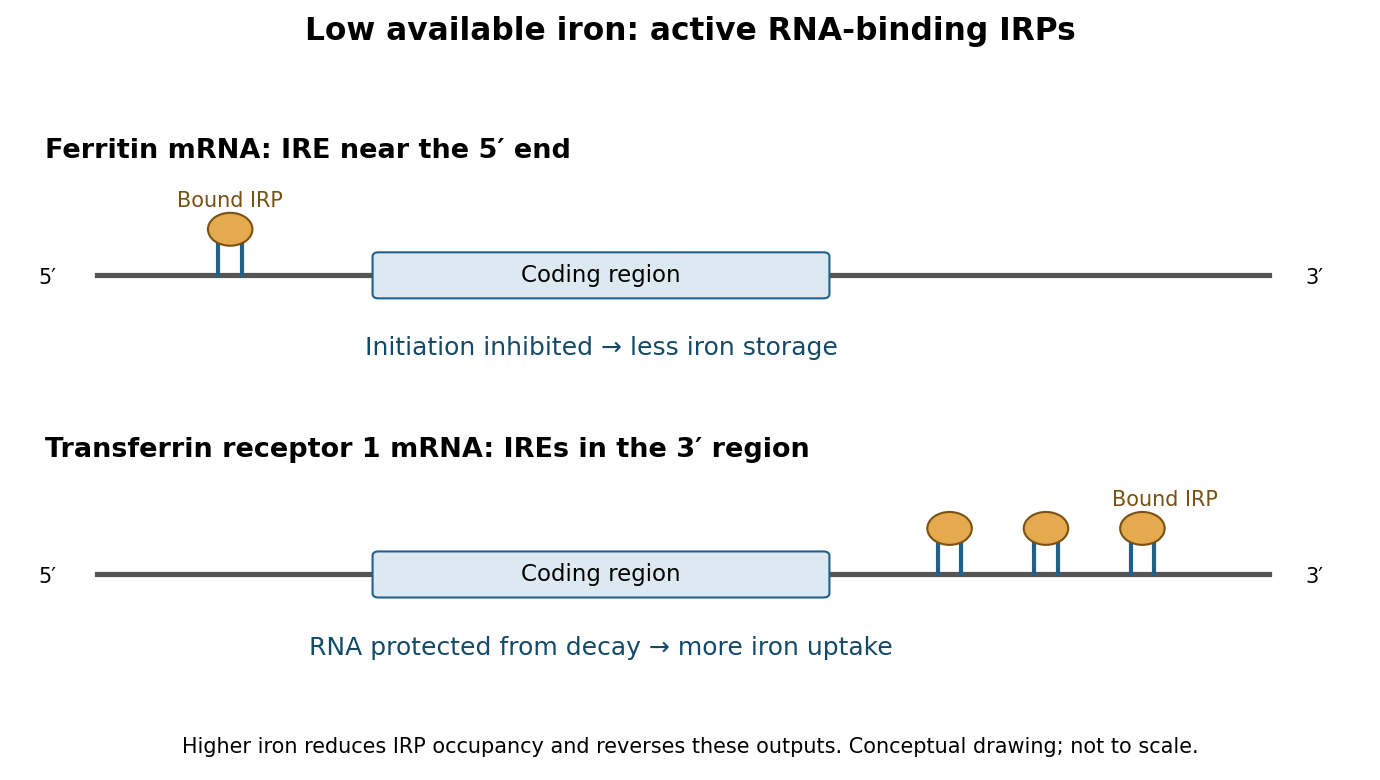
Low iron produces coordinated but opposite outputs from 5′ and 3′ iron-responsive elements
. Details. The network also adjusts use and release. A 5′ IRE in erythroid ALAS2 mRNA couples synthesis of a haem-pathway enzyme to iron availability, helping limit production of porphyrin precursors when metal incorporation is restricted.[ref]
Identification of a novel iron-responsive element in murine and human erythroid delta-aminolevulinic acid synthase mRNA by Thomas Dandekar and others. Erythroid ALAS leader element and iron-dependent translation
IRE-containing ferroportin transcripts can reduce iron export through translational repression during deficiency.[ref]Ferroportin IRE regulation study by Athina Lymboussaki and others. IRE deletion reporters distinguish protein-output regulation from RNA abundance
These are transcript-specific rules, not universal predictions from a gene name: alternative transcripts can lack an IRE, and ferroportin is also regulated at the protein level by systemic signals. The central RNA logic coordinates uptake with conservation, rather than simply maximizing the expression of every iron-related protein.Regulation beyond the mammalian IRE system illustrates a broader eukaryotic principle. In budding yeast, iron deficiency induces the RNA-binding regulator Cth2, which recognizes AU-rich elements in selected 3′ regions and accelerates degradation of messages encoding iron-consuming metabolic functions. This remodels resource allocation rather than protecting an uptake message. Sergi Puig and colleagues combined expression and RNA-turnover analysis with RNA-element recognition to establish targeted degradation during deficiency.[ref]
Coordinated remodeling of cellular metabolism during iron deficiency through targeted mRNA degradation by Sergi Puig and others. Cth2-dependent turnover of iron-utilization transcripts
It is therefore misleading to generalize that iron shortage always stabilizes iron-related RNA: the direction depends on the transcript and its physiological role.To establish these mechanisms experimentally, compare iron chelation with iron restoration, measuring available iron, IRP–RNA binding, transcript half-life and newly synthesized protein separately. Match reporter transcription and mutate recognition features without inadvertently changing general RNA stability. For a stem-loop, a compensatory paired mutation helps distinguish loss of structure from loss of a specific sequence contact. Electrophoretic binding assays with purified protein, cellular crosslinking and rescue with functional versus binding-defective IRP variants connect recognition to regulation. RNA-abundance measurements alone cannot establish translational repression, and chelators can cause broad stress, so unrelated control reporters and viability measurements are needed. The decisive principle is position-dependent RNA control: low iron can repress storage and export biological translation while preserving uptake RNA and reducing expendable iron-consuming programmes.
Past exam of the biochemistry course of the University of Cambridge 2024 Paper 2 Section I 2 Solution by Codex 0 2026-10-20
Codex 0 2026-10-20TFx induction makes it a candidate regulator, not yet a cause of neuronal differentiation. I would test when and where it acts, whether it is necessary or sufficient for a specific developmental transition, and which directly regulated genes explain the phenotype. The initial time-course result could instead reflect increasing neuronal abundance, selective survival, a response to differentiation, or expression of an inactive isoform.
First validate the observation in independent differentiations using quantitative RNA measurements and a specificity-validated protein assay. Examine transcript isoforms, protein abundance and nuclear localization over time, comparing neural precursors, differentiating neurons and other biological cell populations. single-cell RNA sequencing combined with imaging can determine whether TFx increases within a lineage or merely marks a population that becomes more common. Actual sampling times and lineage labels should anchor the analysis: a pseudotime ordering alone does not establish chronology or ancestry. Use independent donors or clones and differentiation batches as biological replicates, while tracking biological cell number, proliferation and apoptosis. A bulk increase caused by loss of another population is different from TFx activation within surviving precursors.
For necessity, reduce TFx using at least two independent perturbations, for example CRISPR interference and an independent guide or RNA interference reagent, or generate a verified gene knockout where viable. Rescue with a perturbation-resistant TFx expressed near its normal level. An inducible degron would provide faster and more sharply timed protein depletion than waiting for an existing pool to disappear after transcriptional inhibition. Perturb before commitment, during neuronal maturation and after maturation, using matched induction controls. A defect confined to the early window suggests a role in commitment; a late defect could instead concern maturation or maintenance. Constitutive loss followed by a late endpoint cannot distinguish these possibilities and can select compensatory biological cells.
The readout must define the affected transition. Count precursor and neuronal states, measure cell-cycle exit, neurite architecture and subtype markers, and assess mature physiology with patch-clamp recording and synaptic assays where appropriate. A marker-positive biological cell is not necessarily a functional neuron. Measure survival alongside fate: fewer neurons after TFx depletion could arise because neurons die rather than because precursors fail to differentiate. Track labelled starting biological cells through the transition and compare proportions as well as absolute output. If TFx changes only a particular neuronal subtype, a generic pan-neuronal assay could miss or misclassify its function.
For sufficiency, induce TFx at controlled levels in competent precursors under subthreshold differentiation conditions, and test whether it accelerates or redirects the transition. Include an empty-vector control, a DNA-binding-defective TFx variant and, if the domain architecture permits, a transcriptional-effector-defective variant. Test a second cellular context rather than assuming that all biological cells possess the required cofactors. Defined transcription-factor combinations can convert mouse fibroblasts into functional neurons, as demonstrated by Thomas Vierbuchen and colleagues, but this is an experimental precedent rather than evidence that the unspecified TFx is one of those factors.[ref]
Direct conversion of fibroblasts to functional neurons by defined factors by Thomas Vierbuchen and others. Factor-combination screen and physiological neuronal validation
Failure of TFx alone to induce neurons would not exclude an essential combinatorial role; strong overexpression could also create an activity absent at endogenous levels.Next identify the direct regulatory programme. Map TFx-associated DNA with an endogenous epitope tag and CUT&RUN or chromatin immunoprecipitation, validating antibody specificity against TFx-depleted biological cells and including appropriate background controls. The targeted nuclease strategy underlying CUT&RUN provides a route to map protein-associated genomic regions at low background.[ref]
An efficient targeted nuclease strategy for high-resolution mapping of DNA binding sites by Peter J. Skene and Steven Henikoff. Targeted nuclease mapping of protein-associated DNA
Combine binding with short-interval nascent-RNA measurements after acute TFx removal, before a large fate or survival shift occurs. Occupancy plus an early expression response prioritizes direct candidates; binding alone does not prove regulation, and a response without detected binding may be indirect or reflect an undetected distal site.ATAC-seq before and after perturbation can test whether TFx changes accessibility, while histone-mark measurements and cofactor interaction assays refine the mechanism. Work on ASCL1 reprogramming showed hierarchical factor action and context-dependent chromatin engagement, motivating tests of whether TFx acts as a pioneer transcription factor or requires already accessible DNA.[ref]
Hierarchical mechanisms for direct reprogramming of fibroblasts to neurons by Orly L. Wapinski and others. Chromatin engagement and hierarchical transcription-factor activity
Increased accessibility after TFx expression alone does not prove direct opening; first establish binding and an early response at that locus.Finally edit a candidate TFx-binding motif at an endogenous enhancer, quantify target expression and differentiation, and compare with a matched neutral edit. Rescue a TFx-loss phenotype with the proposed downstream target or programme, while testing whether that target is required for TFx-induced differentiation. Such genetic ordering connects a molecular target to the biological effect; a plasmid reporter alone omits the native chromatin context. A justified conclusion would specify the transition, biological cell type, timing and direct targets supported by perturbation and rescue, while distinguishing differentiation from survival and maturation.
Pinned article: Introduction to the OurBigBook Project
Welcome to the OurBigBook Project! Our goal is to create the perfect publishing platform for STEM subjects, and get university-level students to write the best free STEM tutorials ever.
Everyone is welcome to create an account and play with the site: ourbigbook.com/go/register. We belive that students themselves can write amazing tutorials, but teachers are welcome too. You can write about anything you want, it doesn't have to be STEM or even educational. Silly test content is very welcome and you won't be penalized in any way. Just keep it legal!
Intro to OurBigBook
. Source. We have two killer features:
- topics: topics group articles by different users with the same title, e.g. here is the topic for the "Fundamental Theorem of Calculus" ourbigbook.com/go/topic/fundamental-theorem-of-calculusArticles of different users are sorted by upvote within each article page. This feature is a bit like:
- a Wikipedia where each user can have their own version of each article
- a Q&A website like Stack Overflow, where multiple people can give their views on a given topic, and the best ones are sorted by upvote. Except you don't need to wait for someone to ask first, and any topic goes, no matter how narrow or broad
This feature makes it possible for readers to find better explanations of any topic created by other writers. And it allows writers to create an explanation in a place that readers might actually find it.
Figure 1. Screenshot of the "Derivative" topic page. View it live at: ourbigbook.com/go/topic/derivativeVideo 2. OurBigBook Web topics demo. Source. - local editing: you can store all your personal knowledge base content locally in a plaintext markup format that can be edited locally and published either:This way you can be sure that even if OurBigBook.com were to go down one day (which we have no plans to do as it is quite cheap to host!), your content will still be perfectly readable as a static site.
- to OurBigBook.com to get awesome multi-user features like topics and likes
- as HTML files to a static website, which you can host yourself for free on many external providers like GitHub Pages, and remain in full control

Figure 2. You can publish local OurBigBook lightweight markup files to either OurBigBook.com or as a static website.
Figure 3. Visual Studio Code extension installation.
Figure 5. . You can also edit articles on the Web editor without installing anything locally. Video 3. Edit locally and publish demo. Source. This shows editing OurBigBook Markup and publishing it using the Visual Studio Code extension. 
- Infinitely deep tables of contents:




All our software is open source and hosted at: github.com/ourbigbook/ourbigbook
Further documentation can be found at: docs.ourbigbook.com
Feel free to reach our to us for any help or suggestions: docs.ourbigbook.com/#contact