Original volume: Examination papers for the Degree of Bachelor of Medicine by the University of Oxford (1864–1867). Open the original paper in the scanned volume, PDF page 79.
Original questions are transcribed from the public-domain edition. Credit: Wellcome Collection; original supplied by Royal College of Physicians, London. Source and public-domain rights statement.
The answers distinguish explanations available in December 1867 from later discoveries. In particular, the historical expressions pneumogastric nerve, eremacausis and Entozoa require interpretation rather than unqualified adoption.
Original question.
Describe the movements of the Thorax during ordinary respiration. Mention the muscles concerned in producing them, describing the part which each takes in effecting the general result.
Ordinary inspiration is active enlargement of the thorax; ordinary expiration is chiefly passive recoil after the inspiratory muscles relax. The movements increase and decrease the vertical, anteroposterior and transverse dimensions together. Their relative prominence varies with posture and the individual; there is no exclusive division into abdominal and costal kinds of breathing.
The principal inspiratory muscle is the respiratory diaphragm. Its peripheral fibres shorten towards the central tendon, lowering and flattening its domes. This increases the vertical dimension of the thorax and displaces the abdominal contents downwards and forwards, making the relaxed abdominal wall project. Its costal attachments also act on the lower ribs: with abdominal support opposing unlimited descent, shortening can lift and expand the lower costal margin. Thus the respiratory diaphragm does more than pull down a piston. Its motor supply is through the phrenic nerves, principally from cervical segments C3–C5.
The external intercostal muscles elevate the ribs and help keep the intercostal spaces from being drawn inwards as pleural pressure falls. The interchondral, or parasternal, portions of the internal intercostal muscles also contribute to inspiration. It would therefore be inaccurate to label every part of the internal intercostal muscles expiratory. Coordinated activity braces the rib cage so that the shortening of one layer moves the ribs rather than simply deforming the spaces between them.
Rotation about the posterior articulations raises the anterior ends of the upper ribs and the sternum, increasing the anteroposterior diameter: the usual pump-handle description. The more laterally directed portions of the lower ribs rise and move outwards, increasing the transverse diameter: the bucket-handle description. These are descriptions of components of an oblique movement, not two independent sets of hinges. The scalene muscles can stabilize and elevate the uppermost ribs, with recruitment varying in quiet breathing. The sternocleidomastoid muscle, when the head is fixed, is an accessory elevator of the sternum in greater inspiratory effort; it is not required for an ordinary quiet breath.
The lungs follow the expanding chest wall because their surfaces remain coupled to it through the thin fluid-filled pleural cavity. More negative pleural pressure increases the distending transpulmonary pressure; enlargement of the alveoli briefly makes alveolar pressure lower than atmospheric pressure, so air enters. At the end of inspiration, airflow stops when these last two pressures equalize. During quiet expiration, relaxation of the respiratory diaphragm and inspiratory intercostal muscles allows the stretched lungs and chest-wall system to recoil towards their resting balance. The domes rise, the ribs and sternum descend, and alveolar pressure becomes slightly positive relative to atmospheric pressure while air leaves. Active abdominal muscles and the interosseous portions of the internal intercostal muscles become important in forced expiration, rather than supplying the main force in quiet expiration.[ref]
22.3 The Process of Breathing. Mechanisms of Breathing and Pulmonary Ventilation; Figures 22.16–22.17
A simple mechanical demonstration uses two elastic bags in an airtight transparent vessel, connected through its stopper to the air, with a movable rubber sheet forming the base. Pulling the sheet down expands the vessel and inflates the bags; letting it return allows deflation. A side-tube manometer can show the associated pressure change. This illustrates expansion by lowering the floor, but its rigid vessel cannot demonstrate the movements of the ribs, the action of the intercostal muscles, or the real geometry of the pleural cavity. Observing lower-chest widening and abdominal displacement during quiet breathing supplies those missing observations without confusing the model with the anatomy.
Original question.
Give an account of the origin, course, and connexions of the Pneumogastric nerves. What results have been observed to follow section of these nerves ?
The two pneumogastric nerves are the right and left vagus nerves, the tenth cranial nerves: mixed sensory, branchial motor and parasympathetic pathways from the medulla oblongata to the neck, thorax and abdomen. A useful description separates their apparent origin at the surface of the brain from the origins and terminations of their component fibres.
Rootlets emerge in the groove behind the inferior olive, between the glossopharyngeal nerve above and the accessory rootlets below. Branchial motor fibres arise chiefly in the nucleus ambiguus; preganglionic parasympathetic fibres arise in the dorsal motor nucleus of the vagus and, particularly for cardiac control, the nucleus ambiguus. Visceral sensory fibres have their cell bodies chiefly in the inferior vagal ganglion and terminate centrally in the nucleus of the solitary tract; somatic sensory fibres associated with the superior vagal ganglion project to the spinal trigeminal nucleus. Consequently a sensory fibre is not accurately described as originating in a motor nucleus simply because its peripheral nerve emerges nearby.[ref]
Neuroanatomy, Nucleus Ambiguus (StatPearls). Introduction and Structure and Function: branchial motor output and cardiac vagal control
Each vagus nerve leaves the skull through the jugular foramen, with the glossopharyngeal nerve and accessory nerve. It bears the small superior vagal ganglion at the foramen and the larger inferior vagal ganglion below it. Connections include communications with adjacent cranial nerves and the cervical sympathetic nervous system. Traditional descriptions assign fibres joining the vagus nerve from the cranial part of the accessory nerve to the latter peripheral nerve; their modern classification is more nuanced and does not change the peripheral branches being described. In the neck the vagus nerve descends in the carotid sheath, behind and between the internal jugular vein and the internal, then common, carotid artery.
Its meningeal and auricular branches supply small sensory territories of the cranial covering and external ear. Pharyngeal branches contribute to the pharyngeal plexus, supplying much of the soft palate and pharynx; the stylopharyngeus muscle belongs to the glossopharyngeal nerve and the tensor veli palatini muscle to the mandibular division of the trigeminal nerve. The internal branch of the superior laryngeal nerve supplies sensation above the vocal folds; its external branch supplies the cricothyroid muscle. The recurrent laryngeal nerve supplies the other intrinsic laryngeal muscles and sensation below the vocal folds. The right recurrent laryngeal nerve loops beneath the right subclavian artery; the left loops under the aortic arch beside the ligamentum arteriosum. Both return towards the larynx near the groove between trachea and oesophagus.[ref]
Neuroanatomy, Cranial Nerve 10 (Vagus Nerve) (StatPearls). Structure and Function; Vagus Nerve Branches; Muscles
In the thorax, both vagus nerves pass behind the roots of the lungs, contrasting with the anterior course of the phrenic nerves. Cardiac branches join the cardiac plexus; pulmonary branches join the pulmonary plexus. The trunks break up into the oesophageal plexus, then collect principally as an anterior trunk derived from the left vagus nerve and a posterior trunk derived from the right. Through the oesophageal hiatus they reach the stomach; hepatic and coeliac branches distribute to abdominal viscera. Their parasympathetic territory includes foregut and midgut derivatives, extending into the proximal colon, rather than every abdominal and pelvic organ.[ref]
Neuroanatomy, Cranial Nerve 10 (Vagus Nerve) (StatPearls). Structure and Function: thoracic course and oesophageal plexus
This extensive distribution explains why the level of experimental section is indispensable information.The main consequences of section can be organized by the interrupted functions:
- Heart: cutting both cervical vagus nerves removes tonic inhibition and usually increases heart rate. The heart continues beating because its rhythm is intrinsic. Electrical stimulation of the peripheral cut end can slow the heart, whereas stimulation of the central end tests reflex effects through the brainstem. Cutting a pathway and stimulating its intact output are different experiments.
- Breathing: bilateral cervical section commonly produces slower, deeper breaths in experimental mammals. Sensory information from the lungs has been interrupted; the respiratory diaphragm is not directly paralysed, because its motor peripheral nerves are the phrenic nerves. Persistence of breathing establishes that the vagus nerves modify the respiratory rhythm rather than being its sole generator. The subsequently named Hering-Breuer inflation reflex provides a later explanation of one component of this feedback; its formal description belongs to 1868, after this examination.[ref]Reflexes from the lungs and airways: historical perspective. Discussion of the 1868 lung inflation and deflation experiments
- Larynx and lungs: section proximal to the recurrent laryngeal nerves can impair opening and closure of the vocal folds, causing altered voice, obstruction and impaired airway protection. Classical bilateral cervical experiments sometimes ended in death with pulmonary congestion, oedema or aspiration changes. Such outcomes must be distinguished from an immediate necessary arrest of breathing: species, age, lesion level and airway management affect them.
- Stomach: bilateral section can reduce vagally driven gastric acid secretion and disturb gastric motor coordination and emptying. Nevertheless digestion and nutrient absorption are not abolished as chemical possibilities: enteric nervous system activity, local stimuli and hormones remain. Likewise, interruption of gastric sensory fibres does not establish that the entire experience of hunger originates in them.
A discriminating historical experiment would record respiratory movements and heart rate before and after section, distinguish unilateral from bilateral section, and compare an intact airway with an airway bypass. A tracheal cannula can separate obstruction at the glottis from changes in the respiratory rhythm, while the central-versus-peripheral stimulation comparison separates incoming reflex traffic from outgoing motor effects. Later separate-lung recordings confirmed that a change following unilateral section could affect both lungs, supporting a central reflex interpretation rather than simple local paralysis.[ref]
A Study of the Hering-Breuer Reflex. Summary and Conclusions, points 1–4; separate recordings of the two lungs, 1927
The gastric distinction is also borne out by measurements of persistent acidity after surgical vagotomy, despite major reductions in stimulated gastric acid secretion.[ref]Twenty-four hour gastric acidity after vagotomy. Abstract: hourly intragastric sampling, seven patients, four years after operation
Original question.
‘In the vegetable kingdom solar force is absorbed in the production of our food ; in the animal kingdom it is liberated by the eremacausis of our fat and glands and muscle.’ Odling, Lect. on Animal Chemistry. Explain and illustrate these statements.
The defensible core of William Odling's statement is a transfer of energy: sunlight helps build reduced food material, and its subsequent oxidation can supply animal heat and work. Here solar force means solar energy, and eremacausis means slow oxidation, rather than a special vital agent. The historical wording, from his 1866 lectures, must be qualified where it identifies the fuel with the substance of the working muscle or contrasts plant and animal metabolism too absolutely.[ref]
Lectures on Animal Chemistry by William Odling. Lecture IV, discussion of solar energy and animal oxidation; see also the concluding recapitulation
In oxygenic photosynthesis, light absorbed by chlorophyll supports conversion of carbon dioxide and water into reduced organic material, with release of oxygen. An overall account, written for one glucose unit, isThis summarizes several stages, rather than describing a single elementary chemical reaction. The evolved oxygen comes from water. The product can furnish starch, other carbohydrates and precursors for fat and protein; the latter additionally requires a source of nitrogen. A human obtains these materials directly from plants or through animals that have eaten plants. Thus a grain meal and the flesh of a grain-fed animal can trace much of their chemical energy to the same original sunlight.[ref]
8.1 Overview of Photosynthesis. Main Structures and Summary of Photosynthesis; photosynthetic products and their passage through food chains
The reverse overall balance for complete aerobic utilization of glucose isSimilarly a representative fatty acid, palmitic acid, has the complete oxidation balanceBoth equations conserve each chemical element. The relevant energy difference belongs to the complete set of reactants and products, including oxygen; merely breaking a bond does not supply net energy. In modern terms, controlled cellular respiration couples part of the available chemical free energy to ATP formation. ATP hydrolysis can then support muscle contraction, active transport and biosynthesis; some energy is transferred to heat. The process occurs through enzyme-mediated steps, without a flame, although complete combustion in a calorimeter can help measure the corresponding chemical energy change.
Glands and muscles continually replace constituents, and their proteins can be degraded. Amino acids can contribute carbon skeletons to energy metabolism, while their nitrogen is disposed of largely as urea. But ordinary muscle contraction does not require proportional destruction and rebuilding of the contractile protein at every stroke. Carbohydrates and fats are major fuels for muscle, and a gland can use those fuels to support secretion. The historical picture of burning the organ's own substance is therefore an inadequate general explanation.[ref]
This correction was already an experimental issue before the exam. Edward Frankland's 1866 discussion compared the mechanical work of climbing with the energy that could be assigned to protein destruction estimated from urinary nitrogen. The latter was insufficient to account for all the work. His combustion measurements also distinguished the heat from burning a sample completely from the smaller amount available when nitrogenous material leaves the animal as still-combustible urea. Measuring external work, urinary products and calorific values gives the hypothesis a quantitative test; it is more informative than simply pointing out that an active animal gets warm.[ref]
On the Source of Muscular Power by Edward Frankland. pp. 394–395, criticism of tissue-consumption doctrine; pp. 401–406, calorific measurements and comparison with climbing work, 1866
Finally, plants themselves carry out cellular respiration, and not every ecosystem obtains its input from sunlight: chemolithotrophy is an alternative. The plant-animal contrast is useful for following energy through an ordinary food chain, but it is not an exhaustive division of biochemical capabilities. Conservation of energy also requires the bookkeeping formfor an idealized interval in which is the metabolizable food input, with energy lost in excreta already subtracted and other material-energy exchanges neglected. Here is external work, is net outward heat transfer, and includes changes in stored chemical and other energy. The living body transforms energy; it does not create it.
Original question.
The liver is both a metabolic processing organ and the producer of bile. These functions must be distinguished: much of what the liver contributes to the body enters the blood, whereas bile is its secretion into the digestive system.
Portal venous blood brings absorbed materials from the alimentary organs to the liver, while the hepatic artery supplies oxygenated blood. Within the hepatic sinusoids, the incoming blood encounters hepatocytes and scavenging Kupffer cells. Hepatocytes take up, transform, store and release substances; their bile canaliculi conduct bile towards the bile ducts. The organ's position therefore makes it a useful regulator between absorption from a meal and supply to the rest of the body.
Its main non-biliary functions include the following:
- Carbohydrate metabolism: after a meal, glucose can be stored as glycogen by glycogenesis; during fasting, glycogenolysis and gluconeogenesis help maintain blood glucose. The liver therefore both removes and supplies glucose, depending on the physiological state.
- Lipid metabolism: the liver handles fatty acids, synthesizes and exports triglycerides in lipoproteins, makes cholesterol, and can produce ketone bodies during fasting. Storage of fat is not its sole function in this domain.
- Amino acid metabolism: transamination and deamination redistribute or remove amino groups; the urea cycle converts much potentially toxic Ammonia nitrogen into urea for excretion by the kidneys. The liver synthesizes major plasma proteins, including albumin and many coagulation factors.
- Storage and regulation: stores include glycogen, several vitamins and iron. The liver also modifies many hormones and administered chemicals. Such biotransformation can aid elimination, but the word detoxification must not imply that every product is less toxic than its precursor.
- Clearance and development: Kupffer cells remove material from the incoming blood by phagocytosis. The fetal liver is an important site of haematopoiesis; this is a developmental function, rather than the ordinary principal source of adult red blood cells.
This modern list extends beyond the knowledge and biochemical vocabulary of 1867. It is consistent with the organ's combined metabolic, storage and clearance roles, rather than treating bile production as the whole explanation.[ref]
Physiology, Liver (StatPearls). Introduction; Mechanism; Related Testing: metabolic, synthetic, storage and clearance functions
Bile contains water, electrolytes, bile salts, phospholipids, cholesterol and bilirubin conjugates. Hepatocytes form the primary secretion; ductal epithelium modifies it. The gallbladder stores and concentrates bile between meals and releases it into the duodenum. Production of bile must therefore be distinguished from its intermittent delivery to a meal.
Its two central functions are assistance with lipid digestion and nutrient absorption, and excretion of selected waste substances. Amphipathic bile salts help disperse dietary fat and form mixed micelles carrying products of lipid digestion to the absorptive surface of the small intestine. They thereby promote uptake of fatty acids, monoglycerides, cholesterol and fat-soluble vitamins. Bile is not itself a source of pancreatic lipase, nor does emulsification chemically hydrolyse triglycerides. Biliary bilirubin, produced through haem breakdown, provides an excretory route; cholesterol and selected other substances also leave in bile. Most secreted bile salts are recovered, especially in the terminal ileum, and return through the portal vein: the enterohepatic circulation conserves the useful detergent.[ref]
23.6 Accessory Organs in Digestion: The Liver, Pancreas, and Gallbladder. The Liver; Composition of Bile; The Gallbladder
Two comparisons reveal why both sorts of function matter. First, compare a fresh liver extract with an extract prepared after an appropriate controlled interval: testing for reducing sugar and isolating glycogen distinguishes a stored precursor from newly liberated glucose. A reliable interpretation needs prompt sampling and controls because post-mortem glycogenolysis can change the result after the animal has died; detecting sugar in an organ alone does not prove where all of it was made. Secondly, in classical biliary fistula observations, diverting bile away from the intestine while collecting it separately allowed its output to be measured and the loss of normal fat assimilation to be examined. Comparing faecal fat with and without delivery of bile distinguishes a digestive contribution from a merely excretory one. The modern explanation is that loss of intestinal bile salts impairs mixed micelle formation, while jaundice from retained biliary pigment is a separate consequence of failed pigment elimination.
Original question.
The fourth ventricle is the cavity of the hindbrain between the dorsal surfaces of the pons and upper medulla oblongata anteriorly and the cerebellum posteriorly. It is continuous above with the cerebral aqueduct and below, at the obex, with the central canal of the spinal cord. Its floor is diamond-shaped when viewed from behind, while its roof rises dorsally towards the fastigium.
The floor, the rhomboid fossa, consists of an upper pontine triangle and a lower medullary triangle. Its upper apex points towards the cerebral aqueduct; its lower apex is the obex; the two lateral angles continue into the lateral recesses of the fourth ventricle. A median groove divides it into right and left halves. On each side, the medial eminence lies medial to the sulcus limitans, which separates predominantly motor territories medially from sensory territories laterally. The striae medullares of the fourth ventricle cross the floor near the junction of its pontine and medullary portions; their extent varies.
Several surface features identify deeper relations. In the pontine part, the facial colliculus overlies the abducens nucleus and the internal looping course of fibres of the facial nerve; it is not the exposed facial motor nucleus. The upper part of the sulcus limitans has the superior fovea, and near its rostral lateral region lies the locus coeruleus. Laterally, the vestibular area overlies the vestibular nuclei. In the lower triangle, the hypoglossal trigone overlies the hypoglossal nucleus, and the vagal trigone, lateral to it, overlies the dorsal motor nucleus of the vagus. The area postrema lies still farther caudolaterally, separated from the vagal trigone by the funiculus separans. These are landmarks and underlying relations, not claims that the cranial nerve nuclei lie loose within the cavity.[ref]
Neuroanatomy, Fourth Ventricle (StatPearls). Introduction; Structure and Function; Surgical Considerations: floor and relations
The upper lateral boundaries are formed by the superior cerebellar peduncles. The lower lateral boundaries relate to the inferior cerebellar peduncles and adjacent dorsal medullary margins. The lateral recess on each side curves around the inferior cerebellar peduncle towards its external opening near the flocculus.
The roof has superior and inferior slopes. Superiorly the thin superior medullary velum spans the superior cerebellar peduncles, with the lingula of the cerebellum on its dorsal surface. Inferiorly, the roof relates to the inferior medullary velum, nodule of the cerebellum and thin tela choroidea. Vascular folds of the tela choroidea carry the choroid plexus of the fourth ventricle, with median and lateral extensions. The cavity has an ependymal lining; the choroid plexus has its specialized epithelial covering and produces cerebrospinal fluid.
There are three important exits to the subarachnoid space: one median aperture of the fourth ventricle through the inferior roof, opening towards the cisterna magna, and two lateral apertures of the fourth ventricle at the ends of the lateral recesses of the fourth ventricle. These must be distinguished from the inlet through the cerebral aqueduct and the inferior continuity with the central canal of the spinal cord. Most cerebrospinal fluid leaving the fourth ventricle reaches the subarachnoid space, not the narrow adult central canal of the spinal cord.
The three-dimensional arrangement explains the method of examination: reflecting the cerebellum and opening the delicate roof exposes the rhomboid fossa, but destroys part of the structure one is trying to describe. Studying an intact roof, then the exposed floor, and using a median section to locate the fastigium avoids interpreting a dissected cavity as naturally open behind. The modern nomenclature is useful here; not every fine landmark or physiological interpretation in this account should be read as an assertion about what an 1867 examiner required.[ref]
Morphometric Assesment of the External Anatomy of Fourth Ventricle and Dorsal Brainstem in Fresh Cadavers. Gross-dissection study: dorsal brainstem, rhomboid fossa and fourth-ventricular approaches
Original question.
Examine into the nature of the forces which assist the Heart's action in maintaining the circulation of the blood.
The heart supplies the principal continuing mechanical input to blood circulation; arterial recoil, respiratory movements and the action of skeletal muscles redistribute and supplement that input. They have different sources of energy and should not all be called independent propulsive powers. The pressure gradient remaining after passage through the capillaries is still a consequence of the upstream pump.
Arterial elasticity. During ventricular systole, ejected blood expands the aorta and large arteries, storing elastic potential energy in their walls. During diastole, recoil continues to drive blood into the peripheral vessels while closure of the aortic valve limits return towards the heart. This reduces the intermittency of flow downstream. The recoil is a temporary store of cardiac work, rather than a fresh source of energy. A vessel that is too rigid provides less of this smoothing; a compliant vessel with an open backward outlet would not provide the same useful result.
The skeletal-muscle pump. Contracting skeletal muscles compress adjacent veins. Venous valves make the displacement preferentially towards the heart; when compression ceases, refilling permits another stroke. Walking therefore aids return from dependent limbs. The source of the added work is muscle contraction, and the valves guide it without producing energy. Merely possessing valves cannot make a stationary column circulate.
The respiratory pump. During inspiration, falling intrathoracic pressure and displacement of the abdominal contents favour a pressure gradient from abdominal and peripheral veins towards the thoracic veins and right side of the heart. The respiratory diaphragm thus participates indirectly in venous return as well as pulmonary ventilation. The exact flow varies with posture, vessel collapse and the phase of the respiratory cycle; positive-pressure inflation does not reproduce the same mechanical circumstances as spontaneous inspiration.[ref]
Venous tone and cardiac filling. Sympathetic contraction of venous smooth muscle can reduce venous storage capacity and transfer blood towards the central circulation. This alters the volume available for filling rather than requiring a wave of venous peristalsis. Cardiac relaxation, recoil and movement of the atrioventricular plane also affect filling pressure. Some traditionally described cardiac suction is therefore a feature of the cardiac cycle and mechanical coupling, not a separate organ pulling blood through every vessel.[ref]
20.4 Homeostatic Regulation of the Vascular System. Neural Regulation of Blood Flow; sympathetic effects on vessels and venous return
Gravity and supposed capillary attraction. Gravity assists a downward column but opposes an upward one. In a complete circuit returning to its original height it supplies no net gravitational work; its important practical effects include hydrostatic pressure differences and dependent venous pooling. Ordinary capillary action in an open narrow tube, driven by a liquid meniscus, is not a general pump in a fluid-filled capillary network. Nor does a vague attraction of blood by tissues replace a measurable pressure gradient. Local vasodilation can increase perfusion by lowering vascular resistance, but that change does not manufacture the necessary pressure source.
For a simple mean-flow description, with the volume flow rate, the driving pressure gradient and the effective vascular resistance,This relation distinguishes a source of driving pressure from a change of resistance. In a lumped compliant arterial reservoir with vascular compliance and no inflow between beats, volume balance giveswhere is the downstream pressure. The exponential decay follows from stored volume leaving through the resistance. It explains continued flow during diastole without claiming that the idealization describes every reflected arterial wave.
A physical demonstration can compare a pulsatile pump feeding a narrow outlet through rigid tubing with the same pump feeding it through an elastic reservoir. Collecting the outflow over the pumping and resting phases reveals the reservoir's smoothing action; a side-arm manometer records its falling pressure. Separately compressing a valved flexible tube shows the role of a peripheral muscle pump. Keeping pump stroke and outlet resistance comparable prevents the apparatus from conflating a smoother flow with creation of extra energy.
Original question.
What views are held with regard to the development of blood cells ; (1) in early embryonic, (2) in later life ?
An answer appropriate to 1867 must distinguish the early nucleated embryonic red blood cells from later populations, and report uncertainty about their origin. Simply stating the modern bone marrow account would miss the historical force of the request for the views then held.
Early embryonic life. In the pre-exam account of William Senhouse Kirkes, a first generation of red corpuscles arose from embryonic cells in the early vascular territory before or around the establishment of blood circulation. The initially colourless cellular material acquired colour, and the early corpuscles were relatively large and nucleated. Division of these early nucleated forms was among the processes considered capable of increasing their number. A second generation subsequently replaced the first. His embryological terminology, including the vegetative layer, should not be silently treated as an exact statement of modern germ layer derivation.[ref]
Later fetal and adult life. A widely taught hypothesis made pale corpuscles in lymph and chyle precursors of the later red blood cells. In this scheme the pale cells entered the blood, acquired colouring matter and underwent changes associated with loss or alteration of the cell nucleus. Lymph nodes, the spleen and, during development, the liver were consequently central candidates for sites of renewal. Other opinions proposed multiplication of pre-existing red corpuscles by division or segmentation. These were competing interpretations of microscopic appearances, not demonstrations that every adult leukocyte can become a red blood cell. The pre-exam historical record explicitly documents both the pale-corpuscle hypothesis and the alternative multiplication hypotheses.[ref]
Recent Advances in the Physiology of Motion, the Senses, Generation and Development by William Baly and William Senhouse Kirkes. p. 132, Development of the Blood: rival explanations of post-embryonic corpuscle formation
The modern correction is successive developmental waves of haematopoiesis, followed chiefly by bone marrow production after birth, with distinct committed precursors. Primitive erythropoiesis begins in extra-embryonic yolk sac blood islands. Definitive blood-forming populations emerge through additional embryonic processes, including haemogenic endothelium in the aorta-gonad-mesonephros region, and expand in the fetal liver. Bone marrow becomes a major site later in fetal development and is the normal main adult site. The sequence is not a single already mature cell population merely migrating unchanged from organ to organ.[ref]
Definitive hematopoiesis is autonomously initiated by the AGM region. Abstract: organ culture distinguishes autonomous generation from interchange through the circulation, mouse study, 1996
[ref]Single cell resolution of human hemato-endothelial cells defines transcriptional signatures of hemogenic endothelium. Abstract and Introduction: endothelial-to-haematopoietic transition; human stem-cell-derived experimental populations, 2018
[ref]18.2 Production of the Formed Elements. Sites of Hemopoiesis; Differentiation of Formed Elements from Stem Cells
During definitive erythropoiesis, erythroblasts divide and accumulate haemoglobin; the late precursor extrudes its cell nucleus, producing a reticulocyte, which matures into a red blood cell. The large primitive mammalian erythroblasts also ultimately undergo enucleation; their transient nucleation does not make them adult leukocytes.[ref]
Enucleation of primitive erythroid cells generates a transient population of pyrenocytes in the mammalian fetus. Abstract: mouse primitive erythroblast maturation and nuclear extrusion, 2008
A mature human red blood cell does not normally divide. Adult haematopoietic stem cells maintain progenitor populations giving rise to red blood cells, leukocytes and megakaryocytes; platelets are fragments produced by the last of these, not equivalent nucleated cells. Lymphocytes have their own differentiation and maturation pathways, including development in lymphoid organs. Thus a precursor's pale or lymphoid appearance under a nineteenth-century microscope is insufficient to identify it as an ordinary mature circulating leukocyte.Two observations explain both the attraction and the weakness of the older theories. Comparing embryonic and adult blood under a microscope reveals genuine differences in corpuscle size, colour and nuclear appearance. But a series of different-looking cells in separate samples is not direct observation of one changing into another. Mixing developmental stages, species or preparations can create a false apparent sequence; reagents that reveal or damage the cell nucleus add another complication. A stronger test samples the proposed blood-forming organs and identifies proliferating precursors there, then connects them experimentally to their descendants. The explicit identification of adult bone marrow as a blood-forming organ in 1868 is therefore a subsequent development, not established background to the December 1867 paper.[ref]
Chronicle of Physiology (January 1869). Part I, Blood-Circulation, item 1: report of the 1868 preliminary communication on marrow and corpuscle formation
Original question.
To what subdivisions of the animal kingdom are the Entozoa referred ? Mention their leading peculiarities of structure and development. Enumerate the species most commonly found in man.
Entozoa is a historical grouping of internal animal parasites, especially parasitic worms; it is not a single natural modern clade. The core human examples belong principally to the flatworms, including Cestoda and Trematoda, and to the roundworms, or Nematoda. The distinction is structural and developmental, not merely between inhabitants of the intestine and inhabitants of other organs.
The nineteenth-century higher arrangement was unsettled. Schemes placed these animals within Vermes, or within annuloid assemblages that included other supposedly related simple invertebrates. For a specifically pre-exam example, the 1865 geological-museum classification put Entozoa in Annuloida, within Annulosa. These historical groups must not be equated with the modern Annelida: an Ascaris lumbricoides is not an earthworm. A more useful subdivision for this answer is the four orders treated in Thomas Spencer Cobbold's 1864 Entozoa, an Introduction to the Study of Helminthology by Thomas Spencer Cobbold: Trematoda, Nematoda, Acanthocephala and Cestoda.[ref]
Catalogue of the Collection of Fossils in the Museum of Practical Geology (1865). p. lxxvi, classification table: Annulosa, Annuloida, Entozoa
[ref]Entozoa, an Introduction to the Study of Helminthology by Thomas Spencer Cobbold. contents and order headings: Trematoda p. 14, Nematoda, Acanthocephala p. 97, Cestoda p. 104
The leading distinctions are as follows:
- Cestoda, the tapeworms, have a flattened adult body with an attachment scolex and, in the familiar human forms, a chain of reproductive proglottids. They lack an alimentary canal and absorb nutrients across the tegument. The proglottids are repeated reproductive units, not annelid segments. Typical development passes through an egg and one or more larval stages, commonly in an intermediate host, before the adult reaches a definitive host. A bladder-like cysticercus and its adult tapeworm are stages of one life cycle.
- Trematoda, the flukes, have flattened unsegmented bodies, attachment suckers and a mouth leading to a usually branched, blind-ending digestive system. Most are hermaphrodites, but Schistosoma has separate sexes. Typical human-infecting flukes use a molluscan intermediate host, undergo multiplication in larval stages there, and produce dispersing cercariae. The route from that stage into the human varies by species; there is no universal requirement to swallow an adult worm.
- Nematoda have cylindrical unsegmented bodies, a resistant cuticle, a fluid-filled pseudocoelom, longitudinal muscles and a complete alimentary canal with mouth and anus. The sexes are usually separate. Development includes moults; some life cycles are direct, while others need a vector or another host. Human infection may follow swallowing infective eggs, penetration by larvae, ingestion of tissue larvae or transmission by a vector, depending on the species.
- Acanthocephala, the thorny-headed worms, possess a retractile hooked proboscis and lack an alimentary canal. Their development normally involves an arthropod intermediate host. They belong in a general survey of historical Entozoa, but are not among the usual common human intestinal worms.
For a British medical answer of this period, the ordinary intestinal list should begin with the following species; the modern life-cycle details clarify their identities rather than claiming that every step was already established in 1867:
- Ascaris lumbricoides, the large intestinal roundworm. Adults inhabit the small intestine; infective eggs are acquired from the environment, and larvae migrate through the liver and lungs before returning to mature in the intestine.[ref]DPDx: Ascariasis. Causal Agents; Life Cycle
- Enterobius vermicularis, historically commonly named Oxyuris vermicularis, the small pinworm or threadworm. Adults chiefly occupy the caecal region; females deposit eggs on the perianal skin, explaining spread through contaminated hands and objects and the usefulness of perianal sampling rather than reliance on ordinary stool samples.[ref]
- Trichuris trichiura, historically Trichocephalus dispar, the whipworm. The thin anterior end is embedded in the caecal or colonic mucosa; eggs passed in faeces must develop outside the host before becoming infective.[ref]DPDx: Trichuriasis. Causal Agent; Life Cycle
- Taenia solium, the pork tapeworm, with an armed scolex, and Taenia saginata, the beef tapeworm, historically often Taenia mediocanellata, with an unarmed scolex. Intestinal adults follow ingestion of tissue cysticerci in infected meat. Their eggs alone cannot reliably distinguish the two species by ordinary microscopy.[ref]
- The broad fish tapeworm, historically Bothriocephalus latus, later Diphyllobothrium latum, now Dibothriocephalus latus. It is especially relevant in freshwater-fish-eating populations and has a life cycle involving a copepod and fish before the human definitive stage. Its attachment organs are longitudinal grooves, rather than the four rounded suckers typical of Taenia.[ref]DPDx: Diphyllobothriasis. Causal Agents; Life Cycle
Important tissue examples extend the answer beyond intestinal adults. The historical Trichina spiralis is Trichinella spiralis: ingested infected meat carries larvae, adults develop in the small intestine, and new larvae migrate to skeletal muscle. This is a distinct developmental pattern from the egg-acquired intestinal Ascaris lumbricoides infection.[ref]
DPDx: Trichinellosis. Causal Agents; Life Cycle
Human hydatids are the larval hydatid cysts of Echinococcus, classically the Echinococcus granulosus complex; the small adult tapeworm is in a canid, while humans accidentally occupy the intermediate host role, commonly with liver or lung cysts.[ref]A crucial distinction is that human Taenia solium cysticercosis follows ingestion of eggs; eating pork containing cysticerci produces intestinal taeniasis. The historical name Cysticercus cellulosae designates a larval form, rather than an additional independent species to add to the list.[ref]
DPDx: Cysticercosis. Causal Agent; Life Cycle
Geographical qualification matters: Ancylostoma duodenale, Dracunculus medinensis and Schistosoma haematobium are important in their endemic settings; Fasciola hepatica is a familiar liver fluke but is not an ordinary common human worm in Britain. An enumeration should not turn this into a claim that all named species are equally common in every population.[ref]
DPDx: Intestinal Hookworm. Causal Agents; Geographic Distribution
[ref]DPDx: Dracunculiasis. Causal Agent; Geographic Distribution
[ref]DPDx: Schistosomiasis Infection. Causal Agents; Geographic Distribution
[ref]DPDx: Fascioliasis. Causal Agents; Geographic Distribution
Conversely, calling only the familiar intestinal adults Entozoa would omit the instructive tissue stages and their transmission.The decisive experimental insight is continuity of development. Comparing eggs, tissue larvae and adults, and tracing transfer between appropriate hosts, demonstrates a life cycle rather than spontaneous generation within a patient. In diagnostic material, an adult attachment organ, a proglottid, an egg and a tissue cyst answer different identification questions. Identification therefore depends on the stage and the material actually supplied.
Original volume: Examination papers for the Degree of Bachelor of Medicine by the University of Oxford (1864–1867). Open the original paper in the scanned volume. Scanned volume: PDF page 77, December 1867.
Original questions are transcribed from the public-domain edition. Credit: Wellcome Collection; original supplied by Royal College of Physicians, London. Source and public-domain rights statement.
Original question.
Dissect the orbit in the head before you.
Expose the orbit (anatomy) in layers, preserving the eye, extraocular muscles, nerves and blood vessels so that their relations can be demonstrated. This is a general explanation of the practical exercise, using present-day anatomy terminology. It does not assert findings in the individual head supplied in 1867. The essential result is an intelligible dissection, with structures traced to their attachments or continuations, rather than a collection of detached parts.
Begin by orienting the head and choosing a superior approach through the anterior cranial fossa. Useful apparatus consists of a small saw or bone-cutting forceps, a fine chisel, fine scissors, dissecting forceps and a blunt probe. The coarse instruments remove bone; the fine instruments separate connective tissue. Remove the dura mater from the orbital part of the frontal bone, then carefully remove the roof of the orbit (anatomy) while leaving the periorbita intact. Extend the exposure towards the apex without driving the instrument into the optic canal or the superior orbital fissure. Preserve the delicate medial wall and its anterior ethmoidal nerve, posterior ethmoidal nerve and accompanying blood vessels. Open the periorbita under direct vision. This establishes a continuous view from the anterior margin towards the apex.[ref]
Dissection of the Orbit by Loyola University Chicago. Step 1, dorsal approach: removal of the roof, preservation of the medial ethmoidal structures, and gentle removal of fat
In the superficial plane, identify the frontal nerve above the levator palpebrae superioris, and follow its division into the supraorbital nerve and supratrochlear nerve. On the lateral side identify the lacrimal gland, lacrimal nerve and accompanying lacrimal artery. On the medial side find the superior oblique muscle and the trochlear nerve entering it. Separate small amounts of adipose tissue with a blunt probe instead of stripping out the contents in a single mass. Preserve the superior ophthalmic vein where recognizable. Before reflecting any structure, show its course in place: removal of an overlying structure should answer a particular question about what lies underneath it.[ref]
Lab Manual - Eye by Texas Tech University Health Sciences Center El Paso. Procedure 6 and opening of procedure 7: superficial nerves, superior ophthalmic vein, and reflection of levator and superior rectus
Next isolate the levator palpebrae superioris and trace it forward towards the upper eyelid. Its expanded tendon reaches the upper tarsal plate and adjacent eyelid tissues; distinguish this from an attachment to the sclera. Divide the levator palpebrae superioris across its belly and reflect the ends, keeping its oculomotor nerve supply attached where practicable. The superior rectus muscle is now exposed. Isolate and reflect it in turn, preserving the superior division of the oculomotor nerve entering the deep surface. This successive exposure demonstrates why the superficial frontal nerve should be located before either muscle is divided.[ref]
Reflection of the orbital muscles by Texas Tech University Health Sciences Center El Paso. Procedure 7 commentary: reflect the levator and superior rectus at their mid-bellies and identify the superior division of III; levator attachment is detailed in the anatomy tables
[ref]Trace the four rectus extraocular muscles from the common tendinous ring at the apex to the anterior sclera, behind the margin of the cornea. Demonstrate the medial rectus muscle and lateral rectus muscle along the corresponding sides of the eye, and the inferior rectus muscle below it. Trace the superior oblique muscle forwards to the trochlea of superior oblique; its tendon turns backwards and laterally beneath the superior rectus muscle to reach the sclera behind the equator of the eye. The inferior oblique muscle is the important exception to an apical origin: it begins on the anterior medial orbital floor of the maxilla, lateral to the lacrimal sac, and passes backwards and laterally beneath the inferior rectus muscle to the posterior lateral sclera.[ref]
Anatomy Tables - Muscles of the Head and Neck by Texas Tech University Health Sciences Center El Paso. Rows for rectus and oblique muscles: origins, insertions, and the superior oblique pulley
The following functional explanation makes the muscle display meaningful. The medial rectus muscle produces adduction of the eye, and the lateral rectus muscle produces abduction of the eye. The superior rectus muscle elevates and the inferior rectus muscle depresses, with additional adduction of the eye; the superior rectus muscle also produces intorsion of the eye, and the inferior rectus muscle produces extorsion of the eye. The superior oblique muscle produces intorsion of the eye, depression and abduction of the eye; the inferior oblique muscle produces extorsion of the eye, elevation and abduction of the eye. Depression by the superior oblique muscle is demonstrated most clearly with the eye adducted. Gentle traction on an isolated tendon, with the eye supported, can illustrate its line of pull in the preparation. This mechanical demonstration does not test living nerve function, and no observed movement in the historical specimen is supplied.[ref]
Dissector Answers - Eye by Texas Tech University Health Sciences Center El Paso. Learning objective 3: rectus and oblique actions and clinical testing positions
In the deeper plane, identify the optic nerve entering the posterior eye and trace it back to the optic canal. Identify the nasociliary nerve crossing above it towards the medial wall; follow the long ciliary nerves towards the eye and the anterior ethmoidal nerve and posterior ethmoidal nerve towards the medial bone. On the lateral side of the optic nerve, between it and the lateral rectus muscle, seek the small ciliary ganglion. Preserve the short ciliary nerves that pass from its anterior aspect to the eye; an indiscriminate sweep through the surrounding adipose tissue would destroy precisely these small connections.[ref]
Anatomy Tables - Eye by Texas Tech University Health Sciences Center El Paso. Nerves table: optic, nasociliary, long ciliary, ethmoidal, ciliary ganglion and short ciliary entries
Trace the inferior division of the oculomotor nerve towards the medial rectus muscle, inferior rectus muscle and inferior oblique muscle, including its connection to the ciliary ganglion. Trace the abducens nerve to the deep surface of the lateral rectus muscle, and the trochlear nerve to the superior oblique muscle. The lateral rectus muscle receives the abducens nerve, the superior oblique muscle receives the trochlear nerve, and the remaining listed extraocular muscles receive the oculomotor nerve. The ciliary ganglion is a relay for fibres of the parasympathetic nervous system carried with the oculomotor nerve towards the sphincter pupillae muscle and ciliary muscle. Sensory fibres connected with the nasociliary nerve, and already postganglionic fibres of the sympathetic nervous system, traverse the ciliary ganglion without another relay.[ref]
Anatomy Tables - Eye by Texas Tech University Health Sciences Center El Paso. Nerves table: oculomotor and nerve to inferior oblique entries; ciliary ganglion and short ciliary entries distinguish the three fibre components
Trace the ophthalmic artery from the optic canal and its continuity with the internal carotid artery, distinguishing it from the optic nerve. Follow recognizable branches: the central retinal artery towards and into the optic nerve, the posterior ciliary arteries towards the posterior sclera, the lacrimal artery towards the lacrimal gland, and the supraorbital artery, anterior ethmoidal artery and posterior ethmoidal artery towards their exits. The usual ophthalmic artery crosses above the optic nerve to reach the medial side, but an individual course must be established by tracing, not imposed on the preparation. [ref]
https://pubmed.ncbi.nlm.nih.gov/6507890/. Variability in relationships of arteries and nerves in the human orbit (1984), abstract: direct dissections found an inferior ophthalmic-artery crossing in some orbits
Follow the superior ophthalmic vein towards the superior orbital fissure and cavernous sinus. The inferior ophthalmic vein also has communications towards the pterygoid plexus through the inferior orbital fissure.[ref]Lab 27: Dissection: Orbit and Ear by Washington State University. Dissection section, ophthalmic artery and superior ophthalmic vein; branching pattern is variable
Finally, show the relations at the apex. The optic nerve and ophthalmic artery pass through the optic canal within the common tendinous ring. The divisions of the oculomotor nerve, nasociliary nerve and abducens nerve enter through the part of the superior orbital fissure enclosed by the common tendinous ring; the frontal nerve, lacrimal nerve and trochlear nerve enter outside it. Keep the eye attached until these relations have been displayed. A final labelled account should distinguish structures actually preserved from structures damaged or too small to demonstrate. That qualification matters in a practical dissection: an expected structure is a guide to search, not an observation.
Original question.
Write a detailed description of the bone on the table ; mention the muscles, ligaments, and tendons attached to it, with their places of origin or insertion.
TODO: The displayed bone is unavailable and unidentified; its name, side and individual attachments cannot be determined from the paper. This leaf remains incomplete.
With the actual bone, first establish its identity and anatomical position from its shape and characteristic landmarks. Where applicable, establish the side by combining several landmarks rather than choosing a side from an isolated prominence. Describe the ends or principal parts, surfaces, borders, processes, grooves and openings in a reproducible order. Identify the surfaces forming each anatomical joint, their partners and any covering of cartilage evident or inferred from the normal anatomy. Do not claim that a dry preparation retains all the soft parts.
Then give each named muscle, its exact attachment landmark and whether this is its muscle origin or muscle insertion. Name the attached ligaments and any passing or attached tendons, distinguishing a groove transmitting a tendon from a true muscle insertion. A tendon is the means by which many muscles attach; it should not be counted as an unrelated second attachment. An appropriate completed record would therefore associate every attachment with a named landmark, rather than merely list neighbouring muscles. No such specimen-specific record can be filled in here without inventing the missing bone.
Original question.
What are the microscopical objects, Nos. i. ii. iii. iv. ?
TODO: Microscopic objects i, ii, iii and iv are unavailable; all four identifications remain undetermined. There are no slide images, observations or preparation details in the supplied paper. This leaf remains incomplete, including each of its four objects.
For each available object separately, begin with low-power microscopy to establish its overall organization, then examine characteristic details at higher power. Record what is visible before assigning a name: the arrangement of biological cells, the proportions and distribution of connective tissue, recognizable boundaries and repeated structures. For a histological section, record its cutting direction and any known histological staining, since orientation and preparation can change its appearance. State the positive features supporting the identification and what further feature would discriminate a close alternative. A drawing should represent the actual field examined, with scale where determinable. None of these observations can be recovered from the labels i–iv alone; the method does not supply the four missing results.
Original volume: Examination papers for the Degree of Bachelor of Medicine by the University of Oxford (1864–1867), scanned PDF page 87. Open the original paper in the scanned volume.
Original questions are transcribed from the public-domain edition. Credit: Wellcome Collection; original supplied by Royal College of Physicians, London. Source and public-domain rights statement.
Original question.
Refer to their respective orders the flowers on the table. State your reasons for the positions you assign to them.
TODO: The flowers displayed for identification are absent. Their identification and the reasons for assigning each specimen to its botanical group cannot be recovered from the scanned paper. This question remains incomplete.
Original question.
The following definitions use current terminology in plant morphology; the examples illustrate the structures named in the historical paper.
A rhizome is a persistent plant stem, usually growing horizontally at or beneath the ground surface. Like other plant stems, a rhizome has stem nodes separated by internodes, with buds and often reduced, scale-like leaves. It can produce upward-growing shoots and adventitious roots. These features identify it as a plant stem, even though it is underground: a root does not bear leaves at stem nodes. The fleshy, branched part of ginger used as a spice is a rhizome, rather than a root.[ref][ref]
Ginger by the Royal Botanic Gardens, Kew. Plant description: branched underground stem and annual growth from it
A spathe is an enlarged, sheathing bract that encloses, at least initially, a flower or inflorescence. A bract is a modified leaf associated with a flower or inflorescence. A familiar example is the pale green hood surrounding the spadix in Arum maculatum, or cuckoo-pint. The spadix is the thickened axis bearing the small flowers; the surrounding hood is the spathe. The spathe therefore belongs to the covering of the inflorescence, rather than being a large petal of one flower.[ref]
Glossary of Botanical Terms by PlantNET. S, entries “spathe” and “spadix”
[ref]Arum maculatum in Plants of the World Online. CATE Araceae description: spathe tube, spathe limb and flower-bearing spadix
The perianth is the floral envelope: the calyx and corolla considered together, or whichever of these is present. The calyx comprises the sepals, and the corolla comprises the petals. When the two sets look alike and are not differentiated into a recognizable calyx and corolla, their individual members are called tepals. A lily provides an example: its six conspicuous tepals form the perianth. The word is especially useful for such an undifferentiated envelope, but its collective meaning also includes a distinct calyx and corolla. The stamens and carpels are separate reproductive organs and are excluded from the perianth.[ref][ref]
Lilium in New South Wales Flora Online. Generic description: six free tepals, followed separately by stamens and ovary
An involucre is a ring, or several rings, of bracts surrounding a flower or inflorescence. In the common daisy, the small green bracts underneath and around the base of the flower head together form its involucre. That apparent single blossom is actually a flower head containing many small flowers, called florets. Its involucre surrounds this entire cluster; it is distinct from the calyx belonging to an individual flower.[ref][ref]
Bellis perennis in New South Wales Flora Online. Description: involucral bracts, white ray florets and yellow disc florets
A sepal is one of the parts making up the calyx, the outer floral envelope. Sepals are commonly green and protect the other parts of a flower while it is in bud. They may remain separate or be joined into a calyx with free lobes or teeth. In a typical buttercup, the five outer sepals surround the inner, usually yellow petals. Thus one part is a sepal, the whole outer set is the calyx, and the calyx plus corolla is the perianth.[ref][ref]
Ranunculus in New South Wales Flora Online. Generic description: usually five sepals and five or more yellow or white petals
An umbel is an inflorescence in which the flower stalks arise together from one point at the top of a common stalk. The stalk of each individual flower is a pedicel; the common supporting stalk is the peduncle. The pedicels spread out like umbrella ribs and are often of roughly equal length, producing a rounded or relatively level cluster. A flowering onion is an example of a simple umbel, with individual flowers at the ends of its pedicels. In a compound umbel, the primary branches instead end in smaller umbels, called umbellets, each carrying its own flowers. The defining feature is where the stalks join: a rounded outline alone does not establish that a cluster is an umbel.[ref]
Glossary of Botanical Terms by PlantNET. T–U, entry “umbel”, figure 17 J; P, entries “pedicel” and “peduncle”
[ref]Allium in New South Wales Flora Online. Generic description: umbellate inflorescence; cultivated examples include onion, Allium cepa
[ref]Glossary of the Flora of Newfoundland and Labrador. Entries “ray” and “umbellet”; figure 14, compound umbel
Original volume: Examination papers for the Degree of Bachelor of Medicine by the University of Oxford (1864–1867). Original scanned volume, PDF page 83. Oxford, December 1867.
Original questions are transcribed from the public-domain edition. Credit: Wellcome Collection; original supplied by Royal College of Physicians, London. Source and public-domain rights statement.
The explanations retain the historical meanings of the names while using modern chemical formulas and atomic weights. Historical analytical methods are distinguished from present water quality assessment.
Original question.
What is meant by a combustible substance ?
A combustible substance can sustain a sufficiently rapid, heat-producing chemical reaction with a suitable oxidizing agent. In ordinary air this usually means combination with molecular oxygen, producing enough heat to maintain the reaction after ignition. A visible flame is not essential: carbon can glow as it undergoes combustion.
Combustibility depends on the surroundings and experimental conditions. A cold block of wax does not ignite merely because it meets air: heating must first provide combustible vapour and overcome the kinetic barrier to reaction. Finely divided iron burns much more readily than a massive bar because more surface is exposed per unit mass. Nor is oxygen universally required: molecular hydrogen can undergo combustion in chlorine. The expression “supports combustion” identifies the surrounding reactant in a particular experiment; it does not establish an absolute division between two kinds of chemical substance.
Original question.
Give an account of the physical and chemical changes which constitute the burning of a candle.
The wick supplies liquid wax by capillary action; wax vapour reacts with atmospheric oxygen, releasing the heat that keeps the supply going. The wick is a delivery system, rather than the principal fuel.
First, an external flame warms the wick and adjacent wax. Melting produces a small liquid reservoir, and capillary action draws it into the spaces between the wick fibres. Near the hot tip, evaporation and some pyrolysis supply gaseous fuel. Melting and evaporation are physical changes; pyrolysis and combustion change the chemical substances present. Once established, the candle flame transfers enough heat back to the wax to maintain this sequence.[ref]
The Chemical History of a Candle by Michael Faraday. Lecture I, melting reservoir, wick, capillary attraction and combustible vapour
The candle flame is a diffusion flame. Its relatively dark inner region contains newly supplied fuel with limited access to oxygen. In the yellow luminous region, incomplete conversion of the hydrocarbons produces small soot particles, which become incandescent. Farther out, mixing supplies enough oxygen to consume most of the remaining fuel, carbon monoxide and soot. Thus the yellow light is largely thermal emission from hot particles, rather than a yellow gas.[ref]
The Chemical History of a Candle by Michael Faraday. Lecture II, deposition of carbon on a cold surface and the luminosity of heated solid particles
For a representative alkane component of paraffin wax, complete combustion has the chemical equationReal candle materials are mixtures, and some, such as fatty materials, already contain oxygen; the displayed chemical equation is an illustrative wax model. The principal products of complete combustion are carbon dioxide and water vapour. Convection carries the hot gases upward and brings fresh air into the flame. A cold collector can reveal condensed water; collecting the gases and passing them into limewater gives a calcium carbonate cloudiness from carbon dioxide.
Removing the oxygen supply, cooling the reaction zone, or cutting off wax delivery extinguishes the flame. The smoking wick immediately afterwards can still emit combustible vapour; without sufficient heat at the mixing zone, that vapour need not sustain a flame.
Original question.
How is bleaching-powder made ? What are its chief properties ?
Bleaching powder is made by passing chlorine over slaked lime, exposing a large surface of the powder to the gas. The contemporary name “chlorinated lime” is more informative than treating it as pure calcium chloride. The 1867 British Pharmacopoeia (1867) describes this manufacture and a dull-white, partly water-soluble powder with bleaching and disinfecting properties.[ref]
British Pharmacopoeia (1867). Calx Chlorata, printed p. 65
An idealized overall chemical equation isThe chlorine undergoes disproportionation: one part becomes chloride, with oxidation state , and another becomes hypochlorite, with oxidation state . Commercial bleaching powder contains residual calcium hydroxide, water and chlorine-bearing solid phases; the chemical equation is a useful composition balance, not a claim that the powder is one pure molecular substance or simply a fully characterized mechanical mixture. The old formula is an abbreviated representation, not the formula of pure calcium hypochlorite, .
Its chief properties follow from the hypochlorite chemistry. The aqueous extract is alkaline and oxidizing. The chemical equilibriumproduces hypochlorous acid on lowering the pH. Oxidation of coloured organic compounds can destroy the chromophores responsible for their colour. This is irreversible chemical alteration of a dye, rather than merely washing it away. The historical description that bleaching works through “nascent oxygen” is not a necessary modern mechanism: hypochlorous acid and related reactive chlorine species themselves react with substrates.
With sufficient hydrochloric acid, the hypochlorite and chloride can generate chlorine:This explains acid-induced gas evolution and why acidification is not an innocuous way to use a bleaching material. Exposure, moisture and heating can reduce the stored oxidizing strength. Its useful strength is measured as available chlorine, rather than inferred from the powder's total mass or its smell; inert material and residual lime do not provide the same bleaching capacity.
Original question.
Write equations expressing the changes which take place when chlorate of potash, nitrate of ammonia, and carbonate of lime, are decomposed by heat ; also when zinc, magnesia, and alumina, are dissolved in hydrochloric acid.
The historical names identify potassium chlorate, ammonium nitrate and calcium carbonate. Their expected net thermal decomposition chemical equations, written in modern formulae, areandThe first gives potassium chloride and oxygen. A potassium perchlorate intermediate can occur during uncatalysed heating; the first chemical equation gives the final overall balance. The second represents the familiar nitrous oxide pathway, with hot water initially as vapour. It does not mean that ammonium nitrate has only one decomposition pathway: conditions, impurities and confinement can produce different gases and violent decomposition.[ref]
Chemical Advisory on Ammonium Nitrate by United States Environmental Protection Agency. Decomposition Chemistry: dependence on heating, contamination and confinement
The third is calcination of limestone to quicklime. Its extent depends on temperature and the partial pressure of carbon dioxide; removing carbon dioxide favours further decomposition. Reversing the process gives carbonation of calcium oxide.
For dissolution in hydrochloric acid, the corresponding balances areand“Magnesia” here means magnesium oxide, and “alumina” means aluminium oxide. Zinc undergoes an oxidation-reduction reaction, reducing hydrogen ions to molecular hydrogen. The oxides instead undergo acid-base reactions: hydrogen ions convert oxide into water, without hydrogen evolution. The chlorides are dissolved species in water, rather than anhydrous molecular salts necessarily existing intact in the solution. Strongly ignited aluminium oxide may dissolve slowly; the balance does not determine the rate.
Original question.
How is the atomic theory related to the laws of definite and of multiple proportions ?
Atomic theory explains fixed composition by fixed numbers of chemical atoms, and multiple proportions by different small whole-number combinations of the same chemical atoms. The two laws are empirical regularities which the theory explains; they are not themselves direct observations of individual chemical atoms.
For the law of definite proportions, suppose a pure chemical compound has composition . If the atomic masses are and , every formula unit contains masses and . Enlarging the sample multiplies both by the same number, soThe mass ratio is therefore independent of sample size or preparation route, provided the same pure stoichiometric chemical compound is obtained. Ordinary water supplies an example: its hydrogen-to-oxygen mass ratio is approximately .
For the law of multiple proportions, compare and . At a fixed mass of A, the masses of B are in the ratioThis is a ratio of integers, and is often a ratio of small integers for simple chemical compounds. With carbon monoxide and carbon dioxide, about mass units of carbon combine with about and of oxygen: the oxygen masses are in the ratio . The chemical formulas explain why adding a second oxygen chemical atom doubles the oxygen contribution without changing the carbon contribution.
The laws alone do not uniquely determine the correct chemical formulas or the atomic weights; independent information is needed to distinguish, for example, one formula unit from a multiple of it. Modern qualifications include variations in isotopic composition and non-stoichiometric compounds. These refine the simple nineteenth-century formulation without removing its usefulness for pure compounds of fixed composition.
Original question.
What are the relative specific gravities of carbonic acid, carbonic oxide, nitrous oxide, air, ammonia, olefiant gas, and the vapours of alcohol and ether ?
Take dry air as unity, and compare each gas at the same temperature and pressure. In the ideal gas approximation, , so the relative density isHere is molar mass. The following values use modern approximate molar masses; rounding and historical atomic weight conventions can shift the last figures.
| Historical name and modern identification | Formula | Relative density, air | |
|---|---|---|---|
| Carbonic acid: carbon dioxide gas | 44.01 | 1.519 | |
| Carbonic oxide: carbon monoxide | 28.01 | 0.967 | |
| Nitrous oxide | 44.01 | 1.519 | |
| Dry air | Mixture | 28.97 | 1.000 |
| Ammonia | 17.03 | 0.588 | |
| Olefiant gas: Ethylene | 28.05 | 0.968 | |
| Alcohol vapour: ethanol | 46.07 | 1.590 | |
| Ether vapour: diethyl ether | 74.12 | 2.559 |
“Olefiant gas” is confirmed as a historical synonym of Ethylene in the NIST Chemistry WebBook, SRD 69. In this gas list, “carbonic acid” means carbon dioxide, not an isolated vapour of aqueous . “Alcohol” and “ether” refer to ethanol and diethyl ether, rather than their entire modern chemical classes.[ref]
The order is therefore Ammonia, then carbon monoxide and Ethylene nearly tied, then air, then carbon dioxide and nitrous oxide nearly tied, then ethanol vapour, then diethyl ether vapour. Nearly equal molar masses do not imply equal chemical behaviour.
If instead molecular hydrogen is chosen as unity, divide each molar mass by about and state that convention. In the older nominal approximation, the corresponding values are about in the source order. These are a different reference scale, not different substances.
The vapour comparisons are not comparisons of the liquid densities. Pure ethanol and diethyl ether must be considered at conditions where they remain gaseous, or by a low-pressure gas approximation. Their liquids can be lighter than water while their vapours are heavier than air.
Original question.
Examples heavier than water are Mercury, glycerol and concentrated sulfuric acid; examples lighter than water are ethanol and diethyl ether. “Heavier” here means greater mass for the same volume, rather than greater total mass in arbitrarily sized samples.
| Liquid | Approximate relative density |
|---|---|
| Mercury | 13.5 |
| Concentrated sulfuric acid | 1.84 |
| Glycerol | 1.26 |
| Water | 1.00 |
| Ethanol | 0.79 |
| Diethyl ether | 0.71 |
These are illustrative values, not exact constants independent of temperature or composition. Dilution changes the density of sulfuric acid, glycerol and ethanol solutions.[ref]
NIOSH Pocket Guide to Chemical Hazards. Glycerin, Sulfuric acid, Ethyl alcohol and Ethyl ether entries, Specific Gravity fields
[ref]A distinction from simply observing a floating layer is essential: ethanol is miscible with water, so it mixes rather than remaining as a permanent upper layer. Diethyl ether has limited water solubility and can form an upper layer. More broadly, gases are also fluids, but the natural comparison intended here is between liquids; air at ordinary conditions is much less dense than liquid water.
Original question.
Steel is an iron-based alloy with controlled carbon content and impurities; its manufacture requires extracting iron from an ore and then adjusting the composition. In a modern plain-carbon classification, steel generally contains less than about carbon by mass, whereas pig iron is more carbon-rich. The historical practical contrast was with low-carbon wrought iron and readily cast but relatively brittle cast iron. Hardening by quenching is characteristic of suitable carbon-rich steels, but not every grade hardens in the same way.
Begin, for example, with an iron oxide ore. Preparation removes some earthy matter and, where appropriate, roasting expels moisture or converts a carbonate-bearing ore to oxide. A blast furnace receives the ore, coke and a flux such as limestone. The hot air blast sustains combustion of coke, generating heat and the carbon monoxide needed for much of the oxide reduction. Representative overall balances areCalcination of limestone provides calcium oxide. Combination with acidic gangue, represented by silica, gives a liquid slag:Actual slag is a multicomponent melt, rather than necessarily pure calcium silicate. The descending iron picks up carbon and other impurities and collects as molten pig iron. This first extraction product is not already finished steel.
Two important routes available in 1867 proceed from this stage. In the older indirect route, refining and puddling oxidize much of the carbon and remove slag-associated impurities to obtain wrought iron. Bars packed with charcoal are then heated for a prolonged period in the cementation process. Carbon enters the bars, giving blister steel; forging and welding can make shear steel, while melting suitable stock in covered crucibles produces more uniform crucible steel. The bar can acquire carbon through gas-mediated surface reactions and diffusion; the process should not be imagined as intact charcoal grains travelling through the iron.
In the Bessemer process, an air blast passes through molten pig iron in a converter. Oxidation of silicon, manganese and carbon supplies heat, while lowering the carbon content. Composition can be corrected by adding a controlled carbon- and manganese-bearing material such as spiegeleisen. Henry Bessemer's 1856 patent describes the central idea of maintaining the conversion by oxidizing constituents of the molten metal, without fuel for reheating during that stage.[ref]
Improvement in the manufacture of iron and steel by Henry Bessemer. US16082A, specification and final claim, 11 November 1856
The early acidic converter demanded suitable feed: it did not solve removal of phosphorus from every ore. Later basic converter practice must not be silently imported into a December 1867 account. Final casting, working and heat treatment determine the steel microstructure and properties as well as composition. The useful manufacturing chain is therefore ore → extracted carbon-rich iron → controlled composition → worked and appropriately heat-treated steel.
Original question.
Combine investigation of the source with several independent analyses; a clear appearance or a single negative chemical test cannot exclude sewage pollution. No sample results are supplied, so the answer is an examination scheme rather than a finding about an actual water sample.
Record where and when the water was collected, recent rainfall, proximity of drains and cesspits, and possible routes from sewage to the supply. Compare with a credible uncontaminated sample from the same geological setting. Use clean, suitably closed vessels and examine promptly; contamination introduced during sampling and chemical changes during storage can both mislead. Separate portions are needed when one analysis alters the sample for another.
For an 1867 investigation, inspect colour, turbidity and sediment, and examine concentrated sediment by microscopy for suspicious organic debris. These observations can motivate further work but cannot identify all disease-causing agents. Determine residue after evaporating a measured volume; carefully heating that residue may reveal charring or a volatile fraction. Loss on ignition is not a specific measure of sewage: mineral constituents can also change on heating, and some organic material can be lost during evaporation itself.
Test a separate aliquot for chloride, for example by silver nitrate precipitation, and compare its quantity with local background. Elevated chloride can accompany sewage but also comes from natural salts. Examine Ammonia or ammonium and oxidized nitrogen, nitrite and nitrate. Organic nitrogen can be converted during decomposition and nitrification, so an old contamination may have little free Ammonia. Conversely, a nitrogen signal alone does not establish its origin.
An oxidant-consumption experiment with a known amount of potassium permanganate, under controlled reaction time, temperature and acidity, supplies another comparison. Residual oxidant is measured against a reagent blank. A large consumption indicates oxidizable material, but reducing inorganic constituents also consume oxidant, while some organic materials react incompletely. It is therefore an operational test, not a direct count of sewage organisms.
A method actually reported in June–July 1867 first distils off free Ammonia, then uses alkaline permanganate to generate further Ammonia from some organic nitrogen. A boiling flask, condenser and successive receiving vessels separate the fractions, whose Ammonia can be compared by colour with standards using Nessler's reagent. The later fraction became known as albuminoid ammonia. It is neither proof of the presence of albumen specifically nor complete conversion of every organic nitrogen compound. Contemporary discussion already disputed how different substances behaved in the preliminary distillation.[ref]
A present-day assessment adds validated microbiological water testing, including a specific Escherichia coli test using culture or an accepted equivalent, together with quality control and a sanitary survey. Escherichia coli is an indicator of faecal contamination, not a label for all sewage-derived pathogens. The Australian drinking water guideline specifies none detected in a sample and notes that the indicator does not reliably represent all enteric viruses or protozoa.[ref]
Escherichia coli in the Australian Drinking Water Guidelines. Guideline; Indicator value and application in practice
The justified conclusion comes from the combined evidence and the detection limits. Chemical indicators can support a sewage-contamination hypothesis, but chemical clarity or a negative result in one aliquot is not a certificate of microbiological safety. This modern distinction should not be attributed to an examiner as a fully developed 1867 laboratory protocol.
Original question.
Describe iodine. Whence is it derived ?
Iodine is a dark, lustrous crystalline halogen which gives a violet vapour; historically, an important source was the ash of iodine-rich seaweeds. Elemental iodine commonly occurs as diatomic . It melts at about and boils at about at ordinary atmospheric pressure. It also undergoes sublimation below its melting point, depositing crystals again on a cooler surface.[ref]
Iodine is sparingly soluble in water, giving a brownish solution, and dissolves more freely in ethanol and in an aqueous iodide solution. In the latter case the chemical equilibriumforms triiodide. Iodine with starch, under suitable conditions, gives a sensitive dark-blue colour; this is a useful recognition test, but oxidants that liberate iodine from added iodide must be distinguished from iodine initially present. Reaction with reactive metals gives iodides; in such compounds the iodine usually has oxidation state .
For the historical seaweed route, collect and burn iodine-accumulating seaweeds to produce kelp ash, then extract the soluble salts with water. Crystallization removes much of the common salt content, leaving an iodide-enriched mother liquor. Oxidation releases iodine, which can be distilled or sublimed and collected on a cool surface. One representative net chemical equation for liberation from iodide isThe manganese dioxide is the oxidizing agent; the seaweed does not contain a supply of ready-made violet iodine crystals. This describes the chemical principle of a historical extraction, rather than prescribing one unique factory recipe.
Iodine also occurs in natural brines and in iodate-bearing deposits associated with nitrate ores. Modern industrial supply relies heavily on brines and iodate ores; it should not be equated with the seaweed source emphasized in nineteenth-century teaching.[ref]
Iodine — Element information, properties and uses. Natural abundance, iodide in seawater and modern brine/iodate sources
Original question.
In what forms does carbon appear ?
The conventional nineteenth-century answer is diamond, graphite and so-called amorphous carbon. These exhibit strikingly different physical properties while sharing the same chemical element, carbon.
Diamond is a crystalline, usually transparent form, very hard and ordinarily an electrical insulator. Its three-dimensional covalent bond network joins each carbon chemical atom to four neighbours. Graphite is a dark, soft, crystalline form with layered bonding; easy shear between layers makes it useful for marking and lubrication. Its electronic structure permits much greater electrical conductivity than that of ordinary diamond.
The historical “amorphous” class includes charcoal, coke, lampblack and soot, as well as carbon-rich materials such as gas-retort carbon. Such names describe preparation and texture as well as structure: restricted-air heating of wood gives charcoal, destructive distillation of coal gives coke, and incomplete combustion of a carbon-bearing fuel produces soot. Modern structural examination shows that these materials can contain small, imperfect graphitic regions. They are not necessarily one distinct pure allotrope. Coal itself is a chemically complex, impure carbon-rich material, rather than a pure form of the chemical element.
A common chemical connection is that pure samples of these carbon forms can yield carbon dioxide upon complete combustion in excess oxygen:Their different hardness, appearance and electrical conductivity arise from structure and impurities, rather than from being different chemical elements. This is the explanatory role of allotropy.
Modern lists also include fullerenes, carbon nanotubes and graphene. These are useful present-day extensions, not discoveries to credit to the 1867 candidate.[ref]
Original question.
Confirm hydrogen cyanide by chemical tests on a suitable separated fraction, then estimate its amount with a defined quantitative assay. Here “hydrocyanic acid” means in water, also historically called prussic acid. Smelling or tasting is not an analytical test: hydrogen cyanide is acutely poisonous, and its characteristic odour is not reliably perceived.[ref]
Hydrogen cyanide emergency response information by NIOSH. Agent characteristics; odor warning and exposure hazards
A coloured or complex liquid can mask the tests or contain interfering ions. Historically, controlled distillation separated volatile hydrogen cyanide from many nonvolatile constituents; a receiving solution captures the analyte. In a modern analytical apparatus, the closed transfer system includes a condenser and an alkaline gas absorber. The receiver converts volatile hydrogen cyanide into less volatile cyanide:The measured fraction must be defined: free hydrogen cyanide in the original liquid is not automatically identical to “total cyanide” released from complexes by a more aggressive preparation. A blank, a known positive control and recovery checks help distinguish a negative sample from failed separation or reagent failure.[ref]
Cyanide in Waters and Extracts, Method 9014 by United States Environmental Protection Agency. §§1–4, scope and interferences; §§8–9, sample handling and quality control
For the historical iron test, make a separate test portion alkaline, introduce fresh iron(II) salt together with an iron(III) source, and finally acidify as specified by the historical procedure. Cyanide first forms ferrocyanide with iron(II); iron(III) then gives the characteristic Prussian blue precipitate. Idealized balances explaining the recognition chemistry areReal Prussian blue can be hydrated and nonstoichiometric; the second chemical equation is a useful idealization. The acid stage also removes obscuring iron hydroxide precipitates. These steps explain the old mixed iron-salt test, rather than assuming that cyanide simply reacts directly with any iron salt to give blue material.[ref]
British Pharmacopoeia (1867). Acidum Hydrocyanicum Dilutum, printed p. 14, mixed ferrous/ferric salt, potash and acid sequence
A second aliquot can be examined with silver nitrate. Silver cyanide is white, but whiteness alone is not specific because silver chloride and other precipitates can interfere:For a purified aliquot, precipitate the cyanide quantitatively with silver nitrate, separate the silver cyanide, wash it free of soluble salts, dry it to constant mass and weigh it. The precipitation conditions must avoid losses as volatile hydrogen cyanide and dissolution as dicyanoargentate; interfering precipitates must be excluded. For complete collection of pure, dry silver cyanide, gravimetric analysis givesThus mass units of silver cyanide correspond to about mass units of hydrogen cyanide. The 1867 pharmacopoeial check was grains of dry silver cyanide from grains of the diluted acid, corresponding approximately to its stated strength by mass.[ref]
British Pharmacopoeia (1867). Acidum Hydrocyanicum Dilutum, printed pp. 13–14, identity, strength and silver-precipitation assay
A fraction or dilution of a sample requires the corresponding scaling back to the original quantity.Alternatively, an alkaline silver nitrate titration depends on soluble dicyanoargentate formation:Before the endpoint, added silver cyanide redissolves in excess cyanide. In the classical version, the first persistent turbidity marks the end of this clear-solution stage. For a clean sample, a standardized silver nitrate molar concentration , a blank-corrected endpoint volume , and an aliquot fraction of the total recovered sample,Here is in litres if is in . Divide by the original sample mass for a mass fraction, or by its original volume for a mass concentration. No numerical sample amount is supplied, so no particular measured concentration can be inferred.
Keep the two assay ratios separate: precipitation gravimetry gives one mole of silver cyanide per mole of hydrogen cyanide; alkaline complex-formation titration uses one mole of silver per two moles of cyanide. A modern version of the latter uses a rhodanine indicator and a blank, as in EPA Method 9014; that indicator procedure is a modern comparison, not a detail assigned to 1867.[ref]
Cyanide in Waters and Extracts, Method 9014 by United States Environmental Protection Agency. §11.3, silver-to-cyanide reaction, rhodanine endpoint and blank; §12.2, calculation
Original volume: Examination papers for the Degree of Bachelor of Medicine by the University of Oxford (1864–1867). Original paper, PDF pages 85–86.
Original questions are transcribed from the public-domain edition. Credit: Wellcome Collection; original supplied by Royal College of Physicians, London. Source and public-domain rights statement.
Original question.
If two parallel forces act upon a rigid body in opposite directions, state how to determine the magnitude and position of their single resultant.
Discuss the case in which the two forces become equal in magnitude.
Let an upward force act along and a downward parallel force along . An equivalent resultant force must preserve both total force and torque. Its signed vertical component is , and taking torques about the first line gives . ThusIts magnitude is and its direction agrees with the larger force. Its line lies outside the two original lines on the side of that larger force; for example, gives . The products of the original forces and their perpendicular distances from this line are equal.
If and , the total force vanishes but the torque is . No single force is equivalent to this system: it is a force couple of magnitude . Its torque is independent of the reference point because translating that point changes the torque by the cross product of the shift and the zero resultant force. Equal opposite forces on the same line instead cancel completely.
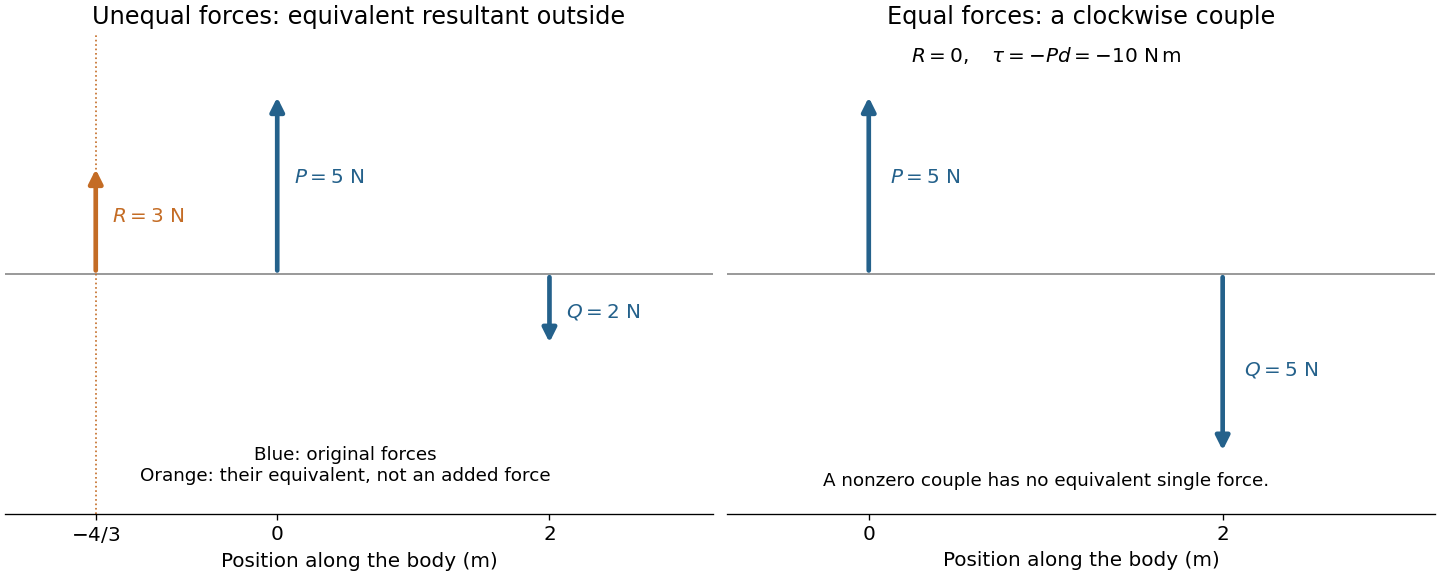

The eye is rotated by six extraocular muscles. The rectus muscles approach it from behind; the oblique muscles have oblique lines of pull, with the superior oblique muscle tendon redirected by an orbital pulley. Attachments on the sclera give lever arms about the rotation centre. In the straight-ahead position their actions are as follows; vertical and torsional contributions vary with gaze direction.[ref]
Neuroscience, 2nd edition edited by D. Purves and others. Chapter 20, “The Actions and Innervation of Extraocular Muscles”: antagonistic pairs, axes and eye-position dependence
| Extraocular muscle | Actions | Innervation |
|---|---|---|
| Medial rectus muscle | Turns the eye inward | Oculomotor nerve, III |
| Lateral rectus muscle | Turns the eye outward | Abducens nerve, VI |
| Superior rectus muscle | Elevates; also turns inward and rotates the upper pole inward | Oculomotor nerve, III |
| Inferior rectus muscle | Depresses; also turns inward and rotates the upper pole outward | Oculomotor nerve, III |
| Superior oblique muscle | Rotates the upper pole inward; also depresses and turns outward | Trochlear nerve, IV |
| Inferior oblique muscle | Rotates the upper pole outward; also elevates and turns outward | Oculomotor nerve, III |
Coordinated contraction selects the desired rotation and balances unwanted components. This illustrates torque control; the full ocular geometry is not simply the parallel-force pair of the first calculation.
Original question.
When a force produces motion in a body, how is the work done expressed in terms of the force and the circumstances of the motion ? Explain generally the appropriateness of this expression.
For constant force and displacement , the work isFor a variable force, . The work-energy theorem explains this choice: Newton's second law for fixed mass gives . The component along the motion changes kinetic energy; a perpendicular force can change direction without doing work. Exerting a force on a stationary object likewise does no work on it.
Walking on level ground still requires muscular work. The centre of mass rises and falls during each step; the limbs gain and lose kinetic energy; and muscles perform positive work that replaces energy dissipated in impacts and tissue motion. Air resistance can add another cost. Returning to the same height makes the net change of gravitational potential energy zero, not every intermediate work contribution.
At ideal no-slip contact with an immovable road, the contact point is instantaneously stationary, so the road's force does no work through that point. The muscles convert internal chemical energy into motion. Zero net gain of height is compatible with positive muscular work and energy expenditure.
Original question.
If a body falls in vacuo, express the velocity, and the distance of the body from the starting point, at any instant in terms of the time which has elapsed since motion commenced.
Account for the comparatively small velocity of a drop of rain when it reaches the earth, and state what is the effective force upon the drop at that time.
Take downward displacement as positive and gravitational acceleration as constant. In a vacuum, , so with initial downward velocity ,For release from rest these become and .
A raindrop encounters an increasing upward drag force as its speed grows. At terminal velocity, gravitational weight is balanced by drag force and the small buoyancy contribution:The drop remains in motion: zero acceleration does not imply zero velocity. In a quadratic drag force model, , giving . That formula assumes a fixed effective area and drag coefficient; sufficiently small drops or deforming large drops need an appropriate model. The question's “effective force” at settled terminal velocity is the approximately zero resultant force, not its nonzero gravitational weight.
Original question.
Describe some experiment illustrating the property, possessed by a fluid, of transmitting pressure applied to it equally in all directions.
Should the pulse occur simultaneously in all parts of the body ? What is the result of experiment on this point ?
Pascal's law concerns a pressure increment imposed on a confined fluid at rest. A water-filled vessel with small openings in differently oriented walls and a close-fitting piston illustrates it: pressing the piston produces jets in different directions, showing that pressure acts normally to each local surface. Compare ports at the same height to remove differences of hydrostatic pressure. Pressure gauges at equally high ports show the same imposed increment after the disturbance settles.
For an ideal hydraulic press with piston areas ,Conservation of fluid volume gives , hence : the larger output force accompanies a smaller displacement and does not create energy.
The arterial pulse is not simultaneous throughout the body. Pascal's law is an equilibrium statement, not instantaneous transmission. An arterial pulse travels through coupled blood and elastic arterial walls. Synchronized recordings at two sites show different arrival times of corresponding waveform features. For effective propagation distance , pulse wave velocity is . It differs from the transport velocity of individual blood cells.[ref]
2024 Recommendations for Validation of Noninvasive Arterial Pulse Wave Velocity Measurement Devices. Transit-time estimation from pressure waveforms; diastolic-foot detection and temporal resolution
A bench demonstration uses an elastic liquid-filled tube, two pressure sensors at known positions, and a short pressure pulse at one end. The delayed second recording tests finite pressure wave propagation independently of the eventual static pressure balance.
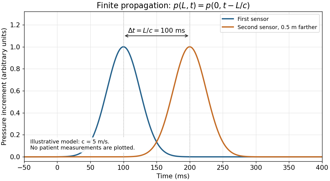
An illustrative pressure pulse reaches the second sensor later; these curves are a model rather than patient data
. Details. 
Original question.
Assuming the lungs to contain 1600 cubic centimetres of air at the termination of an expiration, and 400 cubic centimetres to be introduced by each inspiration, if, after an expiration a vertical tube be placed in the mouth, with its lower end dipping in mercury, and, the nose being stopped, the act of inspiration be performed, to what height will the mercury rise in the tube ? The height of the mercury in the barometer being 760 m.m., and the air in the tube being neglected.
Boyle's law states that a fixed amount of gas at constant temperature obeyswhere is absolute pressure, not gauge pressure. It is an ideal-gas approximation, not an assertion that every rapid compression is isothermal.
The respiratory diaphragm contracts and lowers its dome, increasing thoracic volume. Mechanical coupling through the pleural space lets the lungs expand; alveolar pressure falls below atmospheric pressure, drawing air in. Relaxation of the respiratory diaphragm and elastic recoil drive quiet expiration. These respiratory mechanics describe ventilation, distinct from chemical cellular respiration.[ref]
How the Lungs Work: What Breathing Does for the Body by the National Heart, Lung, and Blood Institute. “Breathing in” and “Breathing out”: diaphragm movement, expansion and recoil
For the stipulated sealed, isothermal gas, initial and final volumes are and . Expressing absolute pressure as a mercury-column height,The mercury column is supported by the atmospheric-to-internal pressure difference:The specified volume change belongs to the historical idealized model. Real tube-air volume, temperature changes and limits on respiratory-muscle pressure generation would affect a physical apparatus.
Original question.
Describe briefly the apparatus in the human ear which, it seems probable, is employed in producing the sensation of quality in musical sounds.
Equal pitch need not mean equal frequency spectrum. For an ideal periodic sound wave of fundamental frequency , a Fourier series has the formChanging relative amplitudes changes the waveform and generally its timbre without changing its fundamental period. Attack, decay and the time evolution of the frequency spectrum also matter for real musical notes.
The middle ear transfers vibration into the cochlea. The basilar membrane supports a frequency-selective travelling wave: higher-frequency responses occur nearer the base and lower-frequency responses nearer the apex. Hair cells in the organ of Corti transduce mechanical stimulation into signals transmitted through the auditory nerve. Different spectral and temporal patterns therefore produce different sensory-activity patterns.[ref]
This supplies a physical basis for timbre discrimination. It does not make the cochlea a set of independent strings, nor does excitation position alone explain every perceptual distinction: the cochlear amplifier and temporal neural coding also contribute.
Original question.
Describe the way in which an image of an object is. formed by a convex lens, and state the relation between the distances of the object and image from the lens.
In what way is the eye enabled to produce, upon the retina, distinct images of objects at different distances from it ?
In the paraxial thin lens approximation, let a converging lens have focal length , real-object distance , and signed image distance , positive beyond the lens. ThenA ray parallel to the axis emerges through the far focus; a ray through the optical centre is approximately undeviated. Their intersection gives a real, inverted image for : reduced for , equal-sized at , and enlarged for . At the emerging rays are parallel. For their backward extensions meet on the object side: and the image is virtual and upright.
The retina remains at an approximately fixed distance from the refracting apparatus. Ocular accommodation changes the optical power. A nearer object increases , so a fixed retinal image distance requires larger .
For near vision, the ciliary muscle contracts and reduces tension in the zonular fibres. The elastic crystalline lens becomes more rounded, increasing its optical power. For distant vision the ciliary muscle relaxes, zonular tension flattens the crystalline lens, and less power is needed. The eye's several refracting surfaces make the single thin lens a qualitative model rather than its full optical prescription.
Original question.
Describe the common thermometer, and the way in which it is graduated.
To what change is a thermometer liable, which produces errors in the indications of the instrument ?
A liquid-in-glass thermometer has a bulb connected to a narrow graduated capillary. Thermal expansion of its liquid relative to the glass raises or lowers the column. A simple linear model iswith capillary area and volumetric expansion coefficients .
For the historical Celsius graduation, establish an ice point in an equilibrium mixture of pure ice and water, and a steam point in saturated steam at standard atmospheric pressure, here a barometer height of . Mark and , then divide the interval into one hundred equal parts. This is a historical two-fixed-point instrument calibration, not the modern definition of the kelvin or of Celsius temperature. Nonuniform capillary area and nonlinear expansion limit exactness.
Bulb contraction raises the column and makes the reading too high; retained expansion makes it too low. Glass volume changes with age and thermal history; heating can cause temporary zero-point depression followed by slow recovery. Rechecking the ice point detects a shifted zero, although it does not diagnose every scale error.[ref]
Original question.
If 100 grammes of iron be boiled for some time in water, when the height of the barometer is 760 m.m., and be then quickly transferred to a vessel containing 150 grammes of water at C, what will be the common temperature of the iron and water ? It being assumed that no heat is lost, that a gramme of hot water adheres to the iron when it is transferred to the cold water, and that the specific heat of iron is 0.114.
Specific heat capacity is heat capacity per unit mass. For a specified thermodynamic process without a phase transition,The constraint matters: constant-pressure and constant-volume heat capacities need not coincide. Here the supplied iron value and water value are treated as constants.
At the stipulated barometer reading, iron left in boiling water reaches . Taking water's specific heat capacity as one in the given units, the iron has heat capacity . The adhering gramme of hot water adds another one. Thus the hot material has total heat capacity , compared with for the initially cool water.
The no-loss calorimetry energy balance issoThe adhering water enters on the hot side, not as extra water initially at . The vessel's heat capacity is neglected, as none is supplied.
Original question.
Explain how a person may be knocked down in a thunderstorm without being actually struck by lightning.
A direct lightning strike is unnecessary. A nearby strike can produce a ground current, putting the feet at different electric potentials and driving current through the body by a step voltage. A side flash from a struck object or conduction along a contacted conductor also provides an indirect current path.[ref]
Lightning Science: Five Ways Lightning Strikes People by the National Weather Service. “Ground Current”, “Side Flash” and “Conduction”: indirect electrical exposure
Rapid air heating by a nearby lightning discharge also produces an expanding pressure disturbance. Its blast can impart a mechanical impulse, causing a fall or blunt injury without current passing through the person.[ref]
The absence of a direct strike therefore does not distinguish electrical incapacity from mechanical displacement. Observations would be needed to identify the mechanism in a particular event.
Original question.
If round an electromagnet, a quantity of insulated wire be coiled, in the same way as the coil of the magnet itself, and the ends of this wire be connected with the terminals of a multiplying galvanometer, describe what takes place, when a current is sent round the coil of the magnet, and when this current is stopped.
The coils form a mutual induction apparatus. Changing primary electric current changes secondary magnetic flux linkage . Faraday's law of induction givesOn closing the primary circuit, increasing electric current produces a transient secondary electric current and galvanometer deflection. By Lenz's law its direction opposes the increasing magnetic flux. Once primary electric current and core magnetization are steady, the derivative vanishes: the galvanometer returns to zero despite the continuing magnetic field.
On opening the circuit, the decreasing magnetic flux induces a secondary electric current tending to sustain that flux. The galvanometer deflects oppositely, then returns to zero. Making and breaking produce opposite transient deflections; steady primary current produces no sustained induced current.
For fixed linear coils, and , with mutual inductance . Opposite equal changes of primary electric current give opposite equal areas under the induced-electromotive force curves in that model. Peak sizes can differ with switching times. Real iron-core magnetic hysteresis and remanent magnetization can also change the flux excursion, so equality of the actual peaks is not required.
Original volume: Examination papers for the Degree of Bachelor of Medicine by the University of Oxford (1864–1867). Open the original paper in the scanned volume, PDF page 81.
Original questions are transcribed from the public-domain edition. Credit: Wellcome Collection; original supplied by Royal College of Physicians, London. Source and public-domain rights statement.
Original question.
TODO: The identities of the chemical substances in A, B and C remain undetermined. The scanned paper supplies the tube labels but no samples, compositions or observations. Each tube therefore requires its own missing experimental evidence; the labels alone do not identify any of the three chemical substances.
Original question.
TODO: The gas in D and the gas in E remain unidentified. Neither bottle nor any observations of its contents are available. There is no basis for assigning a particular gas to either bottle.
Original question.
TODO: Testing F for oxalic acid remains incomplete. The chemical solution and its observed test results are unavailable, so neither the presence nor the absence of oxalic acid can be reported.
TODO: Testing G for antimony or arsenic remains incomplete. The chemical solution and its observed test results are unavailable, so neither antimony nor arsenic can be identified or excluded. The alternatives named in the paper specify what to investigate; they do not establish what G contains.
Original volume: Examination papers for the Degree of Bachelor of Medicine by the University of Oxford (1864–1867). The paper occupies PDF volume pages 101–102 (Oxford, December 1867, No. VII.). Open the original paper in the scanned volume.
Original questions are transcribed from the public-domain edition. Credit: Wellcome Collection; original supplied by Royal College of Physicians, London. Source and public-domain rights statement.
Original question.
Translate :—
Celeres enim, vel acutae passiones, etiam sponte solvuntur, et nunc fortunâ nunc naturâ favente. Quo fit ut ignari homines elati saepe medicos fugiant, et hos proventus incantationibus novis ac ligamentis adscribant, aut fortunae; pelluntur enim (celeres passiones) aut fluore sudoris, aut sanguinis per nares, aut ventris. Chroniae autem, vel tardae passionis morbi, qui jam praejudicio quodam corpora possederint, solius medici peritiam poscunt, cum neque naturâ, neque fortunâ solvantur.
e Caelii Aureliani Morb. Chron. Praefatione.
Translation. For rapidly developing, or acute diseases, also resolve of their own accord, sometimes through favourable chance and sometimes through nature's favour. Consequently, ignorant people, carried away by this, often avoid physicians and attribute these favourable outcomes to new incantations and amulets, or to chance; for these rapidly developing diseases are driven out either by a flow of sweat, by blood discharged through the nose, or by a discharge from the intestines. But chronic diseases, or diseases of slow development, which have already gained possession of the body with a kind of prior claim, call for the skill of the physician alone, since they are resolved neither by nature nor by chance.
Interpretation. Caelius Aurelianus contrasts spontaneous resolution in acute disease with the entrenched course of chronic disease. Here passiones means affections or diseases, not emotions; ligamenta means protective bindings or amulets, not anatomical ligaments; proventus means favourable outcomes. The metaphor praejudicio quodam gives the established disease something like a prior claim on the body. The final assertion is the author's defence of professional medicine, not a present-day law that every chronic disease requires effective medical treatment or that none can remit spontaneously. The account of sweating, nosebleeds and intestine discharge describes his historical explanation, not evidence that deliberately producing these discharges cures acute disease. [ref]
Examination papers for the Degree of Bachelor of Medicine by the University of Oxford (1864–1867). PDF volume p. 101, Latin extract attributed to the preface to Caelius Aurelianus's chronic diseases
Original question.
Translate :—
Κοιλίη, σπλάγχνον πεπτήριον, κάμνει τὴν πέψιν, ὁκότε διάῤῥοια τὸν ἄνθρωπον ἴσχει· καὶ ἔστιν ὑγρῆς τροφῆς ἀπέπτου ἡ διάῤῥοια· ἢν μὴ ἐπὶ σχεδίῳ τῆς αἰτίης γίγνοιτο τόδε ἐς μίαν ἢ δευτέρην ἡμέρην μούνην. Ὅλος ὁ ἄνθρωπος ἀσθενέει, ἀτροφίῃ τοῦ σκήνεος, χρονίη νοῦσος ἡ κοιλιακὴ γίγνεται, ἀτονίῃ τοῦ πέψοντος θερμοῦ, καὶ ψύξει τῆς κοιλίης· εὖτε λύεται μὲν ἐς θερμὸν ἡ τροφὴ, ἀλλ᾽ οὐκ ἐκπέσσει θερμὸν, οὐδὲ ἐς χυμὸν οἰκεῖον τρέπει, ἡμιτελέα δὲ λείπε-
[Printed page break; the preceding word is incomplete in the source.]
ἀδρανείῃ τοῦ τέλεος· ἡ δὲ ἀφεθεῖσα τῆς ἐργασίης ἐς κακὸν ἀλλοιοῦται καὶ χροιῇ, καὶ ὀδμῇ, καὶ συστάσι. Λευκὴ μὲν γὰρ καὶ ἄχολος ἡ χροιή· κάκοδμα δὲ καὶ βορβορώδεα· ὑγρὴ δὲ καὶ ἀσύστατος ἀπραγίῃ, μούνον δὲ ἀρετὴν ἴσχουσα τῆς πέψιος τὴν ἀρχήν· διὰ τόδε φυσώδεες ἐς τὴν γαστέρα ἐρυγαὶ ξυνεχέες κακώδεες.
ex Aretaei de causis et signis morborum diuturnorum Libri Sec. cap. 7ᵐᵒ.
Translation. The stomach, the internal organ responsible for digestion, performs digestion poorly whenever diarrhoea takes hold of a person; and the diarrhoea consists of liquid, undigested food. Unless this happens from a temporary cause for only one or two days, the whole person becomes weak through wasting of the body, and the coeliac affection of Aretaeus becomes a chronic disease, through weakness of the digestive heat and chilling of the stomach. Then the food is indeed broken down under the action of heat, but the heat does not digest it completely or turn it into an appropriate nutritive fluid: it leaves it half-finished (the printed word is incomplete), through failure of the completing power. The food, abandoned by the process, changes for the worse in colour, smell and consistency. Its colour is white and lacking bile; its smell is foul and dung-like; and it is liquid and without cohesion because the process has failed, retaining only the beginning of digestion as its one good quality. Hence there is gaseous, continual, foul-smelling belching in the stomach.
Text and medical meaning. The scan ends PDF volume p. 101 with λείπε- and begins p. 102 with ἀδρανείῃ. The translation marks this defective word rather than silently pretending that the missing ending is visible. The corresponding edited Greek has λείπει, “leaves”, which supplies the sense used here. The Oxford extract also separates the conditional clause from the following sentence more abruptly than that edition; the translation carries its evident qualification forward. [ref]
On the Causes and Signs of Chronic Diseases by Aretaeus of Cappadocia. II.7, opening paragraph: λείπει ἀδρανείῃ τοῦ τέλεος; the edited text also has ἀλλ᾽ and πρὸς δὲ καὶ linking the conditional construction
Aretaeus of Cappadocia uses κοιλίη for the stomach or digestive belly and σκῆνος, literally a tent or dwelling, for the body. ἀτροφίη indicates wasting or failure of nourishment; it need not denote the modern histological concept of atrophy. “Appropriate nutritive fluid” renders χυμὸν οἰκεῖον: the product that the author thinks suitable for bodily nourishment, rather than a modern chemical identification. The pale, foul, loose output is an observed feature; the explanation by weak digestive heat is the author's theory. It is not a modern account of digestion. [ref]
Examination papers for the Degree of Bachelor of Medicine by the University of Oxford (1864–1867). PDF volume pp. 101–102, Greek extract attributed to Aretaeus, Causes and Signs of Chronic Diseases II.7
The historical coeliac affection of Aretaeus cannot establish a modern diagnosis of coeliac disease. Modern coeliac disease involves an immune response to gluten that damages the small intestine; the ancient symptom description alone cannot identify that mechanism. The diagnosis should therefore not be read back into the passage as certain. [ref]
Celiac Disease by the National Institute of Diabetes and Digestive and Kidney Diseases. Definition and Facts: chronic digestive and immune disorder triggered by gluten, damaging the small intestine
Original question.
Give a brief account of the History of Blood-letting from ancient to modern times. Explain the changes of opinion in respect of it.
By 1867 the central change was from confidence in bloodletting as a general remedy to a disputed, increasingly restricted practice tested against clinical outcomes. Bloodletting includes opening a vein, local wet cupping, and the application of medicinal leeches. The history must explain both the theories that supported it and why clinical comparisons weakened that support.
Ancient explanations and qualifications. In the Hippocratic Corpus, disease could be explained through disturbances of bodily fluids. The four-fluid account underlying humorism named blood, humoral phlegm, yellow bile and black bile; restoring their proper mixture offered a rationale for evacuation. This was a historical explanatory system, not a modern description of blood composition. Nor was ancient practice uniformly indiscriminate. Aulus Cornelius Celsus describes bloodletting as a powerful intervention whose suitability depends on bodily strength and whether the condition represents excess rather than exhaustion. Galen likewise distinguishes whether evacuation is needed, whether it should be by venesection, and whether the patient can tolerate it; he discusses the site, quantity and repetition. Thus the ancient debate already included selection and restraint. [ref]
On the Nature of Man in the Hippocratic Corpus. Selection 1, The Elements of Life, paragraphs 1–2: four distinct fluids; translated primary excerpt
[ref] [ref]On Treatment by Bloodletting by Galen. §1, successive questions governing evacuation, tolerance, site and amount
Medieval continuity and the instruments. The Canon of Medicine by Ibn Sina preserves a carefully qualified doctrine of venesection, including cases where removing good blood would leave harmful fluids behind. Its reasoning belongs to humorism; its distinctions should not be mistaken for verified modern indications. In late medieval Europe, bloodletting was also organized by calendars: a surviving Augsburg calendar for 1472 specifies supposedly favourable times according to the patient and the Moon. This documents an additional astrological framework, not evidence of therapeutic efficacy. [ref]
Canon of Medicine by Ibn Sina. Book I, translation §§1019–1022, contraindications, timing and choice of vessels
[ref]Bloodletting calendar for Augsburg (1472). Princeton copy and catalogue description: days selected by lunar phases, patient age, affected part and temperament
The material distinction between general and local removal mattered. Venesection opened a vein with a sharp instrument. Wet cupping combined superficial cuts in the skin with suction under a cup; Celsus describes bronze cups in which burning material reduced the enclosed air's pressure, and horn cups from which air was sucked through a small opening. Dry cupping did not include the cuts and therefore did not itself remove blood. Medicinal leeches provided another means of local removal. These instruments made depletion visibly demonstrable, but the visible removal of blood did not establish that a disease had been removed. [ref]
New physiology did not immediately settle treatment. William Harvey's 1628 account of blood circulation undermined the older picture of blood simply being supplied and consumed in the parts. His observations of venous valves and experiments with arm ligatures showed directional movement: filling below a ligature and the behaviour of compressed veins helped demonstrate return towards the heart. Nevertheless, a circulating blood supply could still be invoked to explain relief through depletion or redistribution. Establishing blood circulation and establishing the benefit of bloodletting were different questions. [ref]
On the Motion of the Heart and Blood in Animals by William Harvey. chapters 8–9, quantitative argument for circulation; chapter 13, ligatures and venous valves
In the early nineteenth century, François-Joseph-Victor Broussais's emphasis on local irritation and inflammation encouraged extensive use of medicinal leeches. This illustrates why changing theory could increase bloodletting: a local inflammatory explanation supplied a fresh reason to remove blood near the supposed seat of disease. The accompanying claims of efficacy still needed comparison with what happened under other treatments. [ref]
Re-imagining Bleeders by Robert G. W. Kirk and Neil Pemberton. section discussing Broussais, local irritation, leeching, and the claim that leeches offered a relatively gentle intervention
Comparative evidence weakened general claims. In 1835 Pierre Charles Alexandre Louis asked whether comparable patients with pneumonia did better when first subjected to bloodletting during days 1–4 than when first bled later. He explicitly considered age and other differences. This numerical method in medicine shifted attention from selected recoveries to outcomes in defined groups. His work restricted confidence in a large, general benefit; it should not be presented as a randomized controlled trial proving that every use was ineffective. In modern terms, these were observational studies: confounding by indication could make the earlier-treated group differ in severity, and surviving long enough to enter the later-treated group could introduce selection bias. Counting cases was an advance, but did not automatically remove these problems. [ref]
Research on the Effects of Bloodletting by Pierre Charles Alexandre Louis. 1835, pp. 70–76; primary passages reproduced by the James Lind Library on timing, age and comparisons between groups
The dispute still alive in 1867. William Pulteney Alison defended earlier experience of benefit, while John Hughes Bennett opposed routine depletion and developed a restorative approach. One defence of the altered practice was the change-of-type theory of disease: formerly vigorous or “sthenic” inflammatory illness was said to have become weak or “asthenic”, so that measures once borne well were now inappropriate. The rival explanation was that understanding of pathology and the evidence about treatment had improved. Alison's 1856 argument and the contemporary 1858 review show that this was an active disagreement, not a completed unanimous rejection. [ref]
Reflections on the Results of Experience as to the Symptoms of Internal Inflammations by William Pulteney Alison. 1856, pp. 769–788, opening response to Bennett and defence of previous clinical experience
[ref]The Bloodletting Controversy (1858 review). opening pp. 1–3, contrasting altered disease type with altered pathological understanding
Bennett's 1864 discussion made the theoretical contrast explicit: an account centred on altered blood and vessels favoured depletion, whereas his account of tissue irritation and inflammatory exudation led him towards the opposite practice. That historical account of inflammation should itself be evaluated historically, rather than adopted as current pathology. The durable methodological lesson is that recovery after treatment is insufficient evidence of recovery because of it: acute disease may resolve naturally, as even the Latin extract acknowledges. Changes in theory, recognition of the patient's limited reserve, and comparative clinical observation together explain the retreat from routine bloodletting. [ref]
Treatment of Inflammation by John Hughes Bennett. 1864, p. 142, opening comparison of vascular and extravascular accounts of inflammation and their therapeutic consequences
Present-day distinction. Contemporary venesection has specific indications: in haemochromatosis, removal of red blood cells helps reduce excess iron; in polycythaemia vera, it can reduce an excessive red blood cell burden. Those uses concern defined abnormalities, rather than correction of historical humorism or a general remedy for fever and inflammation. They are a modern explanatory coda, not knowledge attributed to the 1867 candidate. [ref]
Haemochromatosis treatment by the NHS. Venesection: removal of iron-containing red blood cells and use of iron to replace them
[ref]Erythrocytosis by the NHS. Treatment, Venesection: use in polycythaemia vera and selected other circumstances
Original volume: Examination papers for the Degree of Bachelor of Medicine by the University of Oxford (1864–1867). Open the original paper in the scanned volume.
Original questions are transcribed from the public-domain edition. Credit: Wellcome Collection; original supplied by Royal College of Physicians, London. Source and public-domain rights statement.
Original question.
Write a Report on the cases of [blank in original], and of [blank in original], in [blank in original] Ward, with your Diagnosis, Prognosis, and the Treatment you would propose.
TODO: No clinical case records for the unspecified ward patients. The required physical examination inputs are not supplied in the scanned paper.
Original question.
Describe the Surgical Instruments placed in the Clinical Room, and their uses.
TODO: The displayed surgical instruments are not identified or illustrated. The required physical examination inputs are not supplied in the scanned paper.
Original question.
In the case of Aphonia, in the same room, are the vocal Chords diseased? How have you ascertained their condition?
TODO: No examination findings for the aphonia patient. The required physical examination inputs are not supplied in the scanned paper.
Articles were limited to the first 100 out of 183 total.
Articles by others on the same topic
There are currently no matching articles.